Galectin-3 possible involvement in antipsychotic-induced metabolic changes of schizophrenia:A minireview
2021-11-25MilicaBorovcaninKatarinaVesicMilenaJovanovicNatasaMijailovic
Milica M Borovcanin, Katarina Vesic, Milena Jovanovic, Natasa R Mijailovic
Milica M Borovcanin, Department of Psychiatry, University of Kragujevac, Faculty of Medical Sciences, Kragujevac 34000, Sumadija, Serbia
Katarina Vesic, Department of Neurology, University of Kragujevac, Faculty of Medical Sciences, Kragujevac 34000, Sumadija, Serbia
Milena Jovanovic, PhD Studies, University of Kragujevac, Faculty of Medical Sciences,Kragujevac 34000, Sumadija, Serbia
Milena Jovanovic, Clinic for Nephrology and Dialysis, University Clinical Center Kragujevac,Kragujevac 34000, Sumadija, Serbia
Natasa R Mijailovic, Department of Pharmacy, University of Kragujevac, Faculty of Medical Sciences, Kragujevac 34000, Sumadija, Serbia
Abstract Recently, specific immunometabolic profiles have been postulated in patients with schizophrenia, even before full-blown disease and independent of antipsychotic treatment. Proteomic profiling studies offer a promising potential for elucidating the cellular and molecular pathways that may be involved in the onset and progression of schizophrenia symptoms, and co-occurrent metabolic changes. In view of all this, we were intrigued to explore galectin-3 (Gal-3) as a glycan, and in our previous study, we measured its elevated levels in remission of schizophrenia. The finding may be a consequence of antipsychotic treatment and may have an impact on the onset of inflammation, the development of obesity,and the presumed cognitive changes in schizophrenia. In the animal study, it was shown that downregulation of Gal-3 was beneficial in insulin regulation of obesity and cognitive preservation. Strategies involving plasma exchange are discussed in this review, particularly in the context of Gal-3 elimination.
Key Words: Galectin-3; Schizophrenia; Metabolic syndrome; Insulin resistance; Cognition;Antipsychotics
INTRODUCTION
Clinical practice raises many questions regarding somatic states that accompany or are a consequence of mental illnesses. As schizophrenia is an extremely complex and debilitating mental disorder, overall treatment must take into account the somatic comorbidity of the patients. Although schizophrenia requires special attention and care in terms of lifestyle and antipsychotic treatment, a particular immunometabolic profile has recently been postulated, even before the disease onset[1]. The use of atypical antipsychotics is often associated with undesired metabolic and endocrine side effects including obesity, dyslipidemia, hyperglycemia, and insulin resistance[2].To summarize, patients with schizophrenia most probably could have other comorbidities, regardless of their specific immunometabolic profile and antipsychotic therapy, and the somatic states may also lead to metabolic changes.
The identification of defects in cell biology and molecular phenotype underlying schizophrenia represents a challenging new approach to the study of this complex neurodegenerative disorder. Proteomic profiling studies, in which many proteins are tested for their relevance to the disease, are still in their infancy but the potential for elucidating the cellular and molecular pathways that may be involved in the onset and progression of schizophrenia is promising[3].
Altered protein post translational modifications such as glycosylation have become a new target of investigation in the pathophysiology of schizophrenia[4]. Glycosylation is an enzyme-mediated process in which a carbohydrate or carbohydrate structure, also referred to as a glycan, binds to a protein, lipid, or glycan substrate.Glycosylation is the most common and complex post translational modification and plays a critical role in protein-protein, protein-cell, and cell-cell interactions, including antibody binding, protein degradation, cellular endocytosis, and protease protection[5]. This process regulates nearly all cellular activities and has a critical role in the development and functioning of the central nervous system (CNS). Glycans are involved in many processes, such as neurite outgrowth and fasciculation, synapse formation and stabilization, modulation of synaptic efficacy, neurotransmission, and synaptic plasticity[6]. Altered glycosylation can significantly affect the properties of the glycosylated substrate, resulting in changes in its structure, localization, expression levels, molecular interactions, and/or substrate function.
Aberrant glycosylation has been identified in the serum, cerebrospinal fluid, urine,and postmortem brain tissue of schizophrenia patients[7]. Early evidence of glycosylation abnormalities in schizophrenia reported reduced glycoprotein expression in urine samples from male schizophrenia patients, and was consistent with abnormal glycan composition[8]. Altered monosaccharide composition of attached glycans was also found in the blood serum of the patients[9]. An increased serum glycoprotein level was also confirmed in young schizophrenia patients 13-17 years of age[10].
Abnormalities of N-linked glycosylation in schizophrenia have been observed in neurotransmitter receptor and transporter subunits, subunits from α-amino-3-hydroxy-5-methyl-4-isoxazole propionic acid, kainate, and gamma-aminobutyric acid(GABA)Areceptor families in various brain regions, including the dorsolateral prefrontal cortex, anterior cingulate cortex, and superior temporal gyrus[11-14].Receptors containing abnormally N-glycosylated subunits have also been shown to exhibit abnormal subcellular distribution in schizophrenia, suggesting cellular consequences of abnormal protein glycosylation[15]. Widespread glycosylation abnormalities due to abnormal glycosylation enzyme expression have also been reported in schizophrenia[16-18].
We have recently elaborated on the contrasting roles of the galectin-3 (Gal-3)through the schizophrenia continuance[19]. We also discussed the various somatic states co-occurring in schizophrenia that could be related to Gal-3. In this review, our interdisciplinary team seeks to further elucidate the mechanisms underlying the impact of glycans on early development, and how Gal-3 may further influence subsequent metabolic changes. However, our focus will be on the interplay of Gal-3 with antipsychotics during the course of the disease in an attempt to elucidate specific non-CNS systemic changes. Overall, that may lead to conclusions that allow more selective therapy of schizophrenia in the future.
GAL-3 AND NEURO-IMMUNO-METABOLIC CROSSTALK
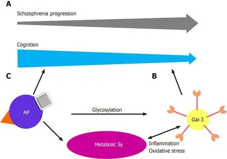
In recent years, an increasing body of evidence has highlighted the involvement of Gal-3 in neurodevelopment and neurodegenerative diseases[20]. Scientific advances during the last decade have led to the discovery that Gal-3 plays a significant role in normal murine brain development, neuroblast migration, oligodendrocyte differentiation, and basal gliogenesis[21-24]. Chronic inflammation, mitochondrial damage and oxidative stress are factors common to neurodegenerative and metabolic diseases,in which sustained responses to inflammation contribute to neurodegeneration and progression of the disease[24,25]. Glial cell dysregulation is the main characteristic of chronic inflammation in neurodegenerative diseases, leading to changes in glycan expression in brain cells[26,27]. Previous studies have shown that inflammatory stimuli upregulate Gal-3 expression in activated microglia, and conversely, Gal-3 has been proposed as a modulator of the inflammatory response through microglial activation, cell adhesion, and cytokine release[28-32]. Recently, Gal-3 was shown to regulate microglial response to promote remyelination[23]. All this leads to the conclusion that Gal-3 is a key player in control of the switch between protective and disruptive microglial effects. In multiple sclerosis, Gal-3 expression is increased in periventricular inflammatory lesions[33]. Nishiharaet al[34] investigated whether anti-Gal-3 antibodies might be a novel diagnostic marker and a possible therapeutic target in patients with secondary, progressive multiple sclerosis. Gal-3 deficiency reduces inflammation and disease severity in experimental autoimmune encephalomyelitis,Alzheimer’s, and Parkinson’s disease[35-37]. We reported elevated levels of Gal-3 in the stable phase of schizophrenia, with the suggestion that this glycan has a proinflammatory effect in the later phase[19] (Figure 1A). All the data indicate that Gal-3 might be a potential biomarker and therapeutic agent in this cohort of neurodegenerative disorders. Gal-3 is not only found in the cells themselves but is also secreted into the extracellular space in kidneys and heart, suggesting its multiple functions[38]. In addition to cell proliferation and differentiation, it promotes oxidative stress and proinflammatory processes and plays an important role in angiotensin II and aldosterone-induced myocardial and kidney fibrosis[39,40]. Studies have shown that elevated levels of Gal-3 are predictors of coronary disease in diabetes mellitus type 2[41]. Gal-3 levels are elevated in maintenance hemodialysis patients, and can be used as a biomarker of vascular calcification, left ventricular hypertrophy, and left ventricular diastolic dysfunction[42-44].
Gal-3 has recently been recognized as an important modulator of biological functions and an emerging participant in the pathogenesis of immune/inflammatory and metabolic disorders[45-47] (Figure 1B). Gal-3 serum levels are elevated in women with polycystic ovary syndrome, especially those with insulin resistance, and those with increased insulin and glucose levels in the glucose tolerance test and it is considered a potential biomarker in prediabetes and diabetes[48-50]. The role of Gal-3 in metabolic disorders and the mechanism by which this lectin modulates excess fat mass, adipose tissue, systemic inflammation, and the associated impairment of glucose regulation, remains to be elucidated. Gal-3 is produced by many cell types, including adipocytes, and increased levels have been confirmed in obese patients[51,52]. Gal-3 is upregulated in growing adipose tissue and during inflammation[53,54]. Gal-3 is an important chemotactic factor for tissue macrophages in adipose tissue[55]. However,the role of Gal-3 in adipose tissue remains disputable because it exerts both deleterious and protective effects. In the general population, levels of circulating Gal-3 correlate positively with age, the prevalence of obesity, diabetes, hypercholesterolemia, and hypertension, markers of inflammation, and target organ damage, indicating a clear association of Gal-3 with metabolic disorders and associated risk factors and complications[50,52,56,57]. Seemingly contradictory results were reported by Ohkuraet al[58],who demonstrated that Gal-3 affected the concentration of insulin more than that of glucose, and that the increase of Gal-3 activity in diabetic patients had a protective effect on insulin resistance.

Figure 1 Galectin-3 and neuro-immuno-metabolic crosstalk.
Obesity may influence not only behavior, cognition, and mood, but also adipose tissue dysfunction and inflammation, trigger impairment of insulin signaling,compromise the storage of triglycerides, and contribute to insulin resistance with high levels of free fatty acids[59]. Moreover, all the processes associated with insulin resistance and chronic hyperglycemia induce oxidative stress and inflammatory responses that lead to neuronal death, cognitive impairment, and neurodegeneration.
Hippocampal insulin resistance is the key factor in cognitive deficits. In an animal model study, insulin signaling in the hippocampus was shown to be affected by a cascade in which obesity induced chronic inflammation and chronic inflammation had role in obesity-related insulin resistance[60]. Moreover, chronic inflammation is suppressed by Gal-3, so Gal-3 directly impacts insulin signaling and might be a targetable link between inflammation and insulin sensitivity. Qinet al[60] suggested that the development of cognitive deficits in obese people could be inhibited through Gal-3 decrement.
Obesity is reported in approximately 50% of patients, metabolic syndrome in up to 40%, glucose intolerance in up to 25%, and diabetes in up to 15% of patients with schizophrenia[61].The increased prevalence of these conditions is multifactorial.Antipsychotics can cause weight gain, glucose intolerance, and other metabolic complications[62] (Figure 1C). A recent meta-analysis of metabolic parameters in patients with first-episode psychosis, which can be described as early schizophrenia,showed increased insulin resistance and impaired glucose tolerance in the patients compared with healthy, matched controls,implying that schizophrenia might share intrinsic inflammatory disease pathways with type 2 diabetes[63]. We have previously discussed our findings of the possibly protective properties of Gal-3 in type-2 diabetes,but triggering metabolic changes and myocardial fibrosis[19].
GAL-3 AND ANTIPSYCHOTIC TREATMENT IN SCHIZOPHRENIA
Relatively few studies have investigated the effects of antipsychotic treatment on the serum glycosylation profiles in schizophrenia patients. Reports examining glycan expression in schizophrenia patients showed that the glycan profile in serum and cerebrospinal fluid of first onset, unmedicated schizophrenia patients differs from the profile of healthy controls[64]. The results showed that some types of sialylated Nglycans derived from low-abundance serum proteins are significantly increased in patients with schizophrenia compared with controls. The study found a two-fold increase in serum glycan levels in male schizophrenia patients, with gender-specific differences also apparent[65]. Glycemic differences have also been reported in patients with acute paranoid schizophrenia before and after 6 wk of treatment with olanzapine,an atypical antipsychotic medication[65]. Olanzapine administration increased galactosylation and sialylation of serum N-glycans, suggesting increased activity of specific galactosyltransferases and increased availability of galactose residues for sialylation. The results indicate that the glycosylation profile of serum proteins can be used to monitor patients with schizophrenia after treatment. Given the confirmed effects of olanzapine on hepatic enzymes, it is possible that the reported changes in glycosylation induced by olanzapine treatment may occur because of the altered activity of hepatic glycosylation-processing enzymes[66].
As schizophrenia may have an evolving, progressive pathology, Narayanet al[67]focused on changes in gene expression and molecular pathways throughout illness progression. They assessed the alterations in patients treated with the typical antipsychotic medication, chlorpromazine, at early (≤ 4 years), intermediate (7-18 years), and late (≥ 28 years) stages of schizophrenia. The results showed that biopolymer glycosylation, protein amino acid glycosylation, and glycoprotein biosynthesis were increased in intermediate-stage patients. Analysis of differences in gene expression revealed that carbohydrate metabolism was dominant in short-term illness,whereas lipid metabolism prevailed in intermediate-term illness. Overall, short-term illness was particularly associated with disruptions in gene expression, metal ion binding, ribonucleic acid processing, and vesicle-mediated transport. Considerably different from short-term illness, long-term illness was associated with inflammation,glycosylation, apoptosis, and immune dysfunction.
A postmortem study compared the effects of atypical (olanzapine and risperidone)vstypical antipsychotics (chlorpromazine and haloperidol) on the livers, various genes, and molecular functions of patients[68]. The results demonstrated that typical antipsychotics affected genes associated with nuclear protein, stress responses, and phosphorylation, whereas atypical antipsychotics increased gene expression associated with Golgi/endoplasmic reticulum, and cytoplasmic transport, suggesting that atypical antipsychotics affect post translational modifications. The study showed that olanzapine treatment increased the expression of theB4GALT1gene in the liver of schizophrenia patients. That gene encodes β1,4-galactosyltransferase I (Gal-T1).Increased expression and activity of the enzyme lead to increased galactosylation of GlcNAc residues in glycans, which is consistent with the results of a study performed by Telfordet al[65]. Genes associated with lipid metabolism were consistently downregulated in the typical compared with the atypical antipsychotic group.
However, dysregulation of adipose tissue homeostasis appears to be a critical factor[69]. An untargeted proteomic analysis of the effect of antipsychotics on adipose tissue was performed in a rat schizophrenia-like methylazoxymethanol acetate model[70].Chronic, 8-wk-long application of three antipsychotics was characterized by differences in the likelihood of inducing metabolic alterations. Olanzapine,risperidone, and haloperidol, caused alterations in protein N-linked glycosylation in adipose tissue, providing further evidence that dysregulated glycosylation in schizophrenia may also be caused to some extent by antipsychotic treatment. Drug-specific effects included upregulation of insulin resistance (olanzapine), upregulation of fatty acid metabolism (risperidone), and upregulation of nucleic acid metabolism(haloperidol). Individual metabolic characteristics might also predispose to a different likelihood of becoming obese after antipsychotic treatment. Gal-3 has been shown to be associated with the onset of schizophrenia, and its elevation could have consequent deleterious effects (Figure 1). In addition, it must be taken into account that our patients were treated with risperidone or paliperidone, which are antipsychotics that may upregulate fatty acid metabolism and have Gal-3-elevating properties[71].
CONCLUSION
In this context, it is necessary and urgent to develop more selective treatment strategies. The phase of the illness also needs to be considered, with a focus on early interventions. The possibility that schizophrenia is secondary to a circulating, large molecular-weight substance has been explored with variable success. However, a double-blind evaluation of plasmapheresis in ten patients with schizophrenia yielded negative results, and the procedure did not lead to a reduction in psychosis[72]. As hypercholesterolemia has been treated with plasmapheresis, and recently the therapeutic usefulness of Gal-3 depletion apheresis has been demonstrated in inflammation-mediated disease, targeting Gal-3 molecule may be a useful way to address immunometabolic problems and cognitive deterioration in schizophrenia in the future[73,74].
The question is whether extrapolations of preclinical and research data are applicable in clinical practice. Gal-3 relevance could be very interesting in further exploration of the genesis of schizophrenia in parallel with the metabolic alterations of the patients. It might be useful for clinicians to become familiar with this molecule and its precise roles in each phase of the disease in order to improve cognition and reestablishing metabolic balance in schizophrenia.
ACKNOWLEDGEMENTS
This review was enriched in valuable interactions by the Center for Molecular Medicine and Stem Cell Research, at the Faculty of Medical Sciences, University of Kragujevac, Kragujevac, Serbia. We would like to thank Bojana Mircetic for language editing.
杂志排行
World Journal of Diabetes的其它文章
- Non-alcoholic fatty liver disease, diabetes medications and blood pressure
- Diabetes patients with comorbidities had unfavorable outcomes following COVID-19:A retrospective study
- Utility of oral glucose tolerance test in predicting type 2 diabetes following gestational diabetes:Towards personalized care
- Diabetic kidney disease:Are the reported associations with singlenucleotide polymorphisms disease-specific?
- Metabolic and inflammatory functions of cannabinoid receptor type 1 are differentially modulated by adiponectin
- Medication adherence and quality of life among type-2 diabetes mellitus patients in India
