多色彩可视化半定量检测方法用于COVID-19患者治疗过程中抗体浓度变化的快速监测
2021-11-15陈仲辉李金秋俞柳敏涂海健蔡宗苇林振宇
陈仲辉,李金秋,林 伟,俞柳敏,涂海健,陈 宇,蔡宗苇,林振宇
(1.莆田学院附属医院,莆田351100;2.食品安全与生物分析教育部重点实验室,福州大学化学学院,福州350116;3.香港浸会大学环境与生物分析国家重点实验室,香港999077)
1 Introduction
Severe acute respiratory syndrome coronavirus 2(SARS-CoV-2),a novel coronavirus,has caused an explosion serious viral pneumonia disease,and brought serious threats to global public health in the world[1-4].During the outbreak of novel coronavirus pneumonia,serological antibody detection can be assisted to investigate the transmission in the general population and assured the COVID-19 patient timely treatment[2,5-7].Given the SARS-CoV-2 specific antibody significant functions,monitoring the concentration of antibody has been added in the“Diagnosis and Treatment Protocol for Novel Coronavirus Pneumonia of China”as one of the standard methods to diagnose COVID-19.And detecting serological SARS-CoV-2 antibody can provide an important assay for diagnosis of COVID-19.Hence,on-site detecting of SARS-CoV-2 viral target is significant for SARS-CoV-2 infection and assessment of treatment results.
Currently,there are many types of diagnostics methods put forward for body fluid SARS-CoV-2 antibody detection,such as lateral flow immunoassay(LFIA)[8―10],chemiluminescent immunoassays(CLIA)[11,12],and enzyme-linked immunosorbent assay(ELISA)[6,13,14].On-site rapid semiquantitative detection of body fluid antibody will be an effective strategy for assessment of coronavirus infection therapeutic effect[8,15].LFIA has advantages of convenient and rapid.However,the gold nanoparticles based LFIA did not have quantification capability.And fluorescence based LFIA can be used to quantitative or semiquantitative detection of the target,but the signal should be reported by the specific device.And CLIA need sophisticated instruments as well.So,those techniques cannot meet the requirement of rapid detection on-site.In comparison with traditional immunodiagnostic methodology,ELISA colorimetric immunoassay is one of the most active strategy for biomarker detection because it can be quantified by ultraviolet-visible spectrometer(UV-Vis)or recognized by naked eye.Traditional ELISA immunoassay has been used for qualitative detection of biomarker attributed to the single-color change,but human eyes are not sensitive to the similar color for optical density variations,the mentioned immunoassay cannot quantify the concentration of target without the aid of a spectrometer.Hence,these shortages obviously confine the accuracy of naked eye visual identification and the practical usage.
Noble metal nanoparticles have attracted great interest in colorimetric bioanalytical field owing to their excellent localized surface plasmon resonance(LSPR)[16].Typically,gold nanorods(Au NRs)have two representative separate surface plasmon resonances(SPR)bands,corresponding to transverse and longitudinal peak.By simply adjusting the aspect ratio of Au NRs,various optical signals can be acquired in a wide range[17―20].Therefore,Au NRs were considered promising indicators in multicolor colorimetric immunoassay because of their independence of complicated equipment and bare-eye-detectable readout.In early studies,our group[20,21]disclosed that TMB2+came from HRP-TMB system can quantitatively and rapidly etch Au NRs to generate vivid color responses,which were coupled with the traditional HRP-H2O2-TMB ELISA system to develop multicolor immunosensors for many disease biomarkers,such as carcinoma embryonic antigen,prostate-specific antigen,glucose,and hydrogen sulfide.The design steps are simple and can be easily handled by untrained person and evaluated application in the on-site detection field.
In this study,a multicolor immunoassay for SARS-CoV-2 antibody concentration detection has been designed and applied to on-site monitoring the treatment results quickly by using naked eyes as readout.The present of antibody(IgM antibody has been used as an example in this study)will affect the concentration of TMB2+produced.Then,the generated various concentrations of TMB2+can quantitatively etch Au NRs to produce rainbow-like solutions.The color of the system relies on the concentration of the generated TMB2+and has some relationship with the amount of SARS-CoV-2 IgM antibody in the biological samples.By monitoring the change of vivid color,it can semiquantitative determine the IgM antibody in the COVID-19 patients’blood serum samples.
2 Experimental
2.1 Material and Reagents
SARS-CoV-2 IgM ELISA test kit and control solutions were purchased from Xiamen Biotime Biotechnology Co.,Ltd.(Xiamen,China).SARS-CoV-2 nucleic acid test kit was purchased from Sun Yat-sen University Daan Gene Co.,Ltd.3,3′,5,5′-Tetramethylbenzidine(TMB color reagent A solution)and peroxide solution(TMB color reagent B solution)were obtained from Shanghai Aladdin Biochemical Technology Co.,Ltd.(Shanghai,China).Ascorbic acid(AA),hydrochloric acid(HCl),cetyltrimethylammonium bromide(CTAB),sodium bromide(NaBH4)and chloroauric acid tetrahydrate(HAuCl4·4H2O)were acquired from Sinopharm Chemical Reagent Co.,Ltd.(Shanghai,China).All polystyrene 96-well ELISA plates were purchased from Sangon Biotechnology Co.,Ltd.(Shanghai,China).All reagents were of analytical-regent(A.R.)grade and directly used for the following experiments without further purification.Au NRs were prepared according to early reported literatures[20,21].Ultrapure water obtained from Direct-Q3 UV system(Millpore,18.2 MΩ·cm)was used in all experiments.All patient samples were acquired through the affiliated hospital of Putian University.The blood samples are collected from medical procedures performed at affiliated hospital of Putian University and stored at-80°C until use.
2.2 Instruments
Tecnai G2 F20 S-TWIN transmission electron microscopy(TEM,FEI Company,USA)was used to study different oxidation stages of the Au NRs.Spark UV-Vis absorption spectra were recorded by Multimode Microplate Reader(TECAN Company,Switzerland).HydroFlex ELISA microplate wells were washed by programmable automatic microplate washer(TECAN Company,Switzerland).All the photos were taken by EOS 600D digital camera(Canon Company,Japan).
2.3 Construction of the SARS-CoV-2 IgMStandard Reference Card
The procedures were followed by the manual of the SARS-CoV-2 IgM ELISA kit.The SARS-CoV-2 IgM standard reference card was prepared by a series of IgM standard solutions.Positive control solutions(100 μL)with different concentrations were added to the wells coated with recombinant SARS-CoV-2 antigen.Then,the HRP-conjugated SARS-CoV-2 antibody(100μL)was added and incubated for 40 min at 37℃.Following,the wash steps were carried out with 250μL of PBST(three times)and 0.9%normal saline(three times)using a programmable automatic well wash machine.Subsequently,TMB substrate solution was added into each well for colorimetric reaction at room temperature.The reaction was stopped by 5 mol/L HCl(20μL)solution.Finally,Au NRs were added into the solution with gently mixed for 3 min.The colors of the constructed standard reference card were recorded by the camera.

Scheme 1 Principle of the proposed multicolor immunoassay for SARS⁃CoV⁃2 antibody semi⁃quantitative visual detection
2.4 Clinical Samples Detection
All patient samples were acquired through the affiliated hospital of Putian University.These samples are collected from medical procedures performed at affiliated hospital of Putian University.The human serum was performed under the institutional guidelines and approved by the ethical committee for biomedical research of affiliated hospital of Putian University.Briefly,blood samples were centrifuged at 3000 r/min for 15 min to obtained serum samples,and then rested under UV irradiation for 30 min and dispensed in biosafety cabinet.Serum sample(100μL)was introduced into 96-well ELISA plates and the following processes were the same as construction of the SARS-CoV-2 IgM standard reference card at room temperature.For semi-quantitative evaluation of sample,the etched Au NRs solution color was recorded with digital camera and compared with the standard reference card.
3 Results and Discussion
3.1 Mechanism of the Proposed Assay
As a proof-of-concept experiment,the chromogenic mechanism for visual detection of SARS-CoV-2 IgM antibody is shown in Scheme 1.The detect strategy contains two parts:antigen antibody recognition and combination,as well as multicolor chromogenic presentation.Firstly,antigen antibody recognition and combination involve a typical ELISA reaction.The control standard substances(or detect serum)and HRP-conjugated antibody(HRP-Ab)were added into the 96-wells coated with SARS-CoV-2 antigen(S-Ag).Then,TMB liquid substrate system was added to product oxTMB(TMB+)because TMB was oxidized to TMB+by H2O2in the presence of HRP-Ab.After HCl was added,HRP can be inactivated and TMB+turns into TMB2+.TMB2+can quickly promote the process of etching reaction from the tops of Au NRs.And the LSPR of Au NRs will change and be accompanied by vivid colors changing of the solution,which can be easily recognized by naked eye.This phenomenon can be used to monitor the concentration of SARS-CoV-2 IgM antibody in the serum samples.Based on this principle,a relationship between the amount of SARS-CoV-2 IgM antibody in the serum and the vivid color change of the sensing platform can be visual semi-quantitative detected.
The feasibility of the proposed method was investigated by several control experiments.Fig.1 shows the SARS-CoV-2 IgM antibody concentration dependent colorimetric responses of the multicolor immunodiagnostic methodology.As depicted in Fig.1(A),the longitudinal plasmon band of Au NRs located atca.751 nm,and the solution color was brown.Then,IgM antibody solutions with different concentrations were added into the proposed immunoassay,resulting the longitudinal plasmon band blue-shifted and the peak intensity decreased[Fig.1(B)—(D)].The distance of peak shift depended on the SARS-CoV-2 IgM antibody concentration.The Au NRs etching processes were further confirmed by TEM.As depicted in the insets of Fig.1,the original Au NRs have an average diameter of 71.5 nm.When different concentrations of target were added,the aspect average diameter almost decreased.These results verify that different concentrations of SARS-CoV-2 IgM antibody lead to proposed immunoassay displayed by the system.Because of this rainbow-like color change can be readily distinguished by solution color,the equipment-free visual semi-quantitative monitoring of SARS-CoV-2 IgM antibody is possible.

Fig.1 UV⁃Vis spectra(A―D)and corresponding TEM images(A′―D′)of Au NRs with addition of different concentrations of SARS⁃CoV⁃2 IgMantibody
3.2 Optimization of the Experimental Variables
Several experimental conditions,such as the concentration of CTAB and HCl,HRP-Ab capture reaction time,and TMB liquid substrate system reaction time were investigated to achieve the optimum performance of the developed multicolor immunodiagnostic methodology.In this study,the LSPR peak blue shift of Au NRs in etching process(Δλ)has been selected as the indicator to assess the effects of the experimental conditions.The CTAB molecule was modified on the Au NRs surface as AuBr2−(CTA)2+complex to facilitate the Au NRs etched by TMB2+[18].TheΔλincreased with the enhancing of the CTAB concentration and arrived a plateau at CTAB concentration of 0.16 mol/L[Fig.2(A)].This phenomenon can be attributed to the decrease of AuBr2−(CTA)2+oxidation potential.Therefore,CTAB concentration of 0.16 mol/L was chosen for the further study.HCl also plays a significant role in the Au NRs etching process because it can be used to inactive HRP and stop the ELISA reaction.As depicted in Fig.2(B),Δλincreased rapidly with the augmenting of HCl concentration,and then kept stable when HCl concentration was higher than 0.2 mol/L.So,0.2 mol/L HCl was chosen as the optimum condition.
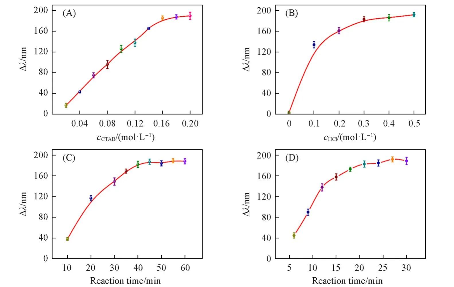
Fig.2 Effects of CTAB concentration(A),HCl concentration(B),the HRP⁃Ab capture reaction time(C)and TMB liquid substrate system reaction time(D)on the LSPR peak blue shift of Au NRs
The amount of HRP-Ab capture time and TMB liquid substrate reaction time affect the performance to the multicolor colorimetric ELISA assay greatly.As shown in Fig.2(C),Δλincreased rapidly with the increase of the HRP-Ab capture time within 10―40 min and reached a plateau after 40 min,indicating that the enzymatic effect was the most outstanding when the HRP-Ab capture time was 40 min,which means 40 min was the optimized reaction time.Therefore,TMB liquid substrate reaction time was directly involved the TMB2+generated.Fig.2(C)and(D)showed the relationship betweenΔλand reaction time.Δλincreased gradually and then reached a maximum when prolonged the reaction time to 21 min.As a result,21 min was selected as the best reaction time for the further study.
3.3 Visual Semi-quantification of SARS-CoV-2 IgMAntibody
Under the optimized conditions,using SARS-CoV-2 antibody quality control materials with different concentrations to investigate the dynamic semi-quantification determination range of the developed method.Fig.3(A)exhibits that the rainbow-like solution color changes can be observed from brown to gray,cyan,blue,violet,and pink with the SARS-CoV-2 IgM concentration increasing from 0 to 1.00,5.00,10.0,30.0,60.0,100 and 200 IU.Those solutions covered the most visible range from 500 nm to 800 nm.Therefore,semi-quantitative detection of SARS-CoV-2 IgM concentration can be realized by naked eye,which provides a basis for visual and semi-quantitative monitoring SARS-CoV-2 IgM concentration changes in the COVID-19 patient therapeutic process.
Besides,theΔλof the solutions blue shifted gradually from 750 nm to 520 nm as the SARS-CoV-2 IgM concentrations increased[Fig.3(B)].Fig.3(C)depicts that the LSPR peak shift was closely related to the SARS-CoV-2 IgM concentration in the 5—200 IU range.The correlation linear equation is Δλ=81.0lgc−0.009(R2=0.9915).The limit of detection(LOD)of this method for SARS-CoV-2 IgM concentration was calculated to be 1.29 IU using the 3σ/slope method.

Fig.3 Plasmonic method color change with SARS⁃CoV⁃2 antibody concentration increased(A),UV⁃Vis spectra change of Au NRs in the oxidation etching process with different SARS⁃CoV⁃2 antibody concentrations presented(B)and the linear relationship betweenΔλand SARS⁃CoV⁃2 antibody concentration(C)
3.4 Specificity of Multicolor Immunoassay
A significant challenge for SARA-CoV-2 IgM antibody detection is the specificity,which is of great significance in exactly analyzing target and eliminating false positives results.Hence,the specificity has been investigated by the H5N1antigen(H5N1Ag),H7N9antigen(H7N9Ag),hepatitis b surface antibody(HBsAb)and hepatitis C virus antibody(HCVAb)quality control materials.As shown in the inset of Fig.4,the change of solution color can be hardly seen with the addition of interference materials,that is,the color is almost the same with that of the blank solution.In the presence of SARS-CoV-2 IgM antibody,the color of solution changes to bluish violet.The difference in solution color can be easily distinguished with naked eye.Fig.4 reveals that an evidentΔλpeak shift was surveyed only when SARS-CoV-2 IgM antibody was added.Contrasting with other virus antibody,theΔλpeak signals were almost the same with the blank sample.Those phenomena were depicted in the histogram ofΔλpeak shift for SARS-CoV-2 IgM antibody and the other virus antibody in this multicolor colorimetric method.Therefore,other virus antigen or antibody did not occur obvious solution color changes.The repeatability of this proposed strategy was evaluated based on coefficients of variation(CVs,n=3).And experimental results show that the CVs of the same batch of Au NRs was 2.42%.These phenomena prove the developed multicolor colorimetric strategy for SARS-Cov-2 IgM antibody concentration detection exhitited a high selectivity,which can be ascribed to the high specificity of the ELISA assay used for antibody recognition.
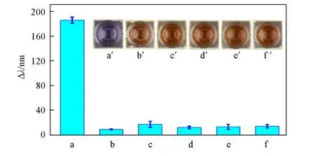
Fig.4 Selectivity of the developed multicolor assay detection of SARS⁃CoV⁃2 IgMantibody
3.5 Clinical Samples Detection
To explore the effectiveness of the method established in this work for clinical applications,clinical sensitivity was determined by healthy volunteers with negative RT-PCR results for SARS-CoV-2 and COVID-19 patient samples that were positive SARS-CoV-2 RT-PCR results.RT-PCR positive results were confirmed by the ORF1/ab and N gene detection.The detection results were summarized in Table 1.Healthy people serum samples can be easily distinguished by reddish brown color,and the normal range is 0—1.00 IU.However,the three COVID-19 patients’serum samples exhibited different colors.The result indicated that the solution colors were in agree with the RT-PCR results.Following,the SARS-CoV-2 antibody IgM concentration of a typical COVID-19 patient has been investigated with prolonging the time and the results are summarized in Table 2.With the COVID-19 patient therapeutic process progressed,the solution color changed from brown to green and blue in one month,and then turned to green and brown.The solution color’s change indicated that the developed multicolor colorimetric strategy can accurately reflect the COVID-19 patient’s serum SARSCoV-2 IgM antibody level in the treatment and rehabilitation process.The spectrophotometry results show that with treatment proceeding,the concentration of SARS-CoV-2 IgM antibody increased at first,then decreased and finally tended to be stable.And those results can be obtained both by visual semi-quantitatively inspection and spectrophotometry quantitative determination.With the assessment of the property,our strategy is beneficial to the future applications in the COVID-19 patients therapy field.In this case,the proposed multicolor colorimetric diagnostic strategy will be presumed to be a more valid and economical method to deal with the clinical diagnosis.
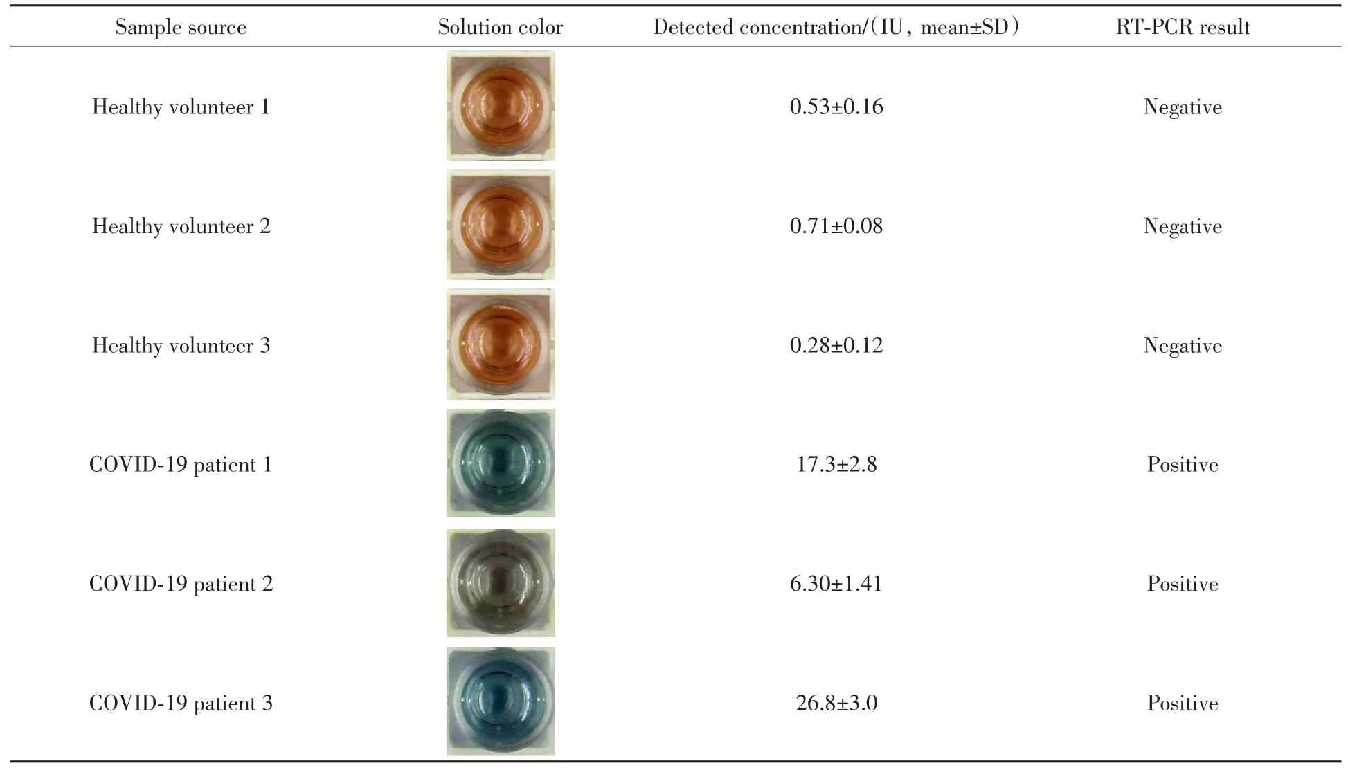
Table 1 Detection of SARS-CoV-2 IgM antibody between healthy volunteers and COVID-19 patients by the proposed method
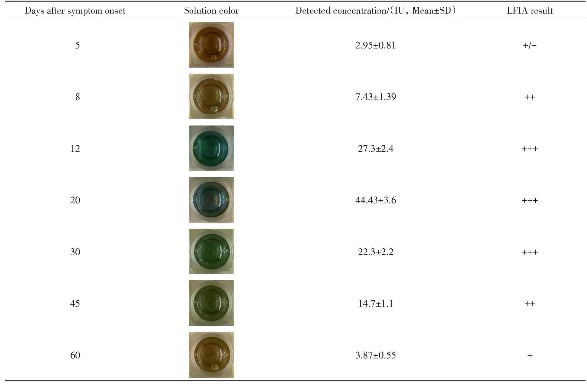
Table 2 Detection of a typical COVID-19 patient SARS-CoV-2 IgM antibody concentration change in therapeutic and convalescent processes by the proposed method
4 Conclusions
A multicolor colorimetric method for high-resolution visual SARS-Cov-2 IgM antibody detection has been designed by using the TMB2+-produced etching of Au NRs as the visual signal reporter.The developed multicolor strategy can be favorably applied to semi-quantitative detecting COVID-19 patients’serum samples by the naked eye during the etching process.Compared with the traditional ELISA immunodiagnostic methodology,the visual semi-quantitative detection of SARS-Cov-2 IgM antibody can be realized according to the vivid rainbow-like multicolor changes without sophisticated equipment.It is believed that the proposed sensitive multicolor colorimetric strategy by direct visualization may be a more efficient solution in a large human population serological surveillance study.
This work is supported by the National Key Research and Development Program of China(No.2019YFC1604701),the National Natural Science Foundation of China(Nos.21675028,21775026),the Natural Science Foundation of Fujian Province,China(No.2021J011370),the Fujian Provincial Health Technology Project,China(No.2019-ZQN-93),and the Fujian Provincial Education and Technology Project,China(No.JAT200504).