Inflammatory abdominal aortic aneurysms treated with leflunomide: an eight-year follow-up case report and literature review
2021-11-15XuePingWUXiaoNingZHAOXiaoQunZHUWeiRenCHEN
Xue-Ping WU, Xiao-Ning ZHAO, Xiao-Qun ZHU, Wei-Ren CHEN,✉
1. Department of Cardiology, the Second Medical Center, National Clinical Research Center of Geriatric Disease, Chinese PLA General Hospital, Beijing, China; 2. Respiration Medicine, the Second Medical Center, National Clinical Research Center of Geriatric Disease, Chinese PLA General Hospital, Beijing, China
The inflammatory abdominal aortic aneurysm (IAAA) is an uncommon variant of aortic aneurysm and represents between 3% and 10% of all abdominal aortic aneurysms(AAA).[1]The pathogenesis and clinical manifestations of IAAA differ from those of AAA, and IAAA can easily be misdiagnosed due to varied clinical symptoms of multiple organs inflammation. IAAA is often complicated with obstructive uropathy which needs to be treated with ureteric stenting,[2]principles of IAAA aim at prevention of aortic rupture and include open-surgical or endovascular therapies. However, open repair of IAAA is challenging and carries a higher perioperative risk, early morbidity, long-term infection rate even in highvolume centers.[3,4]Since pathogenesis of IAAA is now considered to represent aortic lesions of immunoglobulin G4 (IgG4)-related systemic disease,[5,6]and an IAAA case was reported that it was treated with steroid without surgery.[7]Herein, we present an eight-year follow-up case of IAAA in common iliac arteries successfully treated with leflunomide following steroid therapy to avoid ureteric stenting and delay aortic aneurysm surgery. To be best of our knowledge, this is the first case of IAAA treated with leflunomide as a maintenance treatment.
A 68-year-old male presented with left abdominal pain by admission. No direct causes were identified for the pulling pain which was accompanied by sweating and nausea. Abdominal pain was accompanied by increased blood pressure to a maximum of 190/100 mmHg and elevated body temperature up to 37.6 °C. Relief of pain led to normalization of blood pressure and body temperature. Past history revealed gout for six years, hypertension for four years and smoking for over thirty years (20 cigarettes/d). Physical examination revealed tenderness in the left hypochondrium without rebound tenderness, and percussion pain in the left kidney.
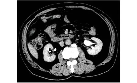
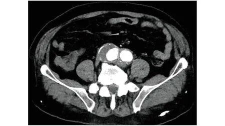
Laboratory tests revealed raised erythrocyte sedimentation rate (ESR, 48.0 mm/h), serum C-reactive protein (CRP, 134.0 mg/L), and creatinine (123.1 μmol/L). Ultrasound B-mode scans showed dilation of the left renal pelvis and ureter. Contrast enhanced computed tomography scan showed dilatation of the lower segment of the abdominal aorta along with multi-aneurysms at the bifurcation of the abdominal aorta and iliac arteries. Aneurysms showed thickened arterial walls (Figures 1 & 2). Medial displacement of the left ureter due to adhension with aneurysm wall was observed. The left kidney showed hydronephrosis (Figure 3). Based on these findings, IAAA of the bilateral common iliac arteries with incomplete obstruction of the left ureter were diagnosed, steroid therapy was started with methylprednisolone at an initial dose of 40 mg/d,which was gradually reduced to a maintenance dose of 6 mg/d over six months. Intensity of abdominal pain was reduced significantly within the first 24 h.No further episodes of abdominal pain occurred.After 20 days of steroid treatment, ESR, CRP, rheumatoid factor (RF), and creatinine levels were normal(ESR: 8.0 mm/h, CRP: 5.6 mg/L, RF: 9.81 IU/mL,and creatinine: 84 μmol/L). Magnetic resonance imaging revealed no dilation of the left renal pelvis or ureter and significant reduction in the exudate surrounding the aneurysms. Two years later, left abdominal pain was recurred due to discontinuing steroids for six months and steroid-related complications. Symptoms totally relieved after reusing steroid. Considering steroid-related complications, the patient refused long-term steroid medication and was given leflunomide (10 mg/d) as an alternative maintenance treatment. During the next six-year follow-up, there were no recurrent symptoms, and the size of aneurysms showed stable (Figure 4).

Figure 1 Three-dimensional-reconstructed computed tomography image shows the bilateral common iliac arteries aneurysms with the maximum diameter being greater than that of the abdominal aorta.

Figure 2 Enhanced computed tomography scan shows bilateral common iliac arteries with thickening aneurysm wall.

Figure 3 Enhanced computed tomography scan shows the left kidney hydronephrosis.
In 1972, Walker,et al.[8]first characterized IAAA as the presence of thickened aneurysm wall, marked peri-aneurysmal and retroperitoneal fibrosis and dense adhesions of adjacent abdominal organs.Pathological manifestations of IAAA include the inflammatory cell infiltrate, which are predominately found in the adventitia and to a lesser extent the media of the aorta. Although the pathogenesis and etiology of IAAA remain unclear, they are suggested to be autoimmune-related.[9,10]Some IAAA patients had autoimmune diseases such as Wegener’s granulomatosis, sclerosing cholangitis, etc.[10]The concurrent occurrence of IAAA and immune diseases may indicate multiple systemic immune disorders.[11]Elevated serum IgG4 levels have been noted in IAAA patients, suggesting an autoimmune cause.[12,13]The pathological changes in IAAA are reportedly consistent with those of idiopathic retroperitoneal fibrosis. These two disorders present with identical clinical symptoms if they involve same abdominal organs. A retroperitoneal fibrosis mass may also develop around the aneurysm of IAAA.Therefore, idiopathic retroperitoneal fibrosis, IAAA,and perianeurysmal retroperitoneal fibrosis are termed as chronic periaortitis, which have common clinical and pathological processes and represent different stages of development of the same disease.[14,15]

Figure 4 Latest computed tomography image shows regression of thickened artery wall, and size of bilateral iliac artery aneurysm has not increased remarkably.
Previous studies have shown that IAAA commonly occurs in 62-68 years old males with a history of smoking. The primary clinical manifestations of IAAA include abdominal or back pain,weight loss, elevated ESR, and hydronephrosis.[16]Computed tomography and/or magnetic resonance imaging in IAAA typically reveal thickening of the aneurysm wall, medial displacement of the ureters. In the present case, our patient was a 68-year-old man who had a history of smoking, presented with severe abdominal pain, had elevated ESR and CRP levels. Imaging studies revealed aneurysm with thickening arterial wall and medial displacement of the left ureter. IAAA was diagnosed based on these characteristic features. The inflammatory changes around the iliac artery aneurysms caused ureteral obstruction. Delayed diagnosis and treatment probably lead to a fibrosis mass, which could lead to further complications. So early diagnosis and management are important.
In general, surgery is indicated when the diameter of an aneurysm exceeds 5 cm. Obstructive uropathy is found in about 20% of patients and is usually treated by aneurysm resection, ureteral stenting, or partial ureterolysis.[17,18]Postoperative followup studies have shown that even after AAA surgery, an involvement of the ureter that did not exist at the time of operation developed and even had marked progression, indicating that the inflammatory process does not terminate the following surgery, and fibrosis persists.[19]There are no controlled treatment trials in the management of IAAA.The rationale of medical treatment is to suppress both vascular and systemic inflammation with appropriate systemic immunosuppression, including corticosteroids and conventional immunosuppressive agents. Leflunomide, is an immunomodulatory agent used in the therapy of rheumatoid arthritis by inhibiting the proliferation of lymphocytes and has been effectively used in other autoimmune diseases,like psoriatic arthritis, Wegener’s granulomatosis,sarcoidosis and others.[20]Recently, leflunomide was demonstrated to be an effective therapeutic option for various vasculitis,[21]and appears to be a fairly well steroid sparing immunosuppressant.[22]Leflunomide was tried in our case and confirmed effective.
In summary, IAAA should be considered in the differential diagnosis of AAA patients with presenting with hydronephrosis. Steroids can effectively alleviate ureteral obstruction and control inflammation, thereby, stent implanting in uteri is not needed,and surgical or endovascular therapy is delayed.Leflunomide can be considered as a long-term good substitute of steroids. Regular follow-up examination is important for guiding the medication appropriately.
ACKNOWLEDGMENTS
All authors had no conflicts of interest to disclose.
杂志排行
Journal of Geriatric Cardiology的其它文章
- The incidence and predictors of high-degree atrioventricular block in patients with bicuspid aortic valve receiving selfexpandable transcatheter aortic valve implantation
- The role of electrocardiographic imaging in patient selection for cardiac resynchronization therapy
- Homocysteine, hypertension, and risks of cardiovascular events and all-cause death in the Chinese elderly population:a prospective study
- Impact of SGLT2 inhibitors on major clinical events and safety outcomes in heart failure patients: a meta-analysis of randomized clinical trials
- Are angiographic culprit lesions true? Disagreement between angiographic and optical coherence tomographic detection
- The misfit mitral valve
