The incidence and predictors of high-degree atrioventricular block in patients with bicuspid aortic valve receiving selfexpandable transcatheter aortic valve implantation
2021-11-15YuanWeixiangOUJingJingHEXuanZHOUGuoYongLIYanBiaoLIAOXinWEIYongPENG1YuanFENG1MaoCHEN1
Yuan-Weixiang OU, Jing-Jing HE, Xuan ZHOU, Guo-Yong LI, Yan-Biao LIAO,Xin WEI, Yong PENG1,, Yuan FENG1,,✉, Mao CHEN1,,✉
1. Department of Cardiac Catheterization Laboratory, West China Hospital, Sichuan University, Sichuan, China; 2. Department of Cardiology, West China Hospital, Sichuan University, Sichuan, China; 3. Department of Radiology, West China Hospital,Sichuan University, Sichuan, China
*The authors contributed equally to this manuscript
ABSTRACT
Bicuspid aortic valve (BAV) is the commonest adult congenital cardiac anomaly and affects approximately half of the younger population requiring surgical valve replacement.[1-3]Transcatheter aortic valve implantation (TAVI) is an increasingly important alternative to surgical aortic valve replacement and has been shown non-inferior or superior to open heart surgery across different risk spectrums by multiple pivotal trials.[4-8]The subsequent trend of expanding TAVI toward the younger population has led to increased use of TAVI in BAV patients.[9]Recent findings from several clinical trials suggest that with contemporary device iteration and strategy optimization, TAVI in BAV population may secure satisfactory outcomes in terms of procedural success, paravalvular leak and 30-day mortality.[10-14]However, the incidence of high-degree atrioventricular block (HAVB) remains relatively high (12.2%-17.9%).[10,12,13,15]Postprocedural HAVB is associated with increased readmission and impaired long-term prognosis, and its minimization in BAV patients is of profound clinical significance in this population with longer life expectancy.[16,17]As most of the major trials have excluded BAV patients, little is known about the mechanism for HAVB in this population. For now,only the classic predictors of HAVB including implant depth and expansion rate have been validated in a few small sample trials.[15,18]However, compared with tricuspid aortic valve (TAV), BAV presents with singularity in anatomy in terms of leaflet(presence of raphe and heavy calcification) and ascending aorta (AAo) morphology. These anatomical features may influence the interaction between stent frame and conduction tissue and therefore are potential risk factors for HAVB.[10,12]To elucidate the post-TAVI HAVB mechanism in the BAV population and therefore further refine the procedural strategy and improve clinical outcome, we characterized the leaflet and AAo morphology in BAV patients through computed tomography (CT) and explored in depth the potential association between these risk factors and HAVB.
METHODS
Study Population and Procedure
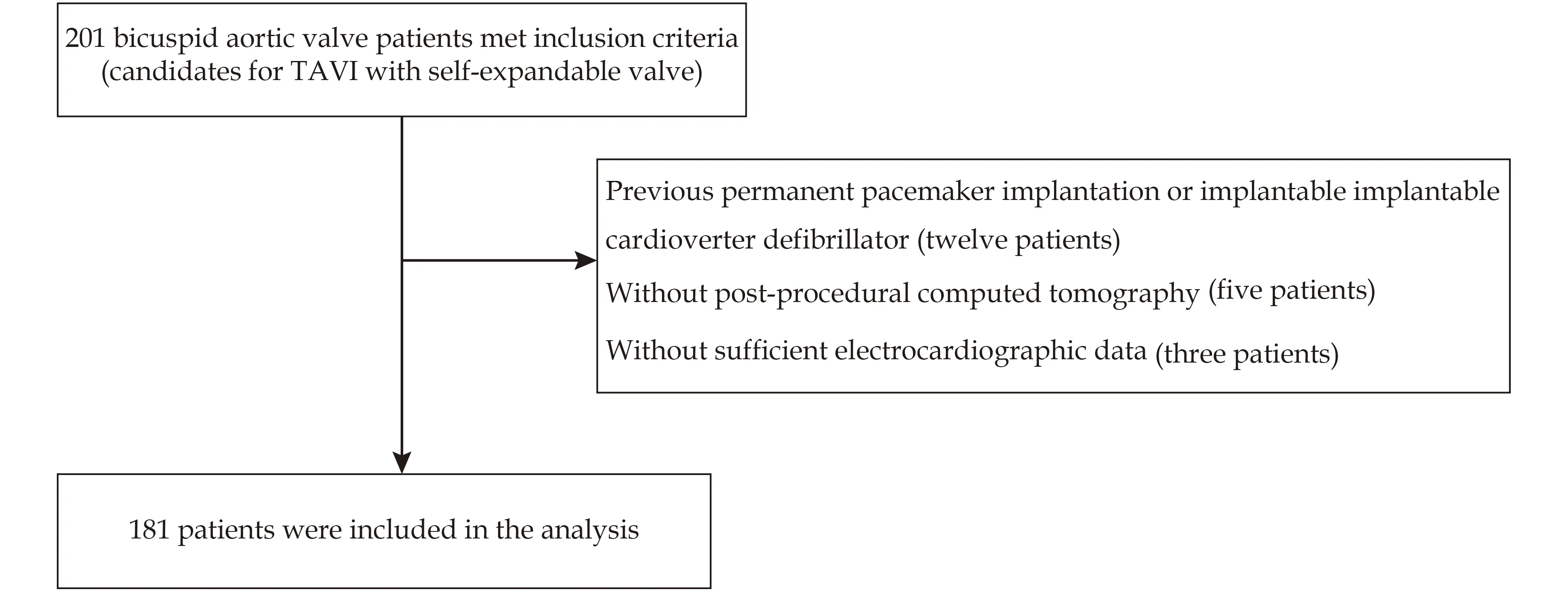
In this study, we included symptomatic severe aortic stenosis patients with bicuspid anatomy treated with transfemoral self-expandable TAVI systems from 2015 to 2019 in West China Hospital,Sichuan University, Sichuan, China. Patients with prior permanent pacemaker implantation, without pre- and post-procedural enhanced CT or CT in poor quality, or without sufficient electrocardiographic data to establish a 30-day HAVB diagnosis,were excluded. As a result, 181 patients were included in the final analysis (Figure 1). Patient data were prospectively collected in West chinA hospiTal of siCHuan university Transcatheter Aortic Valve Replacement (WATCH TAVR) registration study (registered number: ChiCTR2000033419) and retrospectively analyzed. All patients underwent routine work-up including echocardiography, multi-slice CT and standard electrocardiography monitoring.The decision to proceed with TAVI was made upon thorough consult within the multidisciplinary heart team. Patients decided to undergo TAVI procedure were treated with non-retrievable transfemoral selfexpandable devices including VenusA (VENUSMEDTECH, Zhejiang, China), VitaFlow (Micro-Port, Shanghai, China), TaurusOne (Peijia, Suzhou,Jiangsu), CoreValve (Medtronic, Minneapolis, Minnesota), and a self-expandable device with retrievable systems [VenusA-plus, (VENUSMEDTECH,Zhejiang, China)]. The domestic devices have similar stent morphology, deployment procedure, comparable procedural outcomes and similar mechanism in developing HAVB compared to CoreValve systems.[18-20]Valve sizing was individually assessed through CT and angiography of supra-annular structures, as detailed in our previous study.[21]Due to the restrictive supra-annular structure, BAV were normally down-sized to reduce the incidence of distal movement during deployment.[22]During implantation, the double S-curve or the cusp overlap view is used to determine optimal projection view,the prothesis is usually implanted 0-1 mm below the annulus for initial deployment, and the delivery system is held by the operator to avoid apical movement during deployment. Baseline clinical, anatomic,electrocardiographic, echocardiographic and procedural characteristics are presented in Table 1. The study was approved by Clinical Trials and Biomedical Ethics Special Committee of West China Hospital, Sichuan University (No.2020470) in Sichuan,China. All patients gave written informed consent.

Figure 1 Study flow diagram. TAVI: transcatheter aortic valve implantation.
CT Acquisition and Measurement
The patients underwent enhanced CT before and two to three days after TAVI. The CT images were analyzed using FluoroCT 3.0 (Circle Cardiovascular Imaging Inc., Calgary, Canada). Data were recon-structed at the systolic phase at 20%-40% of the R-R interval.

Table 1 Characteristics of study population.

Continued
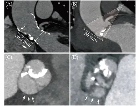
The anatomy of AAo was characterized by AAo angle and AAo perimeter. The AAo angle representing the curvature of AAo was determined by the angle between the certain cross-section of AAo and the plane of annulus as described by previous studies.[20,23]The prosthetic crown-liked outflow was found to interact with AAo usually at the cross-section 35 mm above the annulus and the AAo angle derived from this cross-section was found to perfectly predict device coaxility in previous studies.[20,24]Therefore, in this study, the “35 mm cross-section”was also taken as the certain cross-section and was measured by the plane perpendicularly to the centerline of AAo and passing the point at the outer curvature of AAo 35 mm above the annulus (Figure 2).The AAo perimeter was also measured at the “35 mm cross-section”. The bicuspid anatomy was classified according to Sievers and Schmidtke method as follows: (1) type-0 has two symmetric cusps without evidence of a raphe; (2) type-1 has a single raphe due to left- and right-coronary cusps fusion(L-R) or right- and non-coronary cusps fusion or leftand non-coronary cusps fusion; and (3) type-2 has two raphes.[25]For type-1 BAV, the assessment and definition of calcified raphe was described in details in a previous study.[10]Briefly, bulky or liner calcification extending more than half of the raphe determents the calcified raphe (Figure 2). The leaflet with calcium volume more than median value in the entire cohort was categorized as excess leaflet calcification.[10]Left ventricular outflow track (LVOT)calcium volume is measured from the annulus to 10 mm into the LVOT. Distribution of calcification in membranous septum (MS) and aortomitral curtain is characterized and determined semi-quantitively as none, trivial, moderate and severe.[26-28]MS length was retrospectively measured in the plane bisecting the MS and passing the basal attachment point of left coronary sinus.[29]The prosthetic annular oversizing ratio was calculated as following: (theoretical prothesis perimeter/annular perimeter - 1) ×100%. The implantation depth was derived from post-procedural enhanced CT and was determined as the distance from the stent distal end at non-coronary side to the plane of annular.

Figure 2 Computed tomography measurement of AAo angle and calcified raphe. (A): The stent outflow of a self-expandable valve normally interacts with AAo at 35 mm above annulus (white arrow); (B): AAo angle (a) was defined as the angle between the annulus (white line) and the cross-section (white dash line) at 35 mm above annulus. AAo perimeter was measured at the same cross-section; (C): calcified raphe in a patient with type-1 morphology presenting with left- and right-coronary cusps fusion subtype. White arrow represents the side of membrane septum;and (D): non-calcified raphe in a patient with type-1 morphology presenting right- and non-coronary cusps fusion subtype. AAo:ascending aorta.
Endpoint and Statistical Analysis
The primary endpoint was HAVB within 30-day post-TAVI. HAVB was defined as persistent/paroxysmal third-degree atrioventricular block or Mobitz type II second-degree atrioventricular block,which was established based on post-procedural,ambulatory electrocardiography (5-7 days post-TAVI) and follow-up electrocardiography (one month post-TAVI). Deployment at risk was defined as prothesis pop-out or implantation higher than annulus. Other clinical endpoints were defined based on the Valve Academic Research Consortium-2 consensus document.[30]
For statistical analysis, continuous variables with normal or non-normal distribution are presented as mean ± SD or medians (interquartile range) and compared using Student’st-test or Mann-WhitneyUtest, respectively. Categorical variables are presented as frequencies and percentages, and test for differences was conducted using Pearson’s chisquared test. Variables withP-value < 0.05 on univariate analysis were considered in the multivariate analysis, and forward-logistic regression stepwise method was performed for dichotomous outcomes and forward-liner regression method for continuous outcomes. Receiver-operating characteristic curves were then generated and specific cutoffs using Youden index were produced. Sensitivity,specificity, negative predictive value and positive predictive value were calculated using the specific cutoffs. SPSS 22.0 (SPSS Inc., IBM, Armonk, New York, USA) and R software (version 3.5.2; http://www.R-project.org) were used to perform the computation. Analyses were considered significant at a twotailedP-value < 0.05.
RESULTS
Patients’ Characteristics
A total of 181 patients were included in the final analysis. The baseline characteristics of these patients are reported in Table 1. Of all the patients, the mean age of the population was 73.1 ± 6.2 years, 78 patients (43.1%) were women and the mean Society of Thoracic Surgery Risk Score was 6.3% ± 4.3%.The incidence of 30-day HAVB was 16.0% and the median time to HAVB was three days. Device success was achieved in 144 patients (80.0%). Thirteen patients (7.2%) were implanted a second valve,while moderate or severe paravalvular leakage was found in five patients (2.8%). The details of other clinical outcomes are shown in Table 2. In patients who had HAVB post-TAVI, baseline characters,echocardiography parameters and electrocardiograph characters were distribute similarly to those without HAVB.
As for the valvular morphology, type-1 morphology was found in 79 patients (43.6%) (L-R fusion takes up 79.7%), of which calcified raphe comprised 53%. Significantly higher rate of type-1 morphology (75.9%vs.35.7%,P< 0.001) was found in patients with than without HAVB. In terms of AAo morphology, significantly larger AAo angle was found in patients with than without HAVB, while AAo perimeter distributed equally between two groups.
As for the valvular calcification, the median valve calcium volume of the population was 649.7 (327.0-1 002.9) mm3. The valve calcium volume was significantly lower in patients with HAVB [388.2 (244.7-742.7) mm3vs.698.3 (355.3-1 017.5) mm3,P= 0.019]than those without HAVB. Thus, it is understandable that the incidence of excessive leaflet calcification was also numerically lower in patients with HAVB (31.0%vs.49.3%,P= 0.07). However, the in-cidence of calcified raphe was found to distribute equally between two groups in patients with type-1 morphology. Additionally, the present study explored the association between LVOT calcification and HAVB. The result showed that neither the LVOT calcium volume, nor the calcification distribution in MS or in aortomitral curtain had an impact on HAVB development in BAV population.

Table 2 Clinical outcomes according to raphe type.
For procedural and post-procedural characters,VenusA-plus was implanted in fourteen patients(7.7%), of which HAVB rate was 21.4%, equaling to those treated with first generation device (15.5%,P=0.566). The mean oversizing ratio of the entire cohort was 6.5% ± 9.3%. Patients with HAVB had significantly larger incidence of implantation depth >MS length as compared to those without HAVB(89.7%vs.63.2%,P= 0.005). Other characters including oversizing ratio, post-dilation and type of devices were distributed equally between two groups.
Independent Predictors for HAVB
In the multivariate analysis, AAo angle [odds ratio (OR) = 1.08, 95% CI: 1.01-1.15,P= 0.016], type-1 morphology (OR = 4.97, 95% CI: 1.88-13.16,P=0.001), and implantation depth > MS length (OR =7.12, 95% CI: 1.95-26.09,P= 0.003) were found to be independent predictors for HAVB (Table 3). After including the independent predictors, as well as,baseline right bundle brunch block in the multivariate logistic regression analysis, we produced a strong predictive model of new-onset HAVB with area under the curve of 0.84 (sensitivity = 62.1%,specificity = 92.8%, negative predictive value =92.8%, positive predictive value = 62.1%).
Leaflet Calcification in Type-1 Morphology
In patients with type-1 morphology, L-R fusion was associated with numerically higher incidence of HAVB (31.7%vs.12.5%,P= 0.211). In these patients (type-1 morphology and L-R fusion), calcified raphe showed no additional impacts on HAVB development (28.1%vs.35.5%,P= 0.530), even in the subgroups of implantation depth > MS length(45.0%vs.42.9%,P= 1.00), as compared to noncalcified raphe. Calcified raphe was associated with higher valve calcium volume [747.7 (406.4-1 187.1)mm3vs.553.2 (287.4-776.1) mm3,P= 0.014], while,in patients with type-1 morphology, excessive leaflet calcification was found to associate with signi-ficantly lower incidence of HAVB (13.9%vs.39.5%,P=0.011). This might explain the lack of correlation between calcified raphe and HAVB incidence.

Table 3 Independent predictors for high-degree atrioventricular block in patients with bicuspid aortic valve.
Therefore, after grouping the leaflet calcification based on the presence of excessive calcification and calcified raphe as proposed by Yoon,et al.,[10]we found that HAVB incidence was not significantly different across these subgroups in the overall population, but was significantly higher in the subgroup with no excessive calcification or calcified raphe in the type-1 BAV (versus excess leaflet calcification or calcified raphe versus excess leaflet calcification plus calcified raphe: 45.8%vs.21.9%vs.17.4%,P= 0.03). Incidence of paravalvular leak more than mild was also significantly different among three subgroups (20.5%vs.41.3%vs.43.6%,P= 0.01).
AAo Angle and Device Implantation
Besides the established association with device coaxility, AAo angle was found to negatively correlate with implantation depth in BAV (r= -0.239,P=0.005). Additionally, in univariate liner regression model, sex (P= 0.101), AAo perimeter (P= 0.036),AAo angle (P= 0.005), LVOT perimeter (P= 0.046)and calcified raphe (P= 0.010) were found as potential correlates for implantation depth, while AAo angle, calcified raphe and sex were found to be independent correlates in multivariate linear regression model (Table 4). Furthermore, with median value as the cutoff value (19.2°), large AAo angle was found to be associated with at-risk deployment of the self-expandable valves (9.9%vs.2.2%,P=0.031). Of these eleven patients with at-risk deployment, three patients experienced device pop-out,none of them converted to surgery.
DISCUSSION
This study aims to detect the predictors of HAVB in patients with BAV receiving self-expandable TAVI. Our findings can be summarized as follows:(1) although the proportion of implant below MS in our study is comparable to that found in the TAV population, the post-TAVI HAVB incidence is still relatively high (16.0%); (2) apart from implant below MS, AAo angle and type-1 BAV are also independent predictors of post-TAVI HAVB and comprises a model with powerful predicting power(area under the curve of 0.84); (3) in type-1 BAV, excessive leaflet calcification is associated with lower incidence of HAVB, while calcified raphe is not associated with higher rate of HAVB; and (4) the relatively large AAo angle and calcified raphe synergistically facilitate the high implantation of selfexpandable valve, though the former is associated with at-risk deployment.
With continuous device refinement and strategy optimization, the TAVI outcome in BAV population has secured significant improvement in terms of paravalvular leakage, procedural success and 30-day mortality.[10-13]However, the post-TAVI HAVB incidence is still relatively high.[13,15]As the BAV population requiring intervention is younger, minimizing HAVB is of significant importance in improving long-term prognosis in this population. In our study, the HAVB incidence after self-expandable TAVI among the BAV patients is 16.0%, which is comparable to that of recently reported BAV cohort(15.0%-17.9%), but higher than that in the TAV cohort (6.4%-9.7%) and the surgical BAV cohort(5.4%).[13,15,31-33]Implant below MS is universally acknowledged as an important predictor of HAVB in BAV and TAV population following TAVI.[15,31]The study by Hamdan,et al.[15]compared the MS morphology between BAV and TAV using propensity score matching and demonstrated that compared with TAV, BAV patients have significantly shorter MS length, suggesting that the prosthesis is more likely to be implanted below MS.
Indeed, compared to a previous study in a TAV cohort using the same MS measurement method,the MS length was shorter in the present study.[29,31]However, as a possible result of high implantationand down-sizing strategy, implant below MS among the BAV population (66%) in the present study was in line with that found in TAV cohort (60%-75.2%) in previous studies.[34,35]A similar finding from Hamdan,et al.[15]suggested non-significant difference in the difference between MS length and implantation depth between BAV patients and TAV patients (BAV patients versus TAV patients: 0.62 ±4.3vs.1.27 ± 5.4,P= 0.44). The totality of evidence suggests other potential risk factors, apart from the implant depth, are at play to mediate the development of HAVB.

Table 4 Multiple linear regression model for device implantation depth in patients with bicuspid aortic valve.
From previous studies, BAV morphology is associated with immediate TAVI outcome. Compared to type-0 patients, type-1 patients have lower postprocedural pressure gradients and higher incidence of moderate-severe paravalvular leakage.[12,36]Also, previous studies suggest that type-1 morphology is a potential risk factor for HAVB.[10,12,15]In our study, type-1 BAV, for the first time, is found to be the independent predictor of HAVB. This association may be the result of pressuring from the L-R raphe (which is at the opposing side of MS and comprises 79.7% of type-1 BAV) through the stent frame (Figure 3). Therefore, further down-sizing or adequate pre-dilation may tone down the stentraphe interaction and improve HAVB outcome.
Calcification is another important risk factor for new-onset HAVB. In previous studies, calcification at leaflet landing zone, MS and aortomitral curtain were found to enhance the odds of HAVB in the TAV population.[27,28]Interestingly, in the BAV population, excessive leaflet calcification was found to inversely associate with a lower rate of new-onset HAVB, while neither calcified raphe nor LVOT calcification was associated with the development of HAVB. The anatomic features of BAV leaflets might explain the difference. The restrictive supra-annulus structure and the greater leaflet calcium burden in BAV may limit the expansion of the devices, and further reduce the interaction between the stent inflow and the calcification along with the down-sizing strategy, therefore transforming the role of calcification in the HAVB development.[21]The study of Yoon,et al.[10]demonstrated that classification of leaflet calcification has prognostic implication and provided insight into patient selection. Our study showed that this type of classification can also guide the intra-procedural strategy to reduce the incidence of HAVB. When patient is presented with type-1 calcified raphe BAV, pre-dilation with a large balloon can predispose the risk of annulus rupture.[10]Under this circumstance, more aggressive high implantation may be the more favorable approach to lower the rate of HAVB.

Figure 3 The mechanisms of type-1 morphology in high-degree atrioventricular block development in patients with bicuspid aortic valve. In bicuspid aortic valve patients, the left- and rightcoronary cusps fusion comprised 79.7% of the type-1 morphology. The raphe in this type of valve may push the stent inflow towards membranous septum (A, red arrows) and serve as the fulcrum (B, yellow star), enhances the impact of ascending aorta angle on conduction tissue (white star) through leverage effect(white arrows).
Device non-coaxility is another important mediator of post-TAVI HAVB.[20]In previous studies, potential impact factors of non-coaxial implantation, including the aortoventricular angle and the 50-mm cross section-derived AAo angle, were found to be correlated with post-TAVI conduction disturbance.[23,37]Our previous study suggested that AAo parameters obtained at the specific cross-section 35 mm above annulus, rather than the aortoventricular angle, could affect device implantation and further impact procedural outcomes.[20]In BAV population,the restricted supra-annulus structure may act as a fulcrum and magnify the impact from AAo on MS through the leverage effect (Figure 3). Thus, a direct link between AAo angle and HAVB was established for the first time in the present study, which confirming the path of the “AAo angle-device coaxilityconduction disturbance” in HAVB development.[20]
These novel predictors (AAo angle and calcified raphe) along with implantation depth present two different mechanisms (force and spatial correlation)in interpretation of post-TAVI HAVB. Interestingly,large AAo angle and calcified raphe synergistically promote high implantation. A possible explanation is that, with the coeffect of the calcified raphe, the bent AAo may increase intra-procedural deployment non-coaxility and make the stent inflow closer to MS. The lower edge of stent is more prone to make contact with the ventricular wall and form anchoring, therefore preventing downward migration.[38]With bent aorta or calcified raphe, operators may adopt more aggressive high implantation strategy to implant the device above the lower edge of MS to minimize post-TAVI HAVB. However, it should be noted that greater non-coaxility may precipitate over-high implantation of the stent lower edge at the MS and lead to pop-out. This calls for precise hold and control of the delivery system, and more importantly, proper projection view to accurately decide the implantation depth. The classic double Scurve or the more recent cusp overlap view can help identify optimal angio view at initial implantation.[39]However, these techniques do not allow visualization of the inner and outer curvature of the AAo, rendering the assessment of non-coaxility difficult.[39]When the AAo angle is large, pulling of the delivery system can lead to a significant shift in coaxility, possibly disrupting the co-planar balance achieved under cusp overlap view. Before final releasing, switching to left anterior oblique projection view for further assessment of implantation depth is an important step to prevent pop-out or overhigh implantation in bent aorta (Figure 4).
LIMITATIONS
There are several limitations that must be noted.Firstly, the inherent of the study design, which is a single-center, retrospective study. Thus, caution should be taken when extrapolating the present findings to other cohorts. Future prospective and multi-center studies are warranted to verify the clinical applications of these predictors. Secondly,the mean age of the included population is 74 years and as such, the extrapolation of our findings to the bicuspid patients with younger age should be carefully considered. Last but not least, the present study only included patients treated with selfexpandable device, the predictors for HAVB should be further examined when expanding to patients treated with balloon-expanded devices.
CONCLUSIONS
AAo angle and type-1 morphology are novel independent predictors for HAVB in BAV patients receiving self-expandable TAVI. In type-1 BAV, excessive leaflet calcification is associated with lower incidence of HAVB, while calcified raphe is not associated with higher rate of HAVB. Large AAo angle and calcified raphe are associated with smaller implantation depth, which calls for more progressive high implant strategy, though additional caution should be taken to avoid device pop-out.

Figure 4 Optimal angiographic angulation in bent AAo. Pulling and holding of the delivery system during deployment may shift the coaxility of the stent. (A): In straight AAo, the pulling-associated change in coaxility is minor, therefore at conclusion of the deployment the stent is coplanar with annulus at the cusp overlap view (black arrow heads: the three radiopaque markers at the VenusA valve inflow have same distance to the lower edge, which can be used to determine coaxility); (B): in more bent AAo, as the pull can result in a major shift in coaxility, the cusp overlap view cannot accurately assess the implant depth; and (C): to avoid device pop-out, a switch to left anterior oblique angulation can observe the implant depth from a better view. AAo: ascending aorta.
ACKNOWLEDGMENTS
This study was supported by the National Natural Science Foundation of China (No.81970325 &No.81900348 & No.81901825), and the Science and Technology Support Plan of Sichuan Province(2019YFS0299 & 2019YFS0433). All authors had no conflicts of interest to disclose. The authors gratefully acknowledge Mr. Shi-Quan REN for statistical analysis.
杂志排行
Journal of Geriatric Cardiology的其它文章
- The role of electrocardiographic imaging in patient selection for cardiac resynchronization therapy
- Homocysteine, hypertension, and risks of cardiovascular events and all-cause death in the Chinese elderly population:a prospective study
- Impact of SGLT2 inhibitors on major clinical events and safety outcomes in heart failure patients: a meta-analysis of randomized clinical trials
- Are angiographic culprit lesions true? Disagreement between angiographic and optical coherence tomographic detection
- Inflammatory abdominal aortic aneurysms treated with leflunomide: an eight-year follow-up case report and literature review
- The misfit mitral valve
