Intrahepatic cholangiocarcinoma: Introducing the preoperative prediction score based on preoperative imaging
2021-07-24FinBrtschFelixHhnLuksllerJnineBumgrtMriHoppeLotichiusRomnKloecknerHukeLng
Fin Brtsch ,Felix Hhn ,Luks Müller ,Jnine Bumgrt ,Mri Hoppe-Lotichius ,Romn Kloeckner ,Huke Lng,
a Department of General, Visceral and Transplant Surgery, University Medical Center of the Johannes Gutenberg-University Mainz, Langenbeckst, 1, 55131 Mainz, Germany
b Department of Diagnostic and Interventional Radiology, University Medical Center of the Johannes Gutenberg-University Mainz, Langenbeckst, 1, 55131 Mainz, Germany
Keywords:Intrahepatic cholangiocarcinoma Cholangiocarcinoma Liver surgery Preoperative imaging Survival
ABSTRACT Background:Intrahepatic cholangiocarcinoma (ICC) still has a poor long-term outcome,even after complete resection.We investigated different parameters gathered in preoperative imaging and analyzed their influence on resectability,recurrence,and survival.Methods:All patients who underwent exploration due to ICC between January 2008 and June 2018 were analyzed retrospectively.Kaplan-Meier model,log-rank test and Cox regression were used.Results:Out of 184 patients,135 (73.4%) underwent curative intended resection.Median overall survival(OS) was 22.2 months with a consecutive 1-,3- and 5-year OS of 73%,29%,and 17%.Median recurrencefree survival (RFS) was 9.3 months with a consecutive 1-,3- and 5-year RFS of 36%,15%,and 11%.Site of tumor,parenchymal localization,tumor configuration/dissemination,and estimated tumor volume had significant influence on resectability.Univariate analyses showed that site of tumor,tumor configuration/dissemination,number of nodules,and estimated tumor volume had predictive values for OS and RFS.Together with tumor size the preoperative prediction (POP) score was created showing significance for OS and RFS (all P < 0.001).In multivariate analysis,POP score (HR = 1.779; 95% CI: 1.268-2.495;P = 0.001),T stage (HR = 1.255; 95% CI: 1.040-1.514; P = 0.018) and N stage (HR = 1.334; 95% CI:1.081-1.645; P = 0.007) were the independent predictors for OS.For RFS,POP score (HR = 1.733; 95% CI:1.300-2.311; P < 0.001) and M stage (HR = 3.036; 95% CI: 1.376-6.697; P = 0.006) were the independent predictors.Conclusions:The POP score showed to have a highly significant influence on OS and RFS.The score is easy to assess through preoperative imaging.For patients in the high risk group at least staging laparoscopy or preoperative chemotherapy should be evaluated,because they showed equal outcome compared to the irresectable group.
Introduction
Intrahepatic cholangiocarcinoma (ICC) is the second most common primary malignancy of the liver following hepatocellular carcinoma and its incidence is rising,especially in Western countries [ 1–3 ].Due to late onset of symptoms ICC is often diagnosed in advanced stage [ 4,5 ].Long-term prognosis is poor and complete resection offers the only chance of cure while resectability varies between 50% and 75% [ 4,6–8 ].Therefore,usually major and even extended resections are necessary [9].Both computed tomography (CT) and magnetic resonance imaging (MRI) play important roles in the preoperative assessment and planning of surgical resection [ 10,11 ].Prognosis is related to tumor biology and recurrence.Preoperative prediction of resectability and prognosis in combination with the histopathological results may help to avoid unnecessary explorations and to identify patients who need adjuvant treatment and close follow-up.Comprehensive data on this topic is scarce within the literature.Some studies reported on worse prognosis for factors determined preoperatively through imaging like hilar invasion,multiple lesions,tumor size,necrosis,satellite nodules,and vascular encasement [ 12–14 ].
The aim of this single-center study is to analyze the influence of preoperative imaging features on resectability,survival,and recurrence.
Methods
The data from all patients who underwent exploration due to ICC between January 2008 and June 2018 were prospectively collected in an institutional database.Data were transferred to SPSS(IBM SPSS Statistics for Windows,Version 23.0,IBM,Armonk,NY,USA) for further analyses.Diagnosis was proven through postoperative histological work-up.Patients with other primary or secondary liver malignancies were excluded.
Preoperative work-up
Most patients were referred with suspected diagnosis from secondary care medical centers.For preoperative planning,CT or MRI was both acceptable.Each imaging study underwent thorough rereview in the interdisciplinary tumor board to assess image quality.Tumor board had guaranteed participation of at least one specialized surgeon,radiologist,gastrointestinal oncologist,pathologist,radiotherapist and hematologist.If the image quality provided by the referral center was deemed suboptimal,we performed a high-resolution multiphasic CT of the abdomen and chest in-house.If only abdominal imaging was performed,a CT scan of the chest was added to complete preoperative staging and exclude pulmonary metastases.If necessary,we performed gastroscopy and colonoscopy in order to rule out another primary tumor.Liver biopsy was not performed to confirm diagnosis preoperatively,but some patients had undergone biopsy prior to referral (n= 40).
In advanced disease and expected extended resection,the future liver remnant (FLR) was assessed through radiological volumetric analysis.Our threshold was calculated through 0.5% of the patient’s body weight and transformed grams in volume (millilitre).For example,an FLR of 400 mL would be necessary in an 80 kg patient [80000 g × 0.005 = 400 g (transformed in mL)].This applies only for patients without impaired liver function.
Postoperative parameters and follow-up
Further demographic and surgical factors were collected like age,sex,ASA classification (American Society of Anaesthesiologists) [15],type and extent of resection,vascular and visceral extensions,morbidity,mortality,and common histological parameters.Usually,metastatic disease was a reason to resign from surgery/resection.In a few patients,the histological work-up showed unexpected metastatic disease,which explained why some patients had M1 status.For all histological parameters the actual 8th edition of UICC (Union for International Cancer Control)/AJCC(America Joint Committee for Cancer) staging system was utilized [16].Morbidity and mortality have been graded according to the Clavien-Dindo classification [17].
For assessment of recurrence and survival regular follow-up was performed every three months with alternating ultrasound and CT (or MR) imaging.All patients gave written consent of pseudonymized registration in our database,follow-up within our treatment contract and being part of our university center for tumor disease (UCT).If patients were not able to be followed up at our center due to logistical reasons,the further treating physicians had been contacted.
Radiological screening
Preoperative imaging underwent a detailed re-review regarding the following aspects: site of the tumor (right,left,bilateral),parenchymal localization (central,peripheral),tumor configuration/dissemination (singular,local satellites,intralobular metastases,translobular metastases,diffuse),number of nodules,estimated tumor volume,and character of tumor border (well defined,blurry/infiltrative).Local satellites were defined as lesions within a distance of 2 cm; if distance exceeded 2 cm,they were estimated as intralobular metastases.The radiological data were collected and validated through two radiologists with considerable experience in abdominal oncologic imaging.
Detailed technical information about the CT- and MRI-scanners and the imaging protocols used can be found in the supplement.
Statistical analysis
Only patients with complete datasets were included for statistical investigation.Categorical data were analyzed using the Chisquare test.For univariate survival analysis,Kaplan-Meier analysis with log-rank test were used.The day of resection served as baseline regarding calculation of overall survival (OS).For analyses of OS as well as recurrence-free survival (RFS),perioperative deaths were excluded.Pvalues<0.05 were considered significant.RFS was calculated according to Punt and colleagues [18].
For multivariate analysis,the Cox regression (proportional hazards model) was used with backward selection.For parameters screening those withP<0.05 in the univariate analysis were included in the multivariate analysis.
Results
A total of 184 patients underwent exploration.Out of these,135 patients (73.4%) finally underwent resection.Fourty-nine patients were deemed unresectable.Patient characteristics,types of resections and reasons for irresectability are listed in Table 1.In the resection group 70 patients underwent extended (51.9%),31 major (22.9%) and 34 minor resections (25.2%).Patients with visceral and/or vascular resection and reconstruction during hemihepatectomy (n= 16) were also categorized as extended resections.

Table 1 Patients characteristics ( n = 184).
Morbidity and mortality
In the resection group,71 patients (52.6%) showed no deviation from the normal postoperative course and therefore no morbidity as defined by the Clavien-Dindo classification.Minor morbidity(grade I + II) occurred in 16 patients (11.9%).Thirty-eight patients(28.1%) suffered from severe morbidity with grade IIIa morbidity as most common one (n= 27),followed by grade IVa (n= 6),IIIb(n= 3) and IVb (n= 2).
Mortality (in-hospital) was 7.4% (10/135) because of multi-organ failure (n= 4),sepsis (n= 3),and liver failure (n= 3).
Influence of preoperative imaging parameters on resectability
The distribution of different parameters assessed through preoperative imaging is listed in Table 2.Site of tumor (P<0.001),parenchymal localization (P= 0.001),tumor configuration/dissemination (P= 0.005),and tumor volume (P= 0.003)were associated with resectability.
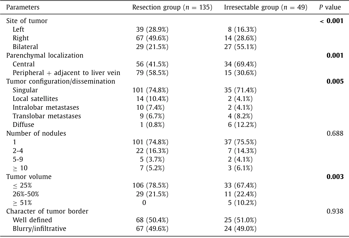
Table 2 Influence of preoperative imaging parameters on resectability.
Survival
In the intention-to-treat analysis,OS of the resection group was significantly better compared to the irresectable group (P<0.001).Detailed survival analysis of the resection group with and without perioperative deaths as well as the different parameters assessed through preoperative imaging are listed in Table 3.Of the 135 patients underwent curative intended resection,median OS was 22.2 months with a consecutive 1-,3- and 5-year OS of 73%,29%,and 17%,and median recurrence-free survival (RFS) was 9.3 months with a consecutive 1-,3- and 5-year RFS of 36%,15%,and 11%.Best OS was demonstrated for tumors of the right liver lobe,singular,peripheral lesions occupying less than 25% of the liver with a well-defined border.
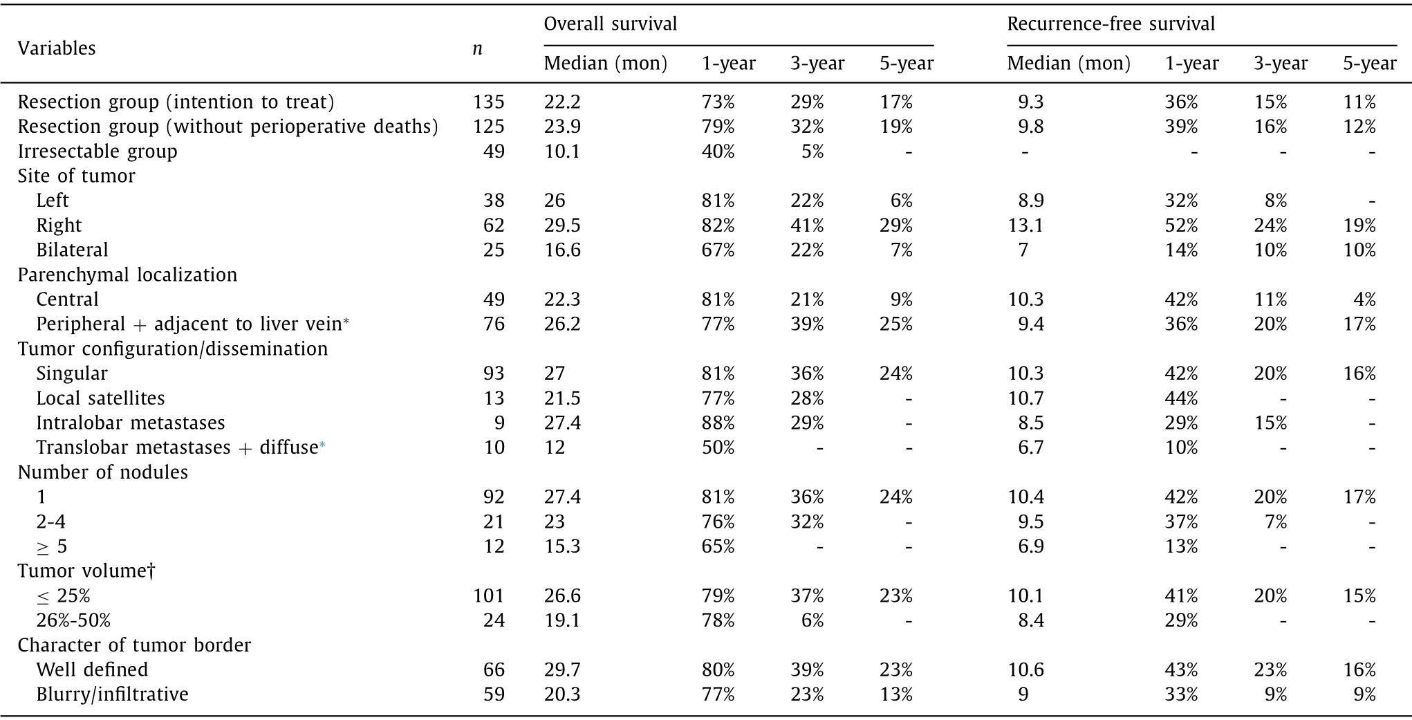
Table 3 Overall survival and recurrence-free survival regarding to the preoperative imaging parameters.
In univariate Kaplan-Meier analyses OS and RFS were tested for the preoperative imaging parameters,general and surgical as well as histopathological parameters ( Table 4 ).Tumor recurrence occurred in 89 patients.
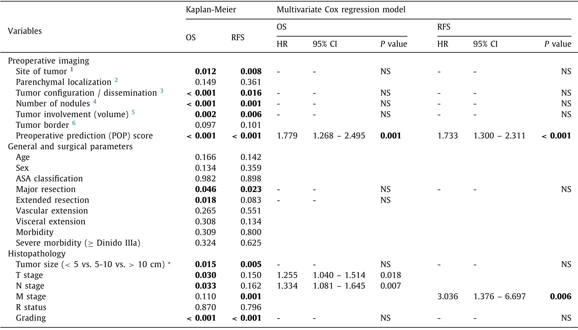
Table 4 Univariate and multivariate analyses.
Regarding OS,site of tumor ( Fig.1 A),tumor configuration/dissemination ( Fig.2 A),number of nodules ( Fig.3 A),tumor involvement (estimated volume; Fig.4 A),major resection,tumor size,T stage,N stage,and grading had significant influence.
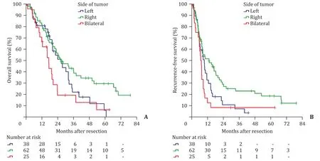
Fig.1.Survival curves for site of tumor assessed through preoperative imaging.A: Overall survival ( P = 0.012).Subgroups: left vs.right,P = 0.087; left vs.bilateral,P = 0.179;right vs.bilateral,P = 0.008; B: recurrence-free survival ( P = 0.008).Subgroups: left vs.right,P = 0.070; left vs.bilateral,P = 0.079; right vs.bilateral,P = 0.004.
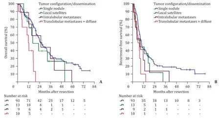
Fig.2.Survival curves for tumor configuration/dissemination assessed through preoperative imaging.Translobular and diffuse dissemination were combined.A: Overall survival ( P < 0.001).Subgroups: single nodule vs.local satellites,P = 0.312; single nodule vs.intralobular metastases,P = 0.431; single nodule vs.translobular metastases + diffuse,P < 0.001; local satellites vs.intralobular metastases,P = 0.709; local satellites vs.translobular metastases + diffuse,P = 0.034; intralobular metastases vs.translobular metastases + diffuse,P = 0.031; B : recurrence-free survival ( P = 0.016).Subgroups: single nodule vs.local satellites,P = 0.0.223; single nodule vs.intralobular metastases,P = 0.156; single nodule vs.translobular metastases + diffuse,P = 0.003; local satellites vs.intralobular metastases,P = 0.985; local satellites vs.translobular metastases + diffuse,P = 0.148; intralobular metastases vs.translobular metastases + diffuse,P = 0.401.

Fig.3.Survival curves for number of nodules assessed through preoperative imaging.A: Overall survival ( P < 0.001).Subgroups: 1 vs.2-4,P = 0.282; 1 vs.≥5,P < 0.001;2-4 vs.≥5,P = 0.008; B : recurrence-free survival ( P = 0.001).Subgroups: 1 vs.2-4,P = 0.097; 1 vs.≥5,P < 0.001; 2-4 vs.≥5,P = 0.057.
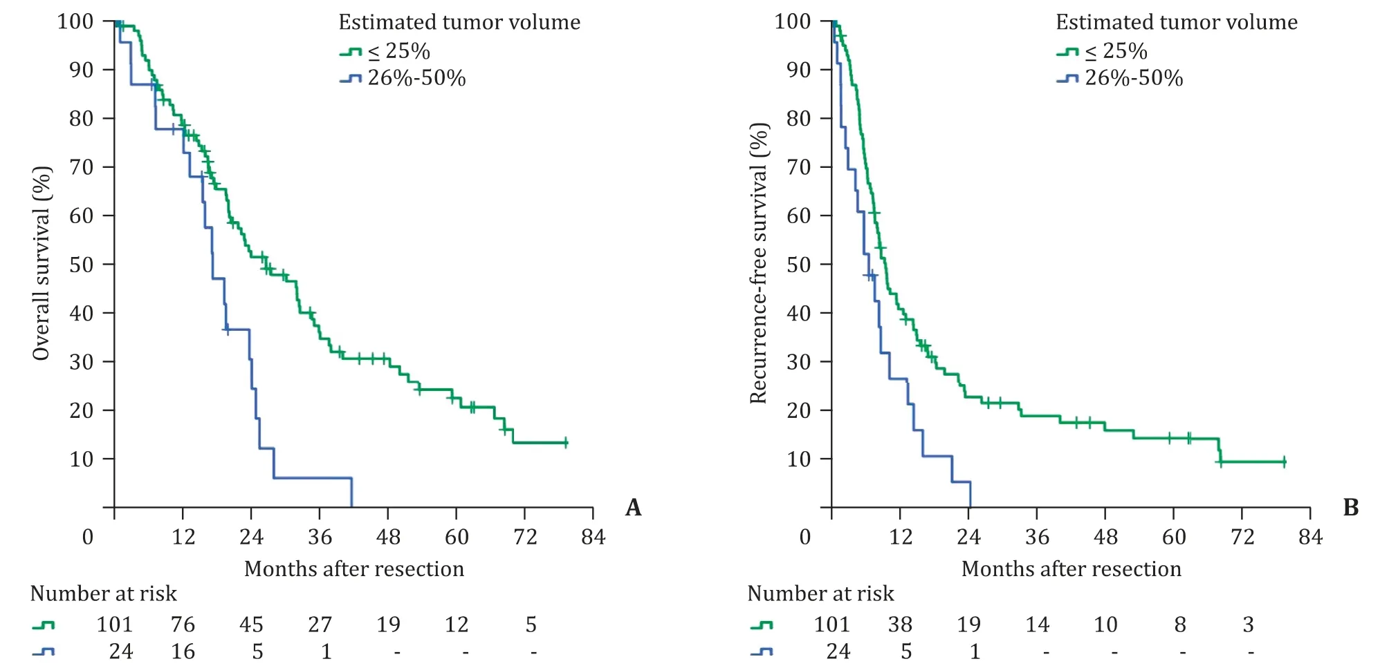
Fig.4.Survival curves for tumor involvement (estimated volume) assessed through preoperative imaging.A: Overall survival ( P = 0.002); B: recurrence-free survival( P = 0.006).
For RFS,site of tumor ( Fig.1 B),tumor configuration/dissemination ( Fig.2 B),number of nodules ( Fig.3 B),tumor involvement (estimated volume; Fig.4 B),major resection,tumor size,M stage and grading had significant influence.
Preoperative prediction (POP) score
The parameters that were significant factors for OS or RFS in univariate analysis were utilized to create the POP score ( Table 5 ).We categorized the patients to a low risk group (0–4 points),an intermediate risk group (5–8 points) and a high risk group (9–15 points).
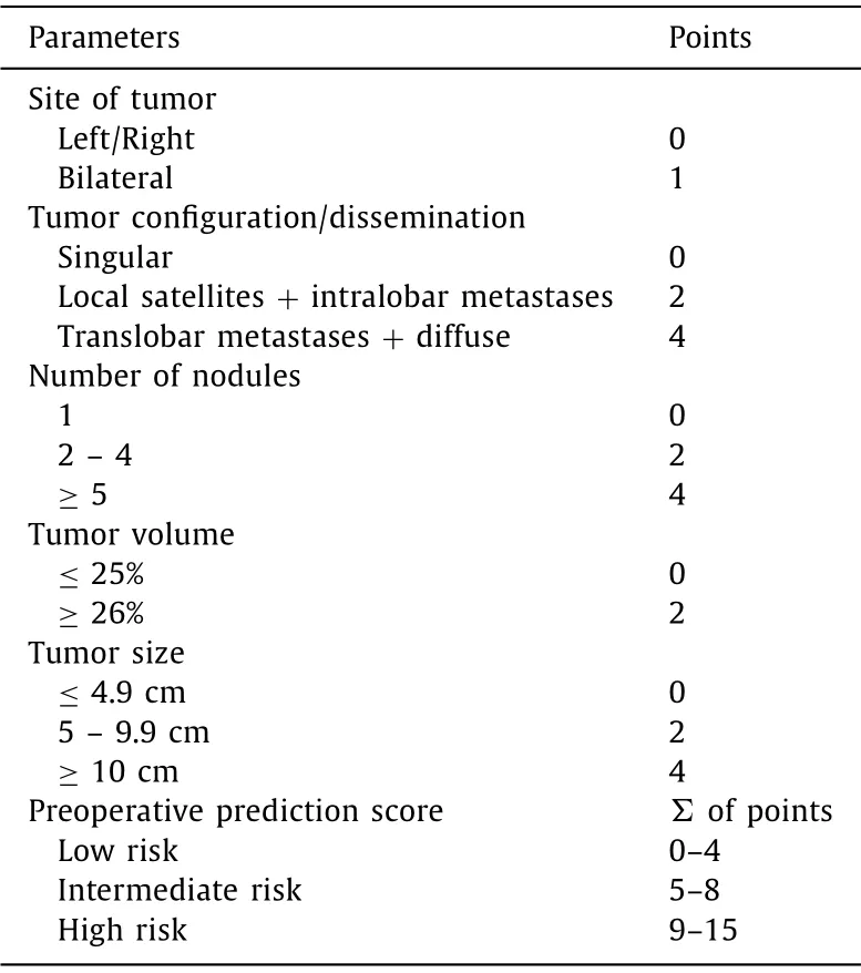
Table 5 Preoperative prediction score.
In the resection group,87 patients (64.4%) were categorized as low risk,30 (22.2%) as intermediate risk and 18 (13.3%) as high risk.In the irresectable group,28 patients (57.1%) were categorized as low risk,9 (18.4%) as intermediate risk and 12 (24.5%) as high risk.The amount of high risk patients was higher in the irresectable group (12/49) compared to the resection group (18/135),but no significant difference could be shown in cross tabulation(P= 0.070).
For OS the POP score had significant influence ( Fig.5 A).The outcome of the high risk group was comparable to the results of the irresectable group (P= 0.841).For RFS the low risk and intermediate risk groups had significantly better outcomes compared to the high risk group ( Fig.5 B).

Fig.5.Survival curves for different risk group according to the preoperative prediction score.A: Overall survival ( P < 0.001).Subgroups: low risk group vs.intermediate risk group,P = 0.069,low risk group vs.high risk group,P < 0.001,low risk group vs.irresectable group,P < 0.001,intermediate risk group vs.high risk group,P = 0.002,intermediate risk group vs.irresectable group,P = 0.001,high risk group vs.irresectable group,P = 0.841; B: Recurrence-free survival ( P < 0.001).Subgroups: low risk group vs.intermediate risk group,P = 0.056; low risk group vs.high risk group,P < 0.001; intermediate risk group vs.high risk group,P = 0.012.
Multivariate analysis of survival
All significant parameters of the univariate OS and RFS analyses were included in a multivariate Cox regression model to identify parameters with independent influence on survival ( Table 4 ).
The results showed that POP score (HR = 1.779; 95% CI:1.268-2.495;P= 0.001),T stage (HR = 1.255; 95% CI: 1.040-1.514;P= 0.018),and N stage (HR = 1.334; 95% CI: 1.081-1.645;P= 0.007) were associated with OS,while POP score (HR = 1.733;95% CI: 1.300-2.311;P<0.001) and M stage (HR = 3.036; 95% CI:1.376-6.697;P= 0.006) were associated with RFS.All other parameters did not show significance and were eliminated in backward selection.
Discussion
Preoperative imaging plays the most important role for diagnosis,estimation of resectability,and resection planning.Our data show that preoperatively collected information has significant influence on resectability and survival.Uni- or multifocal tumors affecting both liver lobes,centrally located lesions,translobular or diffuse spread and an estimated tumor volume ≥51% affected this analysis the most.Especially the invented POP score showed independent influence on OS and RFS.Nevertheless,patients with these negative predictors still have a chance to reach long-term survival after resection.
While the parts of CT and MRI are clear standards for preoperative imaging,the impact of positron emission tomography is still in debate [10].Several different parameters determined through preoperative imaging have already been analyzed and tested regarding their influence on survival [ 12,14 ].Jiang and colleagues went one step further and invented the “Fudan score” including parameters like tumor size,boundary type and multifocalitywhich discriminated prognosis better compared to the 7th edition of the UICC/AJCC staging system [13].This risk estimative score can be calculated preoperatively and works even for irresectable patients.Another known nomogram was invented by Hyder and colleagues including different factors like histological parameters,tumor size and multifocality [19].The Hyder nomogram could therefore only be assessed after resection.The question regarding nomograms and risk scores is always the applicability in the daily clinical routine.These scores are not difficult to collect,but would you resign from surgery in a case where resection is obviously technically possible after preoperative imaging,only because a nomogram or risk score is high? Our own data showed that extended resection including vascular and even visceral resection can lead to long-term survival in selected patients [8].Furthermore,in cases of tumor recurrence repeated resection is an option leading to prolonged survival in selected patients as well [ 20–22 ].For that reason,we follow an aggressive approach regarding resection which is reflected in a high resection rate of 73.4% with>70% of at least major and>50% of extended resections.Thereby,our morbidity (40%) and mortality (7.4%) are comparable to results of other groups [ 4,5,23 ],especially keeping the number of extended resections in mind.We created the POP score based on preoperative imaging and the distinct results were kind of surprising.In univariate analysis as well as multivariate anal-ysis the POP score showed significant influence on OS and RFS.Furthermore,it showed the highest independent significant influence for OS and RFS as well.The comparable outcome of the high risk group compared to the irresectable group is of special importance.We aimed to analyze radiological parameters of the preoperative imaging and test their influence on resectability,recurrence,and survival.Estimating resectability is a difficult task.Especially for borderline resectable ICC curative treatment alternatives are lacking.Further,it is not predictable if chemotherapy for preoperative downsizing performs well.Data on the usage of preoperative/neoadjuvant chemotherapy are scarce.The biggest analysis regarding this special topic was published by Buettner and colleagues in 2017 including 1057 patients out of whom 62 received preoperative chemotherapy [24].In this multicenter analysis including data from 12 different centers all over the world,preoperative chemotherapy was used more often in patients with advanced disease.Short-term outcome was comparable with patients who did not undergo preoperative treatment.Likewise,OS and disease-free survival were equivalent to patients who underwent primary resection.In a single-center analysis from Le Roy and colleagues,out of the analyzed 186 patients,74 had locally advanced disease and 39 underwent secondary resection after preoperative chemotherapy [25].The results were comparable to the multicenter data from Buettner and colleagues [24].Both analyses followed no standardized regimen or length of preoperative chemotherapy.There is still an urgent need of further data to define standard regimens and procedures in case of advanced ICC.If peritoneal carcinomatosis or other distant metastases are detected,biopsy and initiation of palliative therapy are recommended [10].Staging laparoscopy is reasonable in patients with high risk features like enlarged lymph nodes or if peritoneal carcinomatosis cannot be ruled out [ 26,27 ].Nevertheless,the prognostic impact of enlarged lymph nodes in preoperative imaging is questionable and patients benefit from resection anyway [ 28,29 ].Finally,tumor extent and technical resectability with a sufficient FLR are the most important factors.Preoperative therapy might also be applied for borderline or initially unresectable ICC for downsizing and achieving secondary resectability [ 24,25 ].The POP score is easy to assess,based solely on preoperative imaging and especially patients of the high risk group are candidates for at least staging laparoscopy or even preoperative treatment in potentially neoadjuvant/downsizing intention.Several parameters assessed through preoperative imaging had significant influence on resectability as well as the POP score.These results are not that surprising because they correlate with advanced tumor growth and/or poor tumor biology.Even that the character of tumor border did not influence resectability is comprehensible,because it does not influence complete resection in most cases.Interestingly the number of nodules did not affect resectability as well,but this might be related to the number of patients in groups with multifocal disease.Comparable data are difficult to find and therefore it is also difficult to discuss.The POP score is introduced in this study and showed very promising results which may change the surgical and/or interdisciplinary approach for ICC in the future.Further validation and application are needed to strengthen our findings.
For ICC several parameters have influence on OS and results vary between different studies.Most of these studies are retrospective.Analyzed or included factors differ as well.Common and regularly tested parameters are multifocality,stages of the TNM classification [16],residual tumor status (R status) and other histological features like microvascular or macrovascular invasion.Focussing on results of multivariate analyses,multifocality,R status,margin width,N stage,UICC stage,vascular invasion,tumor size,CA19-9 level,and distant metastases were independent predictors for overall survival [ 4,23,30–33 ].Most of these parameters are histological and only available after resection.Our analyses and the POP score are based on parameters which are easy to collect out of the preoperative imaging and provide interesting and distinct results.Site of tumor for example is associated with a strong survival-benefit especially after three and five years ( Fig.1 A).The finding that tumor located in both liver lobes lead to worse outcome had also been expected.But that survival for tumor located within the left liver lobe is not different from tumors located in bilateral liver lobe is something unexpected.Maybe the lymphatic drainage of the left liver lobe is an attempt of explanation.The left liver lobe might drain more often in lymph nodes of the lesser curvature which were not part of the standard lymphadenectomy for ICC in the past.With the 8th edition of TNM/UICC/AJCC classification [16]lymphadenectomy of gastrohepatic lymph nodes is recommended for tumors of the left liver lobe.OS of single nodules,tumors with local satellites or intralobular spread was comparable ( Fig.2 A).The number of nodules was related to poor tumor biology and/or advanced disease was comprehensible ( Fig.3 A).Another interesting finding was the distinct influence of estimated tumor volume on survival ( Fig.4 A).
Tumor recurrence is one of the major problems for ICC and rates go up to 60%-70% [ 20,22,34 ].This is in line with our results showing a recurrence rate of 65.9%.In an international multicenter analysis of Spolverato and colleagues on 563 patients,several predictive factors have been identified influencing recurrence of ICC: multifocality,size>7 cm,microvascular invasion,cirrhosis,grading,and N stage [22].These results are supported by Chinese data and data from our group [ 8,32 ].Data on the influence of parameters collected in the preoperative imaging on survival are scarce,although the vast majority of these patients undergo preoperative cross-sectional imaging.In an analysis of 66 patients Aherne and colleagues found satellite nodules and largest axial size as independent predictors for disease-free survival [14],while data from Japan on 111 patients showed multiple intrahepatic nodules and CA19-9 [12]to be of predictive value.Our data showed site of tumor,tumor configuration/dissemination,number of nodules and estimated tumor volume as significant factors influencing RFS.In multivariate analysis,the POP score was a predictive independent factor together with M stage.
This study contains five main limitations.First,the cohort of 184 patients with 135 resections led to small subgroups,in particular if parameters have several subitems.Second,these analyses were performed retrospectively.Both factors weaken the statistical validity.Nevertheless,we reported on one of the biggest single-center cohorts for ICC and the results of the POP score are promising.As mentioned above further validation through other groups and application is necessary to prove the value of the POP score in the future.The third limitation is the fact that parts of the imaging have been performed by the referral centers.To exclude bias,we thoroughly re-reviewed all external imaging in our tumor-board comprising at least one board certified radiology consultant with extensive experience in abdominal oncologic imaging.A further possible limitation might be the fact that technical improvements in cross-sectional imaging were made during the study period of 10 years.However,although cross-sectional imaging evolved during the last decade,the imaging of cholangiocellular malignancies remained relatively constant.In our institution,CT- and MRI-scanners were in use for most of the study period and imaging protocols have barely changed as specified in the supplement.Lastly,we used CT as well as MRI for assessment of the liver.MRI allows for slightly better lesion-liver contrast,whereas CT is slightly superior to assess extrahepatic spread.However,both imaging techniques are widely accepted for the imaging of cholangiocellular carcinoma [35].
In conclusion,several parameters assessed through preoperative imaging can help to estimate prognosis as well as the risk for recurrence.The POP score is easy to assess,based on preoperative imaging and showed to be a significant independent predictor for OS and RFS.Especially the high risk group showed to have an equal outcome compared with the irresectable group.Therefore,patients categorized as high risk should at least undergo staging laparoscopy prior to open exploration and may be candidates for preoperative chemotherapy in neoadjuvant intention.
Acknowledgments
None.
CRediTauthorshipcontributionstatement
FabianBartsch:Conceptualization,Data curation,Formal analysis,Investigation,Methodology,Project administration,Resources,Validation,Visualization,Writing - original draft,Writing -review & editing.FelixHahn:Data curation,Formal analysis,Writing - original draft.LukasMüller:Data curation,Formal analysis,Investigation,Resources.JanineBaumgart:Data curation,Validation.MariaHoppe-Lotichius:Data curation,Resources.Roman Kloeckner:Conceptualization,Data curation,Formal analysis,Resources,Supervision,Validation,Writing - review & editing.HaukeLang:Conceptualization,Project administration,Supervision,Validation,Writing - review & editing.
Funding
None.
Ethicalapproval
All patients signed informed consent that data and follow-up were collected anonymously and were potentially used for scientific analysis.Regarding the regulations of the federal state law(state hospital law §36 & §37) and the independent ethics committee of Rhineland-Palatinate,no ethical approval was necessary for this study.The work has been carried out in accordance withtheDeclarationofHelsinki.
Competinginterest
No benefits in any form have been received or will be received from a commercial party related directly or indirectly to the subject of this article.
Supplementarymaterials
Supplementary material associated with this article can be found,in the online version,at doi:10.1016/j.hbpd.2020.08.002.
杂志排行

Hepatobiliary & Pancreatic Diseases International的其它文章
- Cross-talk between hepatic stellate cells and T lymphocytes in liver fibrosis
- Diabetes mellitus is a risk factor of acute kidney injury in liver transplantation patients✩
- Hepatobiliary&Pancreatic Diseases International
- Application of machine learning models for predicting acute kidney injury following donation after cardiac death liver transplantation
- Postoperative adjuvant transcatheter arterial chemoembolization improves the prognosis of patients with huge hepatocellular carcinoma
- The effects of stereotactic body radiotherapy on peripheral natural killer and CD3 + CD56 + NKT-like cells in patients with hepatocellular carcinoma