Ureteral calculi secondary to a gradually migrated acupuncture needle
2021-03-26MshiroMtsukiAtsushiWnifuchiRyutInoueFumiysuTkeiYsuhruKunishim
Mshiro Mtsuki , Atsushi Wnifuchi Ryut Inoue Fumiysu Tkei b, Ysuhru Kunishim
a Department of Urology, Hokkaido Social Work Association Obihiro Hospital, Obihiro, Japan
b Medical Incorporated Association Tenshunkai Tokachi Urological Clinic, Obihiro, Japan
Abstract We herein presented a case of calculi secondary to a migrated acupuncture needle.A 74-year-old woman with a history of acupuncture therapy for lumbago was referred to our hospital for treatment of ureteral and renal pelvic calculi.Abdominal multi-detector computed tomography scans showed ipsilateral hydronephrosis and two calculi secondary to a migrated acupuncture needle.First, a percutaneous nephrolithotomy was performed to extract two calculi and fine needle fragments from the pelvis.Subsequently, residual needle fragments and calculi in the ureter were then removed by flexible transurethral lithotripsy using a holmium laser.In the present case,the formation of the calculi was caused by a migrated acupuncture needle.Calculi and needle fragments were removed safely endoscopically because the whole calculi and needle fragments were located in the ureteral lumen.
KEYWORDS Acupuncture needle;Endoscopic approach;Ureteral calculus;Renal calculus;Flexible transurethral lithotripsy
1.Introduction
Acupuncture is a conventional oriental medical treatment for various disorders and is performed in both eastern and non-eastern countries.However, it can cause not only technical complications such as infection [1] and bleeding[2,3],but also late complications associated with remaining needle or fragments that have migrated [4].
We reported a rare case of ureteral calculi secondary to an acupuncture needle that had migrated into the upper urinary tract, both of which could be removed successfully by endoscopic approach.
2.Case history
A 74-year-old woman was referred to our department for the treatment of ureteral and renal pelvic calculi.Although she had no clinical symptoms or complaints, a postoperative abdominal computed tomography (CT) scan following treatment of gastric cancer showed ipsilateral hydronephrosis and two calculi measuring 6 mm and 15 mm in diameter in the left renal pelvis and ureter,respectively.Physical examination, blood tests, urinalysis, and urine cultures were all unremarkable, and there was no administration (e.g., steroids or calcium compounds) that could have led to formation of a urinary calculus.Approximately 10 years earlier, she had undergone acupuncture therapy for the treatment of lumbago.
A CT and X-ray identified two calculi(Fig.1A,red arrows and Fig.1B) which were connected by an unnatural bridge like structure, and buried acupuncture needles primarily in the lumbar region were observed(Fig.1A,white arrows).To confirm the time of calculi formation,we reviewed her previous image data.A CT scan taken 3 years prior showed a left renal calculus with a conjoined acupuncture needle in the left renal pelvis (Fig.2A, red arrow) and many embedded acupuncture needle fragments (Fig.2A, white arrow).Moreover,a coronal CTscan taken 5 years prior indicated that an acupuncture needle had migrated to the left renal parenchyma,and a faint renal calculus was observed at the top of the needle(Fig.2B,red arrow).Therefore,we suspected that the ureteral calculi were due to an acupuncture needle that had gradually migrated into the renal parenchyma.
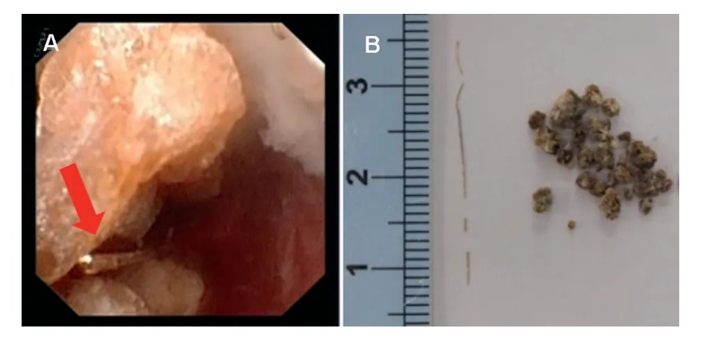
First, a percutaneous nephrolithotomy using 22 Fr rigid nephroscope and holmium laser was performed to extract some calculi and fine needle fragments from the pelvis.Path dilation was performed with NephroMaxTM (Boston Scientific, Natick, NA, USA) until 30 Fr.This is because the relatively big ureteral calculus was near renal pelvis and we would try to extract two calculi with one treatment by pulling out the conjoined calculi.We could extract the renal calculus safely, however, the ureteral calculus was impacted to the wall and the mucosa was so edematous that we were unable to insert a fine guide wire through the ureter and subsequently could not observe the stone even if using a flexible ureteroscope.Therefore, we stopped the procedure after one stone was extracted.After 2 weeks we selected the ureteral approach for a second procedure.As edema of the ureteral mucosa was released, we were able to observe and approach the larger stone using a flexible ureteroscope.Residual fragments of needle and calculi in the ureter were then removed by flexible transurethral lithotripsy using a holmium laser (Fig.3).The calculi were composed of 95% calcium oxalate (Fig.3).Since the procedure, the patient has been calculi free as of the last follow-up at 6 months.

Figure 1 CT and X-ray results.(A) X-ray showing two calculi(red arrows) and many embedded needle fragments (white arrows) in the lumber region; (B) A larger image of the stones and needle fragments.

Figure 2 CT scan results.(A) Abdominal computed tomography (CT) scan taken 3 years prior showing calculus with the needle (red arrow) and many needle fragments (white arrow);(B) Coronal CT scan taken 5 years prior showing the faintly formed calculus with the needle(red arrow)and an embedded needle fragment (white arrow).
3.Discussion
Acupuncture is a traditional treatment same as Chinese herbs in China and commonly used to manage lower back pain and as an alternative therapy performed to alleviate various diseases and symptoms,such as not only pain relief but also chemotherapy-induced nausea/vomiting and idiopathic headache [5].Although acupuncture is a safe procedure in the hands of a competent practitioner, it can be associated with several complications [1-4].It is reported that the most frequent adverse events are pneumothorax and infection [6].However, there are few reports of adverse events related with urinary tracts [7].

Figure 3 Flexible ureteroscope results.(A) Ureteroscopy showing the calculi fragments and a fine needle (red arrow);(B) Removed fine needle fragments and calculi.
There have been few independent reports of a foreign body located in the upper urinary tract.Yuzawa et al.[7]reviewed 49 cases of foreign bodies detected in the upper urinary tract,with the first case being published in 1936.Most of them were embedded acupuncture needles or surgical sutures.Though a calculus around a metal clip as a foreign body after a laparoscopic pyeloplasty was reported[8],there have since been no recent reports of urinary stones that were associated with an acupuncture needle in the upper urinary tract.Furthermore, migration of remaining needles into several organs has also been reported as late complications.Therefore,when we examine a patient with urolithiasis who has previously received acupuncture therapy, it may be necessary to pay attention to calculus formation that may have occurred due to needle migration.
In our case report, the main calculi with an acupuncture needle were located near to the left renal pelvis.Therefore,at first a percutaneous nephrolithotomy was performed.However,because there were residual needle fragments and calculi discovered in the left ureter after the first procedure,we tried to extract them by flexible transurethral lithotripsy as a second procedure.Both were removed safely endoscopically because the whole calculi and all needle fragments were located in the urinary tract.
4.Conclusion
We report a rare case of ureteral calculi secondary to an acupuncture needle that had migrated into the upper urinary tract, both of which could be removed successfully by endoscopic approach.
Author contributions
Study design: Masahiro Matsuki, Atsushi Wanifuchi, Ryuta Inoue, Fumiyasu Takei, Yasuharu Kunishima.
Data acquisition: Masahiro Matsuki, Ryuta Inoue.
Data analysis: Masahiro Matsuki.
Drafting of manuscript: Masahiro Matsuki.
Critical revision of the manuscript: Yasuharu Kunishima.
Conflicts of interest
The authors declare no conflict of interest.
杂志排行
Asian Journal of Urology的其它文章
- Single-port technique evolution and current practice in urologic procedures
- Robotic urologic surgery: Past, present and future
- Magnetic resonance imaging-guided prostate biopsy-A review of literature
- Totally intracorporeal robot-assisted urinary diversion for bladder cancer(part 2).Review and detailed characterization of the existing intracorporeal orthotopic ileal neobladder
- Totally intracorporeal robot-assisted urinary diversion for bladder cancer(Part 1).Review and detailed characterization of ileal conduit and modified Indiana pouch
- The robot-assisted ureteral reconstruction in adult: A narrative review on the surgical techniques and contemporary outcomes
