Novel approach for the diagnosis of occult cytomegalovirus cholangitis after pediatric liver transplantation:A case report
2020-09-15
Ying Liu,Zhi-Jun Zhu,Wei Qu,Liver Transplantation Center,National Clinical Research Center for Digestive Diseases,Beijing Friendship Hospital,Capital Medical University,Beijing 100050,China
Li-Ying Sun,Intensive Care Unit,Beijing Friendship Hospital,Capital Medical University,Beijing 100050,China
Abstract
Key words:Occult cytomegalovirus cholangitis;Biliary complications;Pediatric;Liver transplantation;Bile cytomegalovirus-DNA detection;Case report
INTRODUCTION
Biliary complications represent one of the most prevalent problems after liver transplantation (LT) and are the main cause of liver retransplantation[1,2].Biliary complications post-LT can be the result of many factors,including surgical techniques,local ischemia,infection and immune injury.
Cytomegalovirus (CMV) infection is a common infection in liver transplant recipients.It can occur in the liver,lungs,gut and even present as a systemic infection[3].CMV can be sensitively detected in body fluids and biopsies than in the blood.A study found that some patients were positive for biliary CMV,but negative for serum pp65 and CMV-DNA[4].Therefore,occult CMV infection can be considered a pathogenic factor in the occurrence of biliary complications.In this report,we describe the successful treatment of occult CMV cholangitis after pediatric LT which was achieved by isolating CMV-DNA from bile,which has not been reported previously.
CASE PRESENTATION
Chief complaints
A 7-mo-old baby girl who suffered from aggravated jaundice for 1 mo was admitted to our hospital.
History of present illness
The patient’s symptoms started 1 mo previously with no apparent cause.She had no fever,abdominal pain and other gastrointestinal symptoms.
History of past illness
She received a pediatric LT (donation after cardiac death) at another LT center in China due to bile acid (BA) and cholestasis cirrhosis on November 15,2015.Her initial recovery went well and liver function returned to normal levels.On postoperative day (POD) 22,her liver function appeared abnormal for no apparent reason (Table1).A liver biopsy was performed on POD 26.The results showed hydropic degeneration of the liver,local steatosis and cholestasis,suggesting that ischemia and drug-induced liver injury should be excluded.Moreover,abdominal ultrasound suggested that blood flow in the liver graft was normal with a small amount of effusion and no indication of bile duct dilatation.The doctors at that hospital suspected an acute rejection of her graft;therefore,she was treated with a pulse therapy of methylprednisolone and intravenous gamma globulin.However,she did not recover from aggravated jaundice and was transferred to our hospital.
Physical examination upon admission
The patient’s temperature was 36.4°C,heart rate was 104 beats per min (bpm),respiratory rate was 30 breaths per min,and blood pressure was 91/53 mmHg.Her sclera and skin were severely yellow and no abdominal tenderness was observed.
Laboratory examinations
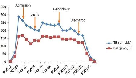
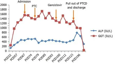
Her liver function tests showed that alanine aminotransferase was 141 IU/L,aspartate aminotransferase was 319 IU/L,alkaline phosphatase (ALP) was 482 IU/L,gammaglutamyl transpeptidase (GGT) was 1349 IU/L,total bilirubin (TB) was 263 μmol/L and direct bilirubin (DB) was 170 μmol/L.Albumin was normal,while cholesterol was 15.78 mmol/L and her tacrolimus trough level was 6.5 ng/mL.CMV-DNA in serum and urine were both negative.
Imaging examinations
Abdominal ultrasound showed intrahepatic and extrahepatic bile duct dilatation.Magnetic resonance cholangiopancreatography suggested an intrahepatic bile duct dilatation and biliary anastomotic stricture.
FINAL DIAGNOSIS
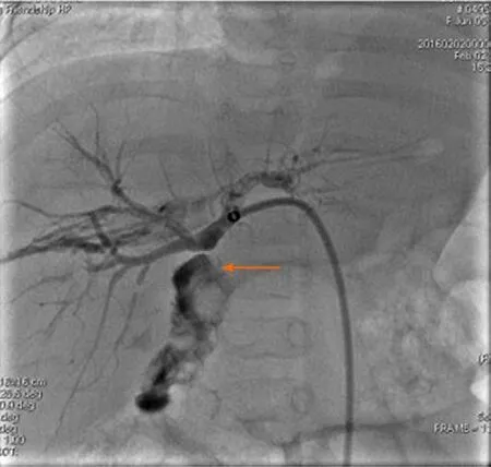
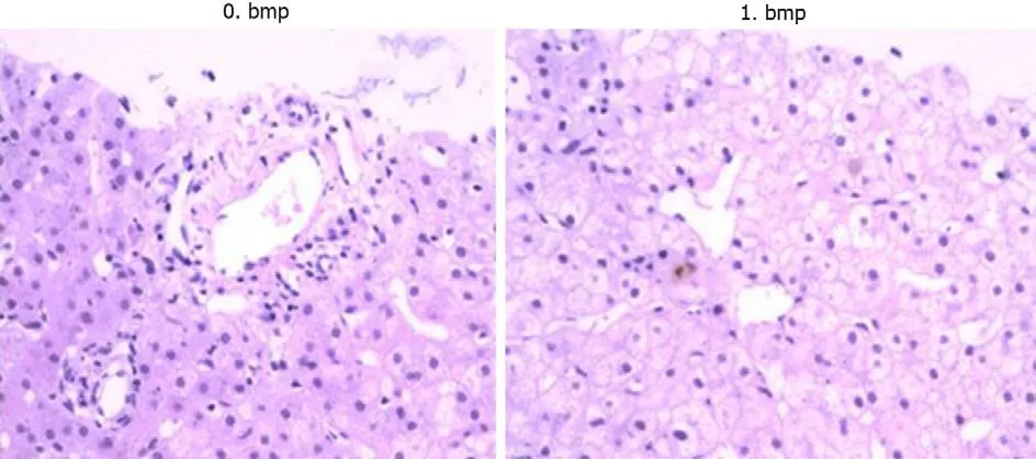
The patient was diagnosed with a biliary complication resulting in jaundice.Consequently,percutaneous transhepatic cholangio drainage (PTCD) was performed on POD 77.As biliary cholangiography revealed biliary anastomotic stricture,biliary drainage was completed after balloon dilatation (Figure1).However,bile drainage volume was quite limited (about 10 mL/d) and its appearance was very pale.Her liver function did not improve after these treatments.TB increased from 200 to 240 μmol/L and DB increased from 130 to 160 μmol/L.Therefore,a liver biopsy was performed on POD 93.The results showed hydropic degeneration and cholestasis of the liver with negative CMV immunohistochemical stains (Figure2).
TREATMENT
Although the serum CMV-DNA was negative,we collected a bile sample from PTCD drainage to test for CMV-DNA and unexpectedly found that bile CMV-DNA was 3 ×10⁶copies/mL.Accordingly,antiviral therapy with ganciclovir was administered at a dose of 10 mg/kg/d.After 2 wk of antiviral therapy,her bilirubin,ALP and GGT began to decline.
OUTCOME AND FOLLOW-UP
After 3-wk of treatment,bile CMV-DNA was tested again and was found to be negative.The child was then discharged after removal of her PTCD tube.Her TB,DB,ALP and GGT all gradually decreased to normal levels in August 2016 (Figures 3 and 4).During the 4-year follow-up period,her liver function has been normal.

Figure1 Biliary cholangiography showing biliary anastomotic stricture (arrow).Biliary drainage was completed after balloon dilatation on postoperative day 77.
DISCUSSION
Biliary complications represent a major problem accompanying LT,with an incidence of 10%-25%[5,6].CMV infection in LT recipients has been associated with many complications and an increased risk of graft loss and death.Indirect complications of CMV include increased risk of hepatic allograft rejection and vanishing bile duct syndrome[7,8].CMV produces inflammatory responses in the vascular endothelium and bile duct,which is related to chronic rejection and biliary complications that can occur after LT[9,10].Although often treated prophylactically,CMV infection and CMV disease still remain a challenge,and continue to be a significant cause of morbidity,economic drain,and fatal disease.
Gotthardt reported that biliary CMV has been detected in LT recipients and was associated with non-anastomotic strictures[4].Occult CMV infection may be associated with the occurrence of biliary complications.Gotthardt suggested that a noteworthy association may exist between occult biliary CMV infection and the development of non-anastomotic biliary complications in orthotopic LT patients.
Chronic CMV latency in bile duct epithelial cells may lead to chronic inflammation,and thus fibrosis of the bile duct system[9].In our case,the patient’s primary disease was biliary atresia.CMV is considered to be a potential cause of the autoimmune destruction of the bile duct in biliary atresia[11].In the study by Gotthardtet al[4],biliary CMV was more common in patients with a recipient-positive status.Therefore,biliary CMV was likely to be a reactivation and not an infection.The detection of pp65 antigen and CMV-DNA in serum lacks sensitivity[12,13].Therefore,bile CMV sampling can be used for diagnosis,especially in patients with jaundice post-LT.As this was a case report,a larger sample size and multicenter investigation are needed in future studies.
CONCLUSION
To the best of our knowledge,this is the first report of the successful treatment of occult CMV cholangitis in a pediatric LT recipient.Our case suggests that detection of CMV in the bile and/or empirical treatment of CMV especially in patients with negative serum CMV-DNA and CMV pp65 may represent a potential option for the treatment of cholestasis post-LT and,in this way,decrease the risk of graft loss.

Figure2 Liver biopsy showing hydropic degeneration and cholestasis of the liver on postoperative day 93.

Figure3 Changes in total bilirubin and direct bilirubin before and after treatment.PTCD:Percutaneous transhepatic cholangio drainage;TB:Total bilirubin;DB:Direct bilirubin;POD:Postoperative day.

Figure4 Changes in alkaline phosphatase and gamma-glutamyl transpeptidase before and after treatment.PTCD:Percutaneous transhepatic cholangio drainage;ALP:Alkaline phosphatase;GGT:Gamma-glutamyl transpeptidase;POD:Postoperative day.
杂志排行
World Journal of Clinical Cases的其它文章
- Assessment of diaphragmatic function by ultrasonography:Current approach and perspectives
- Computer navigation-assisted minimally invasive percutaneous screw placement for pelvic fractures
- Research on diagnosis-related group grouping of inpatient medical expenditure in colorectal cancer patients based on a decision tree model
- Evaluation of internal and shell stiffness in the differential diagnosis of breast non-mass lesions by shear wave elastography
- Real-time three-dimensional echocardiography predicts cardiotoxicity induced by postoperative chemotherapy in breast cancer patients
- Lenvatinib for large hepatocellular carcinomas with portal trunk invasion:Two case reports
