枸橼酸二乙酯、枸橼酸钠和膦甲酸钠对高钙诱导小鼠血管平滑肌细胞钙化的抑制
2020-07-20张冲宇黄玲红欧阳健明
黄 芳 张冲宇 黄玲红 欧阳健明
(暨南大学生物矿化与结石病防治研究所,广州 510632)
0 Introduction
Cardiovasculardisease (CVD)isthe leading cause of mortality and morbidity in patients with chronic kidney disease(CKD),especially in patients receiving long-term dialysis;mortality and morbidity in patients suffering from CVD are mainly attributed to vascular calcification(VC)[1].Arterial media calcification(AMC)is most common in dialysis patients[2].Hyperphosphatemia,hypercalcemia,and high calcium phosphate(Ca×Pi)products are closely related to the occurrence of VC[3].
Increased serum phosphate is considered a major risk factor of cardiovascular events in patients with CKD.However,chronic exposure to high circulating Ca levels is associated with a remarkable increase in the calcification risk among patients with CKD.Villa-Bellosta et al.[3]observed that Ca deposition induced by high Ca is significantly more remarkable than that caused by high phosphate with an equivalent Ca×Pi product.These results indicate the predominant role of Ca over phosphate during calcification.
VC occurs because of the passive precipitation of Ca phosphateand resembles an active process similar to bone formation.Increases in extracellular Ca and phosphate levels stimulate the trans-differentiation of vascular smooth muscle cells(VSMCs)into an osteochondrogenic phenotype,trigger vesicle release,induce apoptosis,and promote further calcification[4].
In this article,we applied high Ca levels to induce the calcification of mouse aortic vascular smooth muscle cells(MOVAS)and added different concentrations of Na3Cit,Et2Cit,and PFA to investigate their effects and possible preventive mechanisms for VC.This strategy also aimed to decrease the morbidity caused by VC.Our study provided new insights into the treatment of patients undergoing hemodialysis and presented a basis for the development of drugs that can act as anticoagulants and inhibit VC.
1 Experimential
1.1 Materials and apparatus
Materials:Mouseaorticsmooth musclecells(MOVAS)were purchased from Shanghai Cell Bank,Chinese Academy of Sciences(donated by Professor Ke Li,Scientific Experiment Center of the Second Affiliated Hospital of Xi′an Jiaotong University).Dulbecco′s Modified Eagle′s Medium(DMEM)was purchased from HyClone Biochemical Products Co.,Ltd.(UT,USA).Fetal bovine serum(FBS)and trypsin were purchased from Gibco Life Technologies(Australia,USA).Alizarin red staining solution,cell lysate and paraformaldehyde were purchased from Xi′an Fiveheart Bio-Tech Co.,Ltd.(Xi′an,China).Bicinchoninic acid(BCA)protein assay kit was purchased from Nanjing Jiancheng Bio-Tech Co.,Ltd.(Nanjing,China).Alkaline phosphatase assay kit,4,6-diamidino-2-phenylindole(DAPI)staining solution and anti-fade fluorescence mounting were all purchased from Shanghai Beyotime Bio-Tech Co.,Ltd.(Shanghai,China).Penicillin and streptomycin were purchased from Beijing Pubo Biotechnology Co.,Ltd.(Beijing,China).Calcium kit was purchased from Beijing biosino Bio-Tech Co.,Ltd.(Beijing,China).Cell proliferation assay kit(Cell Counting Kit-8,CCK-8)was purchased from Dojindo Laboratory(Kumamoto,Japan).Annexin V-FITC/PI(propidiumiodide)apoptosis assay kit was purchased from Becton Dickinson Bioscience Company(San Jose,USA).Phosphonoformic acid was purchased from Chia Tai-Tianqing Pharmaceutical Group Co.,Ltd.(Jiangsu,China).Cell culture plates of 6,24,96-well were purchasedfromWuxiNestBio-TechCo.,Ltd.(WuxiChina).
Et2Cit was synthesized in our laboratory[5].It was characterized and identified by infrared spectroscopy(FT-IR),mass spectrometry(MS)and nuclear magnetic resonance(NMR).Thin-layer chromatography(TLC)and acid value titration tests showed that its mass fraction was 99.27%.
Apparatus:The apparatus used include a microplate reader(Thermo Multiskan MK3,USA),an inverted fluorescence microscope(Leica DMRA2,Germany),light microscope(CKX41,Olympus,Japan),flow cytometer(FACS Aria,BD Corporation,CA,USA).
1.2 Method
1.2.1 Cell culture
4~8 generations of MOVAS were selected as the experimentalobjectwhen calcification occurred[6].Cells were cultured in DMEM supplemented with 10%(V/V)fetal bovine serum(FBS),100 U·mL−1of penicillin and 100 µg·mL−1of streptomycin at 37 ℃ with 5%(V/V)CO2and saturated humidity.
1.2.2 Alizarin red staining
MOVAS calcification was induced as previously described[7-8].A cell suspension(1 mL)with a concentration of 1.5×10⁴cells·mL−1was seeded per well in 24-well plates.The cells were divided into five groups:(A)control group,DMEM culture medium containing 1%(V/V)FBS,0.9 mmol·L−1PI,and 1.8 mmol·L−1Ca2+;(B)high calcium group,DMEM culture medium containing 1%(V/V)FBS and 3 mmol·L−1Ca2+;(C)Et2Cit treatmentgroup,1%(V/V)FBS,3 mmol·L−1Ca2+DMEM culture medium containing 1,4 and 10 mmol·L−1Et2Cit;(D)Na3Cit treatment group,1%(V/V)FBS,3 mmol·L−1Ca2+DMEM culture medium containing 1,4 and 10 mmol·L−1Na3Cit;and(E)PFA treatment group,1%(V/V)FBS,3 mmol·L−1Ca2+DMEM culture medium containing 1,4 and 10 mmol·L−1PFA.The medium of each group was replaced every two days and incubated for 14 d.
After the treatments were administered,the cells were rinsed twice with PBS and fixed with 4%(w/w)paraformaldehyde for 20 min at room temperature.The cells were then washed with PBS twice,incubated with 0.1%(w/w)aqueous Alizarin red solution(pH=8.3)for 30 min,rinsed thrice with PBS,and observed under a light microscope(200×magnification)[9].
1.2.3 Quantification of calcium deposition
The density of seeded cells and experimental grouping as described in Section 1.2.2.After 14 d incubation,cells were decalcified with 0.6 mol·L−1HCl for 24 h at room temperature and calcium content of the supernatants was determined via theo-cresolphthalein complex method using a colorimetric calcium assay kit.After decalcification,cells were solubilized in lysis buffer,and the total protein content was determined using Bicinchoninic acid(BCA)protein assay kit.The calcium content was normalized to the total protein content of the cell lysate.
1.2.4 Morphology observation of apoptotic cells by fluorescence microscope
The density of seeded cells and experimental grouping were the same as those in Section 1.2.2.After 14 d of incubation,the supernatant was removed by suction and the cells were washed thrice with PBS,then,fixed in 4%(w/w)paraformaldehyde at room temperature for 20 min.After the cells were stained with the DAPI solution for 3 min,rinsed thrice times with PBS,and observed under a fluorescence microscope(×400 magnification).
1.2.5 Quantitative analysis of apoptosis rate by Annexin V-FITC/PI double staining assay
Two milliliters of cell suspension with a cell concentration of 1×10⁵cells·mL−1were inoculated per well in six-well plates,experimental grouping was the same as those detected by Alizarin red staining.The specific operation was as follows:collected all cells(included the cells floating on the culture medium).The cells were resuspended and washed with 200µL PBS,followed by centrifugation(1 000 r·min−1,5 min).After that,the supernatant was removed by suction and then 200µL binding buffer was added and mixed thoroughly.The cells were stained with 5µL Annexin V-FITC and mixed thoroughly,then 5µL PI was added to stain cells.After treatment,the cells were detected by flow cytometry.
1.2.6 Cell viability assay
One hundred microliters of cell suspension with a cell concentration of 5×10⁴cells·mL−1were inoculated per well in 96-well plates and experimental grouping was the same as those in Section 1.2.2.Each experiment was repeated in six parallel wells.After incubation for 14 d,the absorbance(A)of each group was detected at 450 nm according to the CCK-8 kit test method.Cell viability was determined using the equation below:
Cell viability=Atreatmentgroup/Acontrolgroup×100%
1.2.7 Alkaline phosphatase(ALP)activity assay
The density of seeded cells and experimental grouping as described in Section 1.2.2.In brief,on day 14 after seeding,alkaline phosphatase(ALP)activity was assessed in the supernatants using an ALP assay kit according to the manufacturer′s instructions.Protein concentrations were quantified using BCA protein assay kit and ALP activity was normalized for cellular protein content,each experiment was repeated in three parallel wells.
1.2.8 ALP staining
The density of seeded cells and experimental grouping as described in Section 1.2.2.After the treatments were administered,the cells were rinsed twice with PBS and incubated with ALP staining according to the manufacturer′s instructions.Stained cells were observed under a microscope(400×magnification).
1.3 Statistical analysis
Experimental data were expressed by mean±standard deviation(±SD).The experimental results were analyzed statistically using SPSS 13.0 software(SPSS Inc.,Chicago,IL,USA).The differences of means between the experimental groups and the control group were analyzed by Tukey′s test.p<0.05 was considered statistically significant.
2 Results
2.1 Ca-induced calcification in MOVAS inhibited by Et2Cit,Na3Cit,and PFA
Alizarin red can chelate with Ca2+and form orange calcium deposits[7,10].Calcium deposition induced by different Et2Cit,Na3Cit,and PFA concentrations was identified by Alizarin red staining after MOVAS were incubated with 3 mmol·L−1Ca2+for 14 d(Fig.1).Calcium deposits were not observed in the control group(NC),whereas calcium deposits were largely detected in the high calcium group(Ca2+).The number of calcified plaques in MOVAS was significantly lower than those in the high Cagroup after Et2Cit,Na3Cit,or PFA was added.This finding indicated that these inhibitors could alleviate VC.These inhibitory effects re-markably increased as the inhibitor concentrations increased.The inhibitory effects of each inhibitor at the sameconcentration exhibited thefollowingtrend:PFA>Na3Cit>Et2Cit.
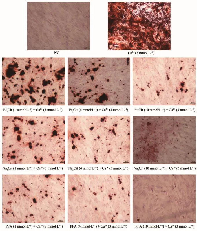
Fig.1 Effect of various Et2Cit,Na3Cit and PFA on high Ca-induced calcium deposition in MOVAS after incubated for 14 d
2.2 Ca-induced calcium deposition in MOVAS reduced by Et2Cit,Na3Cit,and PFA
Calcium deposition induced by different concentrations of the calcification inhibitors were detected through theo-cresolphthalein complex method after MOVAS were incubated with 3 mmol·L−1Ca2+for 14 d.Compared with that in the high calcium group,the amounts of calcium deposited in the Et2Cit,Na3Cit,and PFA treatment groups decreased to varying degrees(Fig.2).Therefore,these inhibitorscould reduce MOVAS calcification in a concentration-dependent manner.Among the amounts of calcium in the three groups,the value obtained in the PFA treatment group was the lowest.Therefore,the strongest ability to prevent VC was observed in PFA,followed by Na3Cit.Among the inhibitors,Et2Cit induced the weakest ability to inhibit VC.These results were consistent with those of Alizarin red staining(Fig.1).
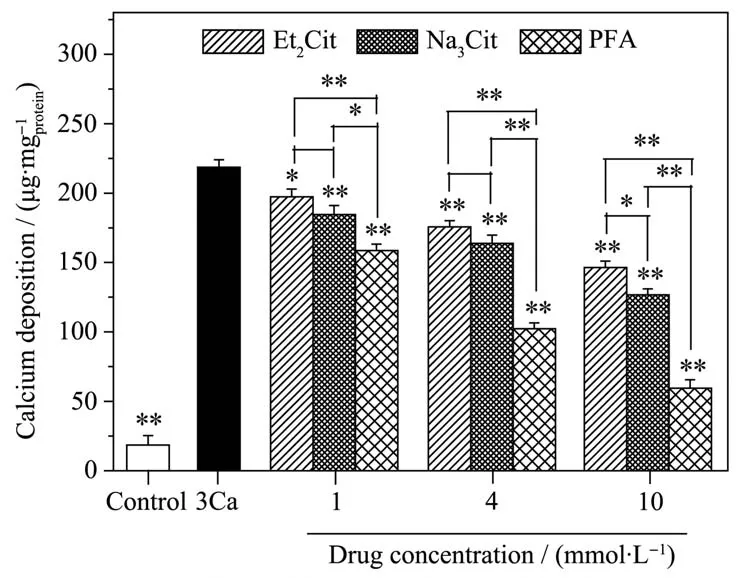
Fig.2 Calcification deposition amount of MOVAS after exposure to various concentration calcification inhibitors for 14 d
2.3 Fluorescence microscopy of the morphological characteristics of cell apoptosis
VSMCs apoptosis is a physiological process that maintains vascular homeostasis,but excessive VSMCs apoptosis is closely related to aneurysm and atherosclerosis[3].The apoptosis of MOVAS treated with different concentrations of the calcification inhibitors for 14 d followed by DAPI staining is shown in Fig.3.

Fig.3 Morphological changes of MOVAS after treatment with varying concentration of calcification inhibitors for 14 d followed by DAPI staining(Arrows show apoptotic cells)
Compared with those of NC,the nuclei in the high calcium group contained condensed chromatin and apoptotic bodies with different sizes and well-preserved but compacted cytoplasmic organelles and nuclear fragments(Ca2+,arrow),which typically characterize apoptotic cells[11].This scenario implied that high calcium concentrations induced MOVAS apoptosis.After low concentrations of Et2Cit(1 mmol·L−1)and Na3Cit(1 and 4 mmol·L−1)were added,the number of broken nuclear fragments significantly decreased.However,high concentrations ofthese inhibitors promoted cell apoptosis.
Different PFA concentrations can promote cell apoptosis.Approximately 10 mmol·L−1PFA stimulated the production of many nuclear fragments and caused cell death because the toxic effects of PFA on cells were more severe than those of the other inhibitors.The number of apoptotic cells in the PFA group was significantly higher than those in the Et2Cit and Na3Cit groups at the same concentration.This finding indicated that PFA failed to alleviate VC by inhibiting apoptosis induced by high Ca.
2.4 Protecting of MOVAS from Ca-induced apoptosis by low concentration of Et2Cit and Na3Cit
The total cell apoptotic ratio(Q2+Q3)of each group was detected by annexin V/PI double staining after the MOVAS were incubated with Et2Cit,Na3Cit,or PFA for 14 d(Fig.4).The apoptotic ratio of the high Ca group(40.17%)was significantly higher than that of NC(12.94%).The cell apoptotic ratios of Et2Cit(1 mmol·L−1)and Na3Cit(1 and 4 mmol·L−1)groups were lower than that of the high calcium group.These results indicated that the three inhibitors could alleviate calcification by inhibiting apoptosis.
The cell apoptotic rates of Et2Cit(4 and 10 mmol·L−1),Na3Cit(10 mmol·L−1),and PFA(1,4,and 10 mmol·L−1)groups were higher than those of the high calcium group.Most of the apoptotic cells were in the late stage possibly because high inhibitor concentrations elicited toxic effects on these cells and thus induced cell death.However,the toxicity of the inhibitors did not prevent calcification.Villa-Bellosta et al.[12]also determined that high concentrations of PFA had greater toxicity on VSMCs.These results are consistent with those illustrated in Fig.3.
2.5 Effects of Et2Cit,Na3Cit,and PFA on cell viability
The cell viability of MOVAS incubated for 14 d under calcification conditions in the presence of different concentrations of calcification inhibitors is shown in Fig.5.Compared with that of NC(100%),the cell viability of the high calcium group decreased to 57.1%,which indicated that high calcium concentrations could induce MOVAS death.
Low concentrations of Et2Cit(1 mmol·L−1)and Na3Cit(1 and 4 mmol·L−1)could significantly reduce the toxic effect of high calcium on the cells,but high Et2Cit(4 and 10 mmol·L−1)and Na3Cit(10 mmol·L−1)concentrations decreased the cell viabilities.
All of the concentrations of the PFA groups(1,4 and 10 mmol·L−1)promoted MOVAS death,and the cell viability decreased to 56.6%,30.5%,and 14.4%,respectively.These results further showed that the toxic effects of PFA on MOVAS were greater than those of the other inhibitors.
2.6 Suppressing Alkaline phosphatase(ALP)expression by Et2Cit,Na3Cit and PFA
An increased ALP activity is an early indicator of VSMCs osteogenic conversion,and this indicator biologically functions in the cleavage of the extracellular calcification inhibitor inorganic pyrophosphate(PPi)and maintenance of Pi/PPi homeostasis and normal bone tissue calcification[7,13].In osteogenesis,ALP is secreted to the extracellular matrix to form a locally high ALP concentration and decrease PPi,which forms an amorphous phosphate crystal and triggers mineralization[14].
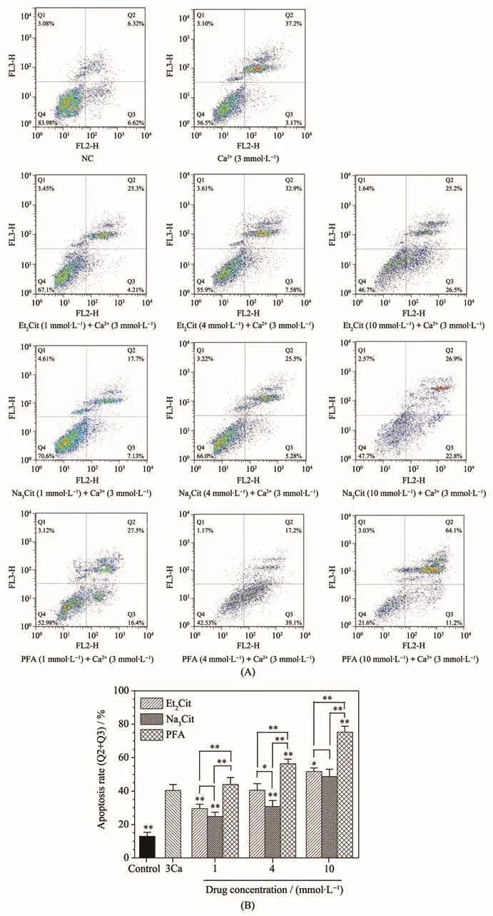
Fig.4 Apoptosis ratio of MOVAS after incubated for 14 d under calcifying conditions in the presence of various concentration calcification inhibitors:(A)Dot plots of cell apoptosis and necrosis detected by flow cytometry;(B)Quantitative analysis of cell apoptosis rate(Q2+Q3)change

Fig.5 Cell viability assay of MOVAS after incubation with varying concentrations of calcification inhibitors for 14 d
An ALP staining kit was used to observe the ALP expression in MOVAS(Fig.6A).Our results showed that the different concentrations of Et2Cit,Na3Cit,and PFA could decrease the ALP expression induced by high calcium in a concentration-dependent manner and inhibit the phenotypic transformation of smooth muscle cells into osteoblast-like cells.These results are consistent with those shown in Fig.6B.The ALP activity in the high calcium group sharply increased compared with that in NC(Fig.6B),and this result indicated that high calcium levels could promote the osteogenic conversion of MOVAS.The ALP activities in the Et2Cit,Na3Cit,and PFA treatment groups were lower than those in the high calcium group.These findings revealed that these inhibitors could inhibit the phenotype transformation of MOVAS and alleviate VC.Thus,their inhibitory effects were concentration-dependent.In particular,the inhibitory effects of these inhibitors at the same concentration exhibited the following trend:PFA>Na3Cit>Et2Cit.
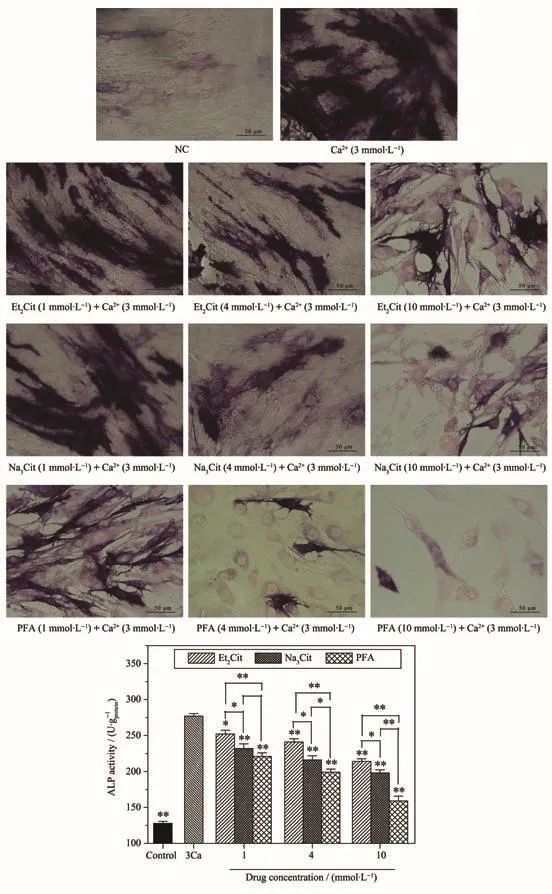
Fig.6 Changes in ALP expression of MOVAS after treated with various concentration calcification inhibitors for 14 d:(A)Observation of ALP expression;(B)Quantitative histogram of ALP expression
3 Discussion
Actually,Na3Cit has been clinically used as anticoagulant[15]since the carboxyl groups in Na3Cit can chelate with calcium ions(Ca2+)to form calcium citrate,so as to reduce the concentration of free Ca2+ions in the plasma,thus achieving the purpose of anticoagulation in vitro[16-18].This indicates that Na3Cit potentially could be used as a calcification inhibitor that it can decrease blood calcium levels by chelating Ca2+,thereby reducing Ca×Pi deposition,and then inhibiting vascular calcification(VC)induce by high Ca.However,considering that Na3Cit has serious side effects such as low calcium,high sodium and acid-base disorders,we converted two carboxyl groups(-COOH)in citric acid into ester groups(-COOC2H5)to form a new anticoagulant diethyl citrate(Et2Cit),which has the following characteristics[5,19,20]:(1)Et2Cit still has oxygen atoms(including oxygen in the ester groups and in the hydroxyl group)that can chelate with Ca2+ions,which can reduce the concentration of Ca2+ions in the blood.(2)Due to the binding of ethyl(-C2H5)near carboxyl oxygen atoms,there is a certain steric resistance in the coordination of Et2Cit and Ca2+ions,which makes the chelating ability of Et2Cit and Ca2+weaker than that of Na3Cit and Ca2+.It also means that the chelate formed by Et2Cit and Ca2+ions exhibited a stronger ability to release Ca2+ions than calcium citrate,which helps to overcome the problems of hypocalcemia and hypercalcemia caused by Na3Cit as an anticoagulant.(3)The ethyl(-C2H5)group has a moderate volume,which means the molecular weight does not increase significantly after it replacing the H atom in-COOH group,thus the passage of Et2Cit through dialysis membrane will not be affected.That is,Et2Cit as an anticoagulant can avoid the occurrence of hypocalcemia,hypercalcemia,hypernatremia and alkalosis of Na3Cit as an anticoagulant,so Et2Cit may become a new anticoagulant[21].However,after converting Na3Cit to Et2Cit,it means that the amount of carboxyl group reduced,so whether Et2Cit has a calcification inhibitory effect and whether its calcification inhibitory effect is significantly different from Na3Cit are one of the goals of our investigation.
Similar to Na3Cit and Et2Cit,PFA also contain carboxyl group that can chelate Ca2+[12,20].Moreover,PFA is a phosphonocarboxylic acid analogous to PPi which is a firmly established calcification inhibitors,it prevents VC by a physicochemical reaction that inhibits the formation and aggregation of calcium phosphate crystals,prevents the transition of amorphous calcium phosphate into hydroxyapatite(HAP),and delays the aggregation of apatite crystals[22-23].Villa-Bellosta et al.[12]revealed that PFA mainly and directly inhibits mineralization and HAP formation,acting similarly to PPi and to bisphosphonate drugs.Their results showed that the chemical structure for preventing HAP formation requires the presence of at least one phosphate group and an acidic group in another position,which can be either a phosphate or a carboxylic group.Bisphosphonates bind to the calcium of HAP by chemisorption with oxygen from each phosphonate group,and PFA acts similarly with the corresponding carboxylic group.However,the mechanisms and differences of Na3Cit,Et2Cit,and PFA in inhibiting high calciuminduced calcification have not been reported.
In our article,we found that low Et2Cit and Na3Cit concentrations decreased VC by inhibiting apoptosis,in contrast PFA promoted cell apoptosis(Fig.3),however,the inhibitory effects of these three drugs on calcium phosphate deposition at the same concentration showed the following trend: PFA>Na3Cit>Et2Cit(Fig.1).The cause of the difference in their inhibition of calcification may be related to their chemical structure(Fig.7).Although PFA contains only one carboxyl group for chelating Ca2+,its calcification inhibitory effect is significantly stronger than that of Na3Cit with multiple carboxyl functional groups.On the one hand carboxylic group in PFA can chelate Ca2+,so that reduce calcium phosphate deposition,on the other hand PFA can bind to the calcium of HAP by chemisorption with oxygen from phosphonate group.Therefore,our findings can provide some inspiration for the development of more effective drugs that inhibit vascular calcification through comparing the calcification inhibitory effects of these three drugs.
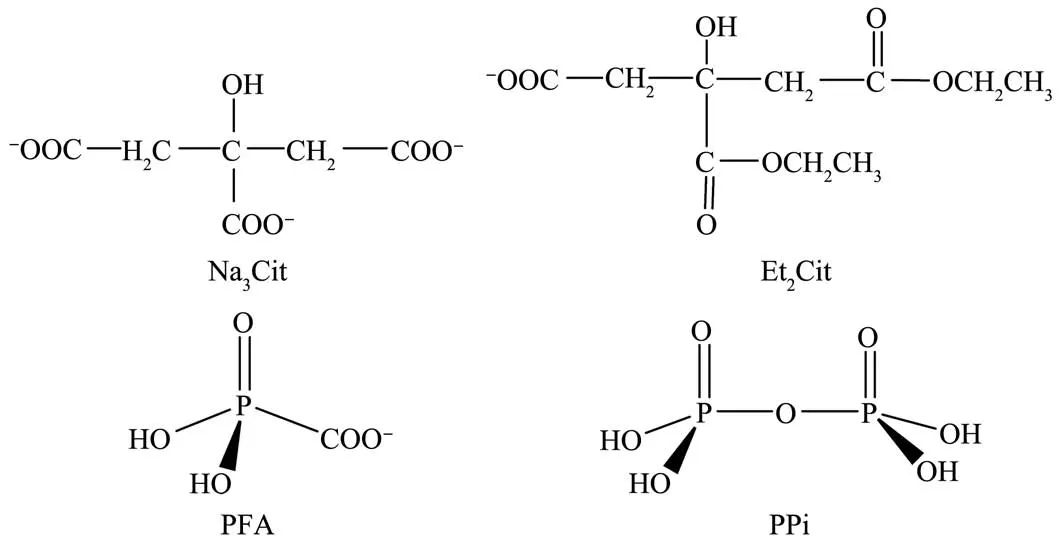
Fig.7 Chemical structure of Na3Cit,Et2Cit,PFA and PPi
3.1 Preventing VC of Et2Cit,Na3Cit,and PFA by inhibiting calcium phosphate deposition
Et2Cit,Na3Cit,and PFA significantly prevented MOVAS calcification induced by high Ca levels,and the inhibitory effects of these substances exhibited concentration dependence.However,the differences in the preventive effects of various inhibitors on VC were attributed to the mechanisms of citrate-series drugs and PFA(Fig.8).
In calcification,Ca2+and PO43−released by apoptotic bodies accumulate in the membrane of matrix vesicles(MVs)or adjacent to the membrane.Ca2+also promotes the release of matrix vesicles of VSMCs,and calcium phosphate minerals precipitate when sufficient Ca2+and PO43−have accumulated within MVs.It is widely believed that apatite crystals are formed through a series of conversions from amorphous minerhydroxyapatiteals,to octacalcium phosphate,to the stable form of highly insoluble HAP[24].

Fig.8 Proposed schematic illustration of the cellular and molecular mechanism of Et2Cit,Na3Cit and PFA inhibiting the calcification of MOVAS induced by high Ca
Na3Cit,Et2Cit,and PFA could reduce the calcification degree of MOVAS exposed to high Ca concentration(Fig.1 and 2).The inhibitory effects of these inhibitors also increased as their concentrations increased.The chemical mechanisms of Na3Cit and Et2Cit for reducing calcification in a dose-dependent manner are due to the fact that they all contain COOH groups(Fig.7)that can chelate Ca2+ions on the surface of HAP crystal,thereby inhibiting the formation and further growth of HAP[25-26].Hu et al.[27]found that adding citrate during the synthesis of HAP significantly reduced the size and thickness of HAP crystals,and the size of crystals decreased with the increase of citrate concentration.PFA also contain COOH group for chelating Ca2+ion,moreover,PFA mainly and directly inhibits mineralization and HAP formation,acting similarly to PPi,that is PFA can bind to the calcium of HAP by chemisorption with oxygen from phosphonate group,and in the range of 0.01~5 mmol·L−1,PFA inhibits high-phosphate induced calcification in a concentration-dependent manner[12].
3.2 Osteogenic conversion of MOVAS which inhibited by Et2Cit,Na3Cit and PFA to alleviate VC
The osteochondrocytic conversion of VSMCs plays a key role in calcification.Clinical and in vitro studies have shown that high phosphate and high calcium levels induce the trans-differentiation of VSMCs into osteoblast-like cells and mineralization of the extracellular matrix[24].
ALP,which is an important marker of osteogenic differentiation,can enhance calcification.Its upregulation can degrade calcification inhibitors,such as PPi,and consequently promote calcification development[28].Sortilin regulates the loading of tissue-nonspecific alkaline phosphatase(TNAP)calcification protein into extracellular vesicles,thereby leading to high mineralization competence in the extracellular milieu[29].Normal VSMCs are composed of low ALP concentrations,but calcified vessels and heart valve tissues contain significantly increased ALP concentrations[13].
Different concentrations of Na3Cit,Et2Cit,and PFA strongly inhibited the ALP activity induced by high calcium levels(Fig.6A and 6B).This finding indicated that the three inhibitors decreased the gene expression associated with the transdifferentiation of MOVAS into osteoblast-like cells to prevent VC.The inhibitory effects of PFA on the osteogenic conversion of MOVAS were significantly higher than those of Et2Cit and Na3Cit at the same concentrations.Research shows that Calcium deposits promote the transformation of VSMCs into osteoblast-like cells[22],and preventing Calcium deposits can inhibit osteogenic differentiation[30].Therefore,we speculate that Na3Cit,Et2Cit,and PFA reduce the osteogenic conversion of MOVAS through inhibiting Ca phosphate deposition.
3.3 Decreasing VC of low concentrations of Et2Cit and Na3Cit by inhibiting apoptosis
Low concentrations of Et2Cit and Na3Cit significantly decreased the high-calcium-induced apoptosis of MOVAS(Fig.3 and 4).Fig.5 also suggests that low Et2Cit and Na3Cit concentrations can alleviate the toxic effects of high calcium levels on cells,whereas high Et2Cit and Na3Cit concentrations promote cell apoptosis.
The uptake of extracellular calcium stimulates VSMC apoptosis and produces mineralization-competent MVs.These bodies are similar to MVs secreted by chondrocytes during bone development,and their properties include the absence of calcification inhibitors,formation of nucleation sites,and accumulation of matrix metalloproteinase-2[31].MVs and apoptotic bodies are produced by apoptotic cells,and these small membrane-bound bodies form a nidus for Ca phosphate deposition in the form of HAP;apoptotic cell death releases more Ca that may stimulate further apoptosis to accelerate calcification[2,32].Sodium citrate can effectively decrease the protein expression of CCAAT/enhancer-binding protein homologous protein to inhibit endoplasmic reticulum stress in rats with adenineinduced CRF[33].Ciceri et al.[34]also found that iron citrate reduces high phosphate-induced VC by inhibiting apoptosis.A sudden increase in citrate concentrations inside the cells may immediately stimulate the Krebs cycle and induce apoptotic cell death[35].However,Zavaczki etal.[36]found that the toxic effect of H2S on VSMCs increases in a concentration-dependent manner,whereas the preventive effect of VC becomes increasingly evident as H2S concentration increases.
Our results showed that Et2Cit,Na3Cit,and PFA effectively inhibits MOVAS calcification induced by high calcium,while heparin,as an anticoagulant,does not affect VC in rats with CKD and secondary hyperparathyroidism[16].In addition,our previous results have indicated that Et2Cit,Na3Cit,and PFA can also prevent the high Pi-induced calcification of MOVAS[37].Thus,Et2Cit and Na3Cit can be widely used in clinical applications with low-cost effectiveness compared to the high cost-effectiveness of drugs,such as lanthanum carbonate,sevelamer,and calcium-sensing receptor agonists,for the treatment of patients with CKD and VC[38-39].Although the calcification inhibitory effects of different inhibitors in a concentration-dependent manner,high concentrations of the three inhibitors are largely toxic on cells,which possess a certain reference value for blood anticoagulant and calcification treatment.
Although Et2Cit,Na3Cit,and PFA can effectively inhibit the ALP expression in MOVAS induced by high calcium and low concentrations of Et2Cit,Na3Cit can inhibit apoptosis induced by high calcium.However,the specific role of the three inhibitors in specific cellular pathways is unclear.
The strong cytotoxic effects of high concentrations of these inhibitors on MOVAS should be considered.Calcificationin vitroand in patients with CKD substantially varies[40].Therefore,the roles of Et2Cit and Na3Cit should be elucidated.
4 Conclusions
Et2Cit,Na3Cit,and PFA can decrease the high calcium-induced calcification of MOVAS.The inhibitory effects of different inhibitors at the same concentration showed the following trend:PFA>Na3Cit>Et2Cit.Low Et2Cit(1 mmol·L−1)and Na3Cit(1 and 4 mmol·L−1)concentrations prevented VC mainly by reducing the high-Ca-induced apoptosis.High concentrations of Et2Cit(4 and 10 mmol·L−1)and Na3Cit(10 mmol·L−1)and varying PFA concentrations mainly inhibited the phenotypic transformation of VSMCs into osteoblastic cells.The inhibitory effects of these substances were positively correlated with their concentrations.Therefore,further investigations should be performed to obtain additional mechanistic insights into the mode of action of these inhibitors in VC prevention.This study may provide a basis for novel clinical applications of anticoagulant drugs and a theoretical foundation for the prevention and treatment of VC in patients with CKD.
