Effect of Milbemycin oxime on Toxocara canis Eggs and Larvae
2020-07-15HuWanjunDingLiangjunChenChunliXuJiaandLiJichang
Hu Wan-jun, Ding Liang-jun, Chen Chun-li, , Xu Jia, and Li Ji-chang, *
1 College of Veterinary Medicine, Northeast Agricultural University, Harbin 150030, China
2 College of Life Sciences, Northeast Agricultural University, Harbin 150030, China
3 Heilongjiang Key Laboratory for Animal Disease Control and Pharmaceutical Development, Harbin 150030, China
4 Raohe Customs District P. R. China, Shuangyashan 155700, Heilongjiang, China
Abstract: Toxocara canis (T. canis) is one of the most important zoonotic parasites of dogs. The aim of the present study was to perform an in vitro analysis of the effect of Milbemycin oxime on T. canis eggs following exposure to a concentration gradient of the drug and to determine the in fl ammatory reaction produced by the infective T. canis larvae in mice. The present study was undertaken using the model nematode, T. canis, to investigate the effect of Milbemycin oxime on T. canis eggs and larvae. T. canis eggs were exposed to a concentration gradient of Milbemycin oxime in vitro, the higher concentration of Milbemycin oxime was, the lower percentage of infective stage larvae was. Light micrographs showed that Milbemycin oxime induced eggs dissolved and eggshell broken. Histological analyses of mice that stained with hematoxylin and eosin (H&E) showed in lower dosing (10-7 and 10-8 g · mL-1)drug-treated groups, atrophy of alveolar space and interalveolar septum thickening appeared, in fl ammatory in filtrates accompanied with erythrocytes around blood vessels and bronchioles presented. In higher dosing (10-6, 10-5 and 10-4 g · mL-1) drug-treated groups,low-grade or no pathological changes occurred, indicating that Milbemycin oxime could obviously decrease the in fl ammatory reaction produced by the infection of T. canis larvae in vivo.
Key words: in vitro, in vivo, Milbemycin oxime, Toxocara canis
Introduction
Toxocara canis(T. canis) is an intestinal parasite that infects dogs, foxes and other canid species,with zoonotic potential for human beings. It has indomitable vitality. This ubiquitous and prolific nematode has a complicated life cycle and is widely distributed throughout the world (Nijsseet al., 2016;Banethet al., 2015).Toxocariasisis a worldwide public health problem (Chenet al., 2018; Fakhriet al.,2018), because the presence of dogs in public areas,for example, parks and play-grounds, creates the infection conditions that can cause zoonosis, due to widespread fecal contamination of these places(Fankhauseret al., 2016).Milbemycin oximeis a member of macrolide antiparasitic antibiotics produced through fermentation byStreptomyces hygroscopicussubspecies aureolacrimosus (Genchiet al., 2004), which has been shown to be effective to be used alone (Böhmet al., 2014).In vivo, it has been shown to be highly effective at a recommended dosage of 0.5-1.0 mg · kg-1once a month against canine heartworm (Genchiet al., 2004). To the best of our knowledge, there is not enough data available in the literature onin vitroeffect ofMilbemycin oximeon parasites, only Lee and Terada (1992) examined the neuropharmacological mechanisms underlying the action ofMilbemycin oximeon the motilities ofAngiostrongylus cantonensisandDirofilariain vitro.However, the effect ofMilbemycin oximeonT. caniseggs is not known. The objective of the present study was to perform anin vitroanalysis of the effect ofMilbemycin oximeonT. caniseggs following exposure to a concentration gradient of the drug and to determine the inflammatory reaction produced by the infectiveT. canislarvae in mice.
Materials and Methods
Collection and maintenance of T. canis eggs
Female worms ofT. caniswere collected from the intestines of naturally infected dogs in a vertical laminar fl ow cabinet under sterile conditions and maintained in sterile phosphate buffered saline (PBS, pH 7.0) and 2%glucose, supplemented with 10% fetal bovine serum(FBS), 50 μg · mL-1gentamicin and 50 IU · mL-1, then were held in an incubator at 37℃ in a 95% carbon dioxide (CO2) atmosphere. To collect eggs, the female roundworms were removed to a new culture flask every 24 h or 72 h until they exhausted. The harvested solutions includingT. caniseggs were centrifuged at 1 500×g for 10 min, washed for 5 times with distilled water and the solution containing 10 000 eggs per mL was obtained. All the experiments performed with animals were in accordance with standard of the local ethics committee in China.
Effect of drug on embryogenesis of T. canis eggs
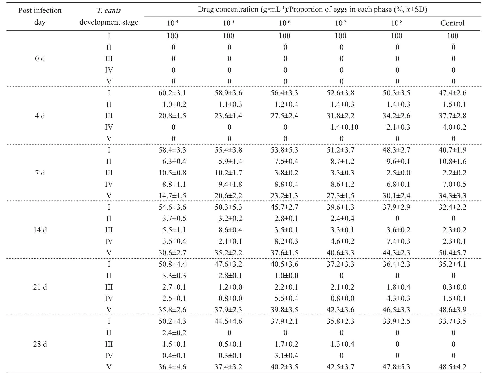
The obtainedT. caniseggs were randomly divided into the control group and five treatment groups. Every group contained approximately 2 000 eggs per mL and housed in 6-well tissue culture plates containing PBS and 2% glucose supplemented with 50 μg · mL-1gentamicin and 50 IU · mL-1, then equal-volume different concentrations ofMilbemycin oxime(>98%purity, Zhejiang Hisun Pharmaceutical Co., Ltd., China)dissolved in dimethyl sulfoxide (DMSO) were added into the five treatment groups, so that the final drug concentrations were 10-4, 10-5, 10-6, 10-7and 10-8g · mL-1.The control group medium only contained DMSO, but noMilbemycin oxime. All the plates were incubated for 28 days at 29℃ supplemented with 5% CO2. The culture medium was replaced with fresh medium on 4, 7, 14, 21 and 28 days after the drug exposure and embryogenesis ofT. caniseggs was assessed by counting the number of parasites, respectively.The relative portions of eggs at different phases of developments (I-V) were recorded by microscopic examination of 10 µL culture samples placed on clean glass slide and covered with cover slips. All the assays were performed in triplicates and all the data were presented as the mean±standard deviation.
Effect of drug on infective T. canis larvae in mice
The eggs ofT. caniswere obtained as above and maintained in PBS (pH 7.0) containing 1% formalin at 27℃ until development to the infective stage,the second-stage larvae were hatched, according to previous study (Savigny, 1975). These larvae with normal morphous and viability observed under microscopy were treated withMilbemycin oxime(the final drug concentrations were 10-4, 10-5, 10-6, 10-7and 10-8g · mL-1) for 24 h, they were washed 5 times with distilled water, each followed by successive sedimentations by centrifugation at 1 500×g for 5 min pending the assay of infectivity in mice. Seventy BALB/c mice with 18-22 g weight were given a standard commercial diet with free access to water and maintained on a cycle of 12 h light and 12 h dark at room temperature. An acclimation period of one week was observed prior to initiation of the trial.Animals were randomly allocated into seven groups.Five groups were orally infected with approximate 1 000 infective drug-treated larvae in 0.5 mL PBS (1%formalin, pH 7.0) per mouse by gavage. One group(positive control) was orally infected with approximate 1 000 infective no drug-treated larvae in 0.5 mL PBS (1% formalin, pH 7.0) per mouse by gavage.One group only oral 0.5 mL PBS (1% formalin, pH 7.0) per mouse by gavage was served as the negative control. Each group was consisted of 10 mice (five males and five females). After 5 days post-inoculation,mice in each group were killed by cervical dislocation with ether anesthesia. The lungs were removed to record the macroscopic injury immediately, then pieces of the lungs were fixed in 10% formalin, embedded in paraffin,sectioned and stained with hematoxylin and eosin (H&E)for pathological examination under light microscopy.
Results
Effect of drug on embryogenesis of T. canis eggs
Table 1 showed embryograms from the control andMilbemycin oxime-treatedT. caniseggs during 28 days of culture. At the end of the study (the 28th day),percentages of V-stage larvae were (36.4±4.6)%,(37.4±3.2)%, (40.2±3.5)%, (42.5±3.7)% and (47.8±5.3)%, respectively in the drug-treated groups at concentrations of 10-4, 10-5, 10-6, 10-7and 10-8g · mL-1and percentage of V-stage larvae of the blank control was (48.5±4.2)%. Light micrographs (LM) of V-stage embryogenesis ofT. caniseggs after exposure to a concentration gradient ofMilbemycin oximeis shown in Fig. 1. Light micrographs showed thatMilbemycin oximebroke the eggshell, destroyed the larvae (Figs. 1B, C and D) and dissolved larvae in the eggshell (Fig. 1E and F).

Table 1 Effect of Milbemycin oxime on T. canis eggs

Fig. 1 Light micrographs (LM) of V-stage embryogenesis of T. canis eggs after exposure to Milbemycin oxime (magni fication, ×400)
Effect of drug on infective T. canis larvae in mice
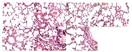
No abnormality was observed in any of the treated groups within 2 days after larvae infection; however,mice exhibited poor appetite and piloerection later.On the 5th day, the most remarkable finding at necropsy was red hepatization and hemorrhagic lung consolidation in the positive control group. Histopathological examinations showed dose-dependent in agreement with the macroscopic observation (Fig. 2).Pieces of the lungs were stained with H&E, pulmonary structure of the negative control is shown in Fig. 2A.The positive control showed atrophy of alveolar space and interalveolar septum thickening appeared, with scattered hemocytes in it and in fl ammatory in filtrates accompanied with erythrocytes around blood vessels and bronchioles presented (Fig. 2B). Compared with the control, the groups treated with higher dosingMilbemycin oxime(10-4, 10-5and 10-6g · mL-1) showed no obvious pathological changes in lungs (Figs. 2C, D and E). Compared with the control, the group treated withMilbemycin oximeat concentration of 10-7g · mL-1showed extensive areas of hemocytes and acidophilia anachromasis materials presented in bronchi and alveolar space (Fig. 2F) and the group treated withMilbemycin oximeat concentration of 10-8g · mL-1showed in fl ammatory in filtrates with the presence of erythrocytes around blood vessels and bronchioles (Fig. 2G).

Fig. 2 Pathological examination of mouse lungs after 5 days post-inoculation (magni fication, ×40)
Discussion
Gastrointestinal nematodes commonly cause infections in dogs, which are of particular concerns of public and veterinary health issues (Jesuset al., 2015). Among these nematodes,T. canishad zoonotic potential(Trasviña-muñozet al., 2017) and was found in the intestine of dogs (Banethet al., 2015). In the development and using of more effective and less toxic chemotherapeutic agents, the examination and characterization of their actions in a simplerin vitrosystem is important. The present study usedT. canisto investigate the effect ofMilbemycin oximeonT. caniseggs and larvae. The results were in line with Schnitzleret al. (2012) thatMilbemycin oximereducedToxocaraeggs in feces up to 99.9%. Fankhauseret al. (2016)demonstrated that 98% efficacy ofMilbemycin oximewith afoxolaner in dogs infected withT. canisafter 7 days of treatment. The results indicated the effects of the drug had dose-dependent on the embryogenesis ofT. caniseggs. The results showed thatMilbemycin oximenot only could lead to the death of the eggs,but also could prolong the eggs development, which was also in agreement with the effects of ivermectin and moxidectin on nematodes (Ardelliet al., 2009;Tompkinset al., 2010). Previous reports demonstrated the anthelmintic ability ofMilbemycin oximein combination with other anthelmintic compounds or ectoparasiticides or using alone to kill the gastrointestinal parasites, such asT. leonina,T. canis,A. caninumandA. braziliense(Nolan and Lok, 2012; Schnitzleret al.,2012; Bienhoffet al., 2013). The present study also scrutinized the effects ofMilbemycin oximeagainstToxocara caniseggs and its larvae infection. The pathological findings indicated thatMilbemycin oximecould obviously decrease the inflammatory reaction produced by the infection ofT. canislarvaein vivo.
Conclusions
In conclusion, the study reported the effect ofMilbemycin oximeonT. canis, which showed thatMilbemycin oximenot only could block embryogenesis ofT. caniseggsin vitro, but also could decrease the inflammatory reaction produced by the infection ofT. canislarvaein vivo. To better understand the effect ofMilbemycin oximeagainstT. canis, future studies should include the examination of its molecular drug targets, particularly the glutamate-gated chloride ion channels.
杂志排行
Journal of Northeast Agricultural University(English Edition)的其它文章
- Evidence-based Landscape Design of University Campus: Relationship Between Characteristics of Outdoor Environments and Actual Uses of Students
- Design and Experimental Evaluation of a Spiral Feeding Device Based on Friction Characteristics of Wheat Straw
- Study on Rice Yield Estimation Model Based on Quantile Regression
- AcrAB Efflux Pump in Fluoroquinolone Resistant Salmonella gallinarum Induced by Cipro fl oxacin Selective Pressure
- Localization of RanBP1 in Early Embryonic Development of Mice
- Expression, Puri fication and Structural Characterization of Mouse Talin-1 Head Δ139-168
