Effects of electroacupuncture on the behaviors and expressions of hippocampal neurotransmitters and Bax/Bcl-2 proteins in rat models of anxiety disorder
2020-04-21ZhaoFeiyi赵非一YanHaixia燕海霞ZhaoYingxia赵英侠XuHong许红HongYufang洪钰芳MaQiayi马恰怡XuYan徐燕FuQiangqiang付强强
Zhao Fei-yi (赵非一), Yan Hai-xia (燕海霞), Zhao Ying-xia (赵英侠), Xu Hong (许红), Hong Yu-fang (洪钰芳),Ma Qia-yi (马恰怡), Xu Yan (徐燕), Fu Qiang-qiang (付强强)
1 Department of Nursing, School of International Medical Technology, Shanghai Sanda University, Shanghai 201209, China
2 School of Basic Medical Sciences, Shanghai University of Traditional Chinese Medicine, Shanghai 201203, China
3 Shanghai Municipal Hospital of TCM Affiliated to Shanghai University of Traditional Chinese Medicine, Shanghai 200071, China
4 Shanghai Changning Tianshan Traditional Chinese Medicine Hospital, Shanghai 200051, China
5 Research Department, Yangpu Hospital, Tongji University School of Medicine, Shanghai 200090, China
Abstract Objective: To investigate the effects of electroacupuncture (EA) on the behaviors of rat with anxiety disorder, and the expressions of hippocampal neurotransmitters including 5-hydroxytryptamine (5-HT), norepinephrine (NE) and dopamine (DA), and the expressions of hippocampal B-cell lymphoma-2 (Bcl-2) and Bcl-2 associated X (Bax).Methods: Forty-six male Wistar rats were randomly divided into a control group (n=10), a model group (n=12), an EA group (n=12), and a drug group (n=12). Except the control group, the other three groups were established into rat models of anxiety disorder using uncertain empty bottle stimulation. Rats in the EA group and the drug group received corresponding interventions for 15 consecutive days [EA group was given EA at Baihui (GV 20) and Sanyinjiao (SP 6); the drug group was given aqueous solution of alprazolam via intragastric administration]. After intervention, all four groups received open-field test (OFT) and elevated plus-maze (EPM) for behavioral evaluations. The expressions of 5-HT, NE and DA in hippocampus were determined by fluorescence spectroscopy (FS) while the expressions of Bcl-2 and Bax proteins in hippocampus were determined by Western blot (WB).Results: The OFT horizontal scores in the control group, EA group and drug group were significantly higher than that in the model group (all P<0.05), and the difference between the EA group and the drug group was statistically insignificant(P>0.05); the OFT vertical scores in the model group, EA group and drug group were significantly lower than the score in the control group (all P<0.05). The EPM percent of open-arm entries (OE%) in the control group, EA group and drug group was higher than that in the model group (P<0.05), and the differences among these three groups were statistically insignificant (P>0.05); though the percent of open-arm total time (OT%) in the EA group was lower than that in the control group (P<0.05), the difference was statistically insignificant when compared with the drug group(P>0.05), and it was significantly higher than that in the model group (P<0.05). The expression of 5-HT in the EA group was higher than that in the control group (P<0.05); the expression of 5-HT in the EA group was significantly lower than that in the model group (P<0.05); the difference between the EA group and the drug group was statistically insignificantly (P>0.05). The expression of NE in the model group was significantly higher than that in the other three groups (P<0.05), and there was no significant difference among these three groups (P>0.05). The expression of DA in the EA group was significantly higher than that in the control group and the drug group (both P<0.05), while the difference between the EA group and the model group was statistically insignificant (P>0.05). The expression of Bax in the model group was significantly higher than that in the other three groups (all P<0.05), whereas the expression of Bcl-2 in the model group was significantly lower than that in the other three groups (all P<0.05), and the differences in both Bax and Bcl-2 among the other three groups were statistically insignificant (all P>0.05). Bax/Bcl-2 in the EA group was significantly higher than that in the control group (P<0.05) and lower than that in the model group (P<0.05), and the difference was statistically insignificant when compared with the drug group (P>0.05).Conclusion: EA shows promising effects in attenuating rats’ anxiety disorder, which may be achieved by the down-regulation of the expressions of 5-HT and NE in the hippocampus and/or inhibition of hippocampal neuronal apoptosis.The efficacy is comparable to that of intervention with alprazolam.
Keywords: Acupuncture Therapy; Electroacupuncture; Behavior, Animal; Neuroregulators; 5-hydroxytryptamine;Apoptosis; Anxiety Disorders; Rats
Anxiety is defined as a persistent sense of fear, or a feeling of disaster and apprehension, accompanied by strong tension and uneasiness[1]. Anxiety disorder refers to a group of mental disorders characterized by psychological and physical anxious symptoms and behaviors without organic brain diseases or other mental disorders[2]. With an overall lifetime prevalence of 24.9%, anxiety disorder is one of the most common health complaints in industrialized nations worldwide[3].Due to the inevitable side effects and drug dependence of anti-anxiety agents, quite a few patients have turned to complementary and alternative medicine (CAM)therapy. In America, 42.7% of the adults suffering from anxiety reported having sought help from CAM in the past few years[4]. In addition, Davidson JR, et al[5]found that 25.3% of the patients in the USA and UK visiting CAM centers met the criteria for at least one anxiety disorder. Despite acupuncture, a representative therapy of CAM, has been increasingly used in the management of anxiety and anxiety disorders[6], its anti-anxiety mechanism is still indistinct, which arouses our interest in investigating the underlying mechanism of acupuncture in emotion regulation via animal experiment.
In this study, uncertain empty bottle stimulation was introduced to establish rat models of anxiety disorder[7].After receiving electroacupuncture (EA) or anti-anxiety agent, the rats’ behavior was evaluated using open-field test (OFT) and elevated plus-maze (EPM) tests. Changes in hippocampal neurotransmitters related to anxiety,including 5-hydroxytryptamine (5-HT), norepinephrine(NE) and dopamine (DA) were detected by fluorescence spectrophotometry (FS). Apoptosis has been reported widely involved in the pathogenesis of neuropsychiatric diseases[8-9]. Considering both B-cell lymphoma-2 (Bcl-2)and Bcl-2 associated X protein (Bax) play important roles in the regulation of apoptosis[10], the expressions of hippocampal Bax and Bcl-2 were determined as well in order to explore if apoptosis was also involved in EA regulation of anxiety, which is also the innovation point of this research.
1 Materials and Methods
1.1 Animals and grouping
A total of 46 male Wistar rats [8 week-old, cleangrade, specific pathogen free, weighing (230±10) g]were provided by Shanghai SLAC Laboratory Animal Co.,Ltd. (No. 2015000501998). They were randomly divided into 4 groups: a control group (n=10), a model group(n=12), an EA group (n=12) and a drug group (n=12).The rats were housed separately at (23±2) ℃ and 40%-60% humidity, with a 12-hour day/night cycle, with free access to standard rat chow and sterile water. The experiment commenced after 7-day adaptive feeding.
1.2 Modeling
The control group received regular feeding without any other interventions while the other three groups were established in anxiety disorder models using uncertain empty bottle stimulation[7]: the animals were given water for 10 min every day from 9:00 a.m. to 9:10 a.m. and 9:00 p.m. to 9:10 p.m., and the water bottles were removed for the rest of the time. This process lasted for 7 d and the stress experiment started on the 8th day and ended on the 21st day. During the stress experiment, empty bottle stimulation was given randomly either during 9:00-9:10 a.m. or 9:00-9:10 p.m.and water was fed in the other period.
1.3 Interventions
1.3.1 Control group
Rats in the control group were normally reared (free drinking and feeding allowed).
1.3.2 Model group
Rats in the model group were established as rat models of anxiety disorder by receiving uncertain empty bottle stimulation.
1.3.3 EA group
Acupoints: Baihui (GV 20) and Sanyinjiao (SP 6).
Method: After successful modeling, each rat was restrained in a cylindrical plastic restrainer which only allowed the rat’s hind legs to extend out. Standard disposable stainless steel sterilized needles (0.25 mm in diameter and 25 mm in length, Jiajian Medical Instrument Co., Ltd., China) were inserted into Baihui(GV 20) and Sanyinjiao (SP 6) for 3-5 mm. According to the Practical Animal Acupuncture Manual[11]and the Experimental Acupuncture Science[12]: Baihui (GV 20) is located above the apex auriculate, on the midline of the head; Sanyinjiao (SP 6) is located on the medial side of the hind leg, 10 mm above the tip of the medial malleolus.
Sanyinjiao (SP 6) was selected alternately from the two sides in each intervention [Sanyinjiao (SP 6) on the left side was selected on the odd number days whereas Sanyinjiao (SP 6) on the right side was selected on the even number days]. The handles of the needles were attached to electrodes from an electrical stimulator(G6805-2, Shanghai Huayi Medicinal Instruments Co.,Ltd., China), with sparse-dense wave at 2 Hz/15 Hz and 2 mA (causing slight vibration of muscles around the acupoints). The treatment lasted for 20 min each time and was conducted once a day for 15 consecutive days.
1.3.4 Drug group
After successful modeling, rats in the drug group were given aqueous solution of alprazolam (Shanghai Sine Pharmaceutical Co., Ltd., China, 0.4 mg/tablet,60 tablets/box, batch number: 01150701) via intragastric administration, 0.12 mg/(kg·bw) per day for 15 consecutive days. Alprazolam was dissolved in pure water at a concentration of 0.012 mg/mL. The alprazolam solution was stored at 4 ℃, and was mixed before use.
1.4 Observation items
1.4.1 Behavioral test
Behavioral evaluation was completed using the OFT and EPM. The laboratory was required to be quiet, the room temperature was controlled at 20-23 ℃, and the light was dim but remained constant.
OFT: The test box was a cubic wooden box(100 cm×100 cm×50 cm) with the walls and the bottom surface all in black. The bottom surface of this box was divided into 25 squares of equal area by white lines. The behavior of each rat was recorded for 3 min using the ZH-ZFT video analysis system. After each test, the bottom of the box would be cleaned with 10% alcohol,and then wiped with dry gauze before the next test. The observation indicators of OFT included the number of horizontal movements (the number of grids that rat crossed on the bottom) and the number of vertical movements (the number of times that rat’s hind limbs erected).
EPM: Before the test, the rat to be tested was placed in a white plastic box (60 cm×60 cm×35 cm) for 5 min to adapt to the new environment, and then the rat was placed in the center of the EPM rack with its head facing the side of the open arm. After the rat was released, the times of open-arm entries (OE), closedarm entries (CE), open-arm time (OT) and closed-arm time (CT) of the rat within 5 min were recorded by the camera system and the PMT-100 behavior system. The percent of open-arm total entries (OE%) and percent of open-arm total time (OT%) were calculated. OE% = OE ÷(OE+CE) × 100%; OT% = OT ÷ (OT+CT) × 100%.
1.4.2 Biomarker tests
Neurotransmitters: After the behavioral test, the rat was immediately sacrificed to collect its brain. The bilateral hippocampi were separated and then rapidly stored in liquid nitrogen for cryopreservation, waiting for the detection of neurotransmitters including 5-HT,NE and DA using FS. During the detection, 10 times of cold acidified n-butanol was added to the hippocampus tissues followed by homogenization, shaking and centrifuge. The detection steps were as follows. First,2.5 mL supernatant with 5 mL n-heptane and 1 mL 0.1 mol/L HCl was shaken and centrifuged. Then, 0.4 mL aqueous phase was added with 0.1 mL 0.05% cysteine,3 mL OPT and 0.13 mL 0.02% NaIO4, and boiled for 5-10 min. After the sample cooled, 5-HT fluorescence value was measured at 365/480 nm. Afterwards, 0.5 mL aqueous phase was added with 1.5 mL acetate buffer,0.4 mL EDTA buffer, 0.2 mL 0.1 mol/L I2and 0.4 mL alkaline NaSO3, and boiled for 2 min. After the sample cooled down, the NE fluorescence value was measured at 365/480 nm, and the DA fluorescence value was measured at 310/370 nm.
Bax and Bcl-2: The expressions of the target proteins Bax and Bcl-2 were detected by Western blot (WB) and the detection steps were as follows. Extracted the total protein from hippocampus and determined its concentration by bicinchoninic acid (BCA) assay.Prepared SDS-PAGE. Loaded the sample (sample load of 100 μg). Electrophoresis at 100 V for 1 h. Placed the PVDF membrane in the transfer tank with a constant current of 200 mA for 40 min. Rabbit anti-Bax antibody[specification: 1 mL, No. ab7977, Abcam Trading(Shanghai) Co., Ltd., China] and rabbit anti-Bcl-2 antibody [specification: 1 mL, No. ab7973, Abcam Trading (Shanghai) Co., Ltd., China] were added into the immobilon-P transfer membrane (No. IPVH0010,Millipore Corp., USA), followed by incubation at 4 ℃for 12 h, and then goat anti-rabbit IgG (H&L) secondary antibody (specification: 0.1 mL, No. BA1054, Boster Biological Technology Co., Ltd., China) was added followed by incubation at 4 ℃ for 4 h. The membrane was then rinsed 3 times by TBS/T for 5-10 min each time. Developed by chemiluminescence (No. P0018,Beyotime Biotechnology Co., Ltd., China) for 5 min, the film [Super RX, FUJIFILM (China) Investment Co., Ltd.,China] was then rinsed and air dried after fixed for 10 min. The image obtained by scanning the film was analyzed by Quantity One analysis software to obtain the gray value of the target brand, which was then compared with that of the internal parameter (β-actin)to obtain the relative grey value of the target brand.
1.5 Statistical methods
IBM SPSS 23.0 statistical software was used for statistical description and inference after the original data were inputted using Excel 2010. Data in normal distribution were expressed as mean ± standard deviation (x ±s). The data comparison among different groups was analyzed by one-way ANOVA, followed by multiple comparisons with least significant difference(LSD). The significance level was set at 0.05.
2 Results
2.1 Behavioral tests
2.1.1 Comparison of OFT score
The OFT horizontal score declined significantly in other three groups compared with that in the control group (all P<0.05). The score in the model group was significantly lower than that in the EA group and the drug group (P<0.05). The difference in the OFT horizontal score between the EA group and the drug group was insignificant (P>0.05). The OFT vertical score in the control group was significantly higher than that in the other three groups (all P<0.05). There was no significant difference in the horizontal score among these three groups (P>0.05), (Table 1).
2.1.2 Comparison of EPM results
Compared with the control group, there was a significant decline in both OT% and OE% in the model group (both P<0.05), indicating the successful modeling of the anxiety rat model. The OT% in the EA group was significantly lower than that in the control group(P<0.05); there was no significant difference in OT%between the drug group and the EA group (P>0.05). No significant difference in OE% was found among the control group, the EA group and the drug group(P>0.05), (Table 2).

Table 1. Comparison of OFT score (x ±s, time)
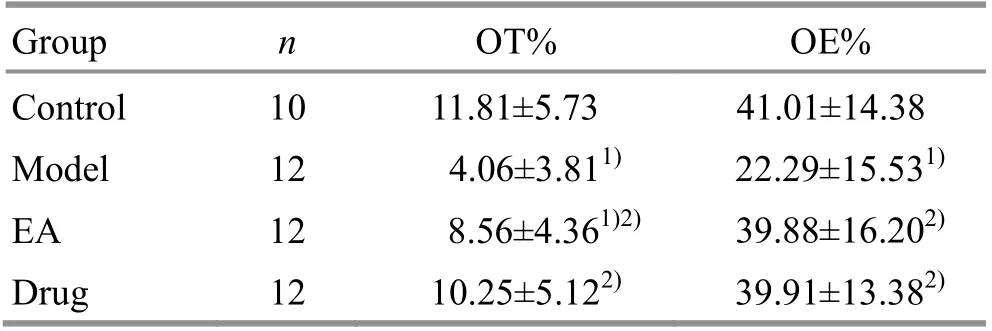
Table 2. Comparison of EPM results
2.2 Biomarkers
2.2.1 The expressions of neurotransmitters
Compared with the control group, the expressions of 5-HT, NE and DA increased significantly in the model group (all P<0.05). The expression of 5-HT in the EA group was markedly lower than that in the model group(P<0.05) and was insignificantly different from that in the drug group, but was significantly higher than that in the control group (P<0.05). There was no significant difference in the expression of NE among the control group, the EA group and the drug group (P>0.05). There was no significant difference in the expression of DA between the control group and the drug group (P>0.05).The expression of DA in the control group and the drug group was significantly higher than that in the EA group(P<0.05) and the model group (P<0.05). There was no significant difference in the expression of DA between the model group and the EA group (P>0.05), (Table 3).
2.2.2 The expressions of Bax and Bcl-2
Compared with the control group, the expression of Bax and the level of Bax/Bcl-2 increased significantly while the expression of Bcl-2 decreased significantly in the model group (P<0.05). There were no significant differences in the expressions of Bax and Bcl-2 among the control group, EA group and the drug group(P>0.05). The level of Bax/Bcl-2 in the EA group was higher than that in the control group (P<0.05), but was lower than that in the model group (P<0.05) and insignificantly different from that in the drug group(P>0.05), (Table 4 and Figure 1).
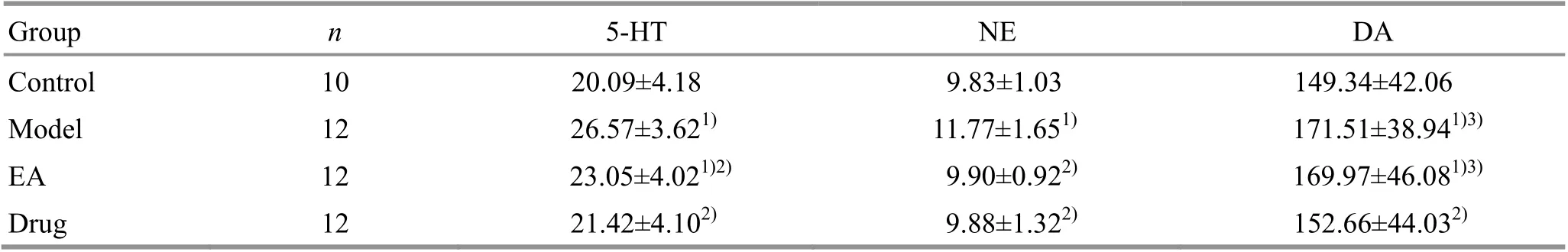
Table 3. Comparison of the expressions of 5-HT, NE and DA (x ±s, ng/g)

Table 4. Comparison of the expressions of Bax, Bcl-2 and Bax/Bcl-2 (x ±s)
3 Discussion
Uncertain empty bottle stimulation, a modeling approach that has been proved effective in triggering the pathogenesis of generalized anxiety disorder, was used to establish the anxiety rat model in this study[7].Meanwhile, both OFT and EPM were introduced to evaluate the anxiety behaviors of rats on the basis of its ambivalence, that is, rodents are afraid of the open and elevated space but prefer to explore new objects[13-15].
According to the results, following the uncertain empty bottle stimulation, rats in the model group showed poor performance in behavioral experiments,including the decline in the horizontal and vertical scores in OFT (P<0.05) and the decrease in OT% and OE% in EPM (P<0.05). The findings suggested that the model was a success, i.e., the rats showed significant anxiety before intervention. With the change of behaviors, the levels of hippocampal 5-HT, NE and DA were significantly increased (all P<0.05), indicating that the anxiety performance of the rats might be associated with the elevation of these three neurotransmitters.Our results were consistent with the previous research.Neurotransmitters including benzodiazepine-GABA, DA,NE, and 5-HT are directly linked to the biological changes in anxiety[16-17]. Specifically, these transmitters are distributed in various areas of the brain, and they directly or indirectly participate in the regulation of nearly all aspects of anxiety, including attention, waking,fear and sedation[16-17]. Studies on anxiety and 5-HT receptors have shown that if the 5-HT1A receptor is knocked out in each brain region, the activity of the raphe nucleus will be increased, followed by the activation of 5-HT2A receptor in the vertebral cells,which eventually leads to an increase in anxiety[18]. The basic function of NE in cortical system is to modulate alertness and observation behavior through adjusting attention to novel environmental stimuli and wakefulness. Therefore, NE in the central system is essentially associated with the stress response, and its disorder is involved in the pathogenesis of anxiety and depression as well[19]. Moreover, the synergistic effect of 5-HT and NE in central nervous system has also been confirmed[20-21]. Though there is no sufficient evidence revealing the direct correlation between DA and anxiety,psychotropic inhibitors have been found to alleviate anxiety by down-regulating the DA level. DA thereby is presumed to be involved in the formation and regulation of anxiety[22]. In summary, 5-HT, NE, and DA are potential therapeutic targets for suppressing anxiety,which has also been confirmed in our experiment. After receiving EA or alprazolam, anxiety-induced high levels of 5-HT and NE decreased significantly (P<0.05).Moreover, EA was comparable to alprazolam in decreasing levels of 5-HT and NE (P>0.05). A dramatic decline in hippocampal DA was found following gavage administration of alprazolam (P<0.05), while the decrease in DA was not significant after EA (P>0.05).The above results demonstrated that 15 consecutive days of EA at Baihui (GV 20) and Sanyinjiao (SP 6) could improve anxiety disorder in rats by down-regulating the expressions of hippocampal 5-HT and NE, and this effect was comparable to that of alprazolam. However,the evidence was no enough to prove that the regulation of anxiety disorder by EA was achieved by down-regulating the level of hippocampal DA.
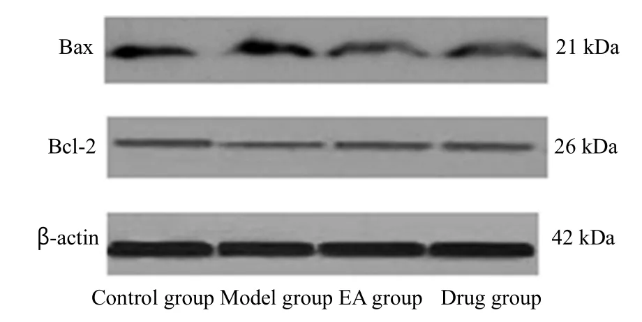
Figure 1. The expressions of Bax and Bcl-2
Papez's circuit is the pathological basis of multiple neuropsychiatric disorders, particularly related to cognition, memory and emotions such as anxiety[23-24].The anxiety caused by neuronal apoptosis is related to Papez's circuit[23,25]. Apoptosis is the process of cellular self-destruction, and genes such as Bcl-2 and Bax are known to inhibit and promote apoptosis,respectively[26-28]. Therefore, Bcl-2 and Bax are hypothesized to be involved in the pathogenesis of anxiety-like behaviors as well[10,29]. In term of our study,the relative grey value of hippocampal Bax increased significantly (P<0.05) while the value of Bcl-2 decreased significantly (P<0.05) in rats with anxiety disorder. After receiving EA or alprazolam, the anxiety-induced high level of Bax decreased significantly (P<0.05) while the anxiety-induced low level of Bcl-2 increased significantly(P<0.05). Moreover, EA was comparable to alprazolam in regulating the relative grey values of Bax and Bcl-2(P>0.05). These findings suggest that EA-induced attenuation of anxiety disorder may be achieved by inhibiting the apoptosis, and this effect should be comparable to that of the alprazolam.
To sum up, EA shows promising effects in attenuating rats’ anxiety disorder caused by uncertain empty bottle stimulation, which may be achieved by the downregulation of the expressions of 5-HT and NE in hippocampus and/or the inhibition of the hippocampal neuronal apoptosis. Furthermore, this effect is comparable to that of alprazolam.
Conflict of Interest
The authors declare that there is no potential conflict of interest in this article.
Acknowledgments
This work was supported by National Natural Science Foundation of China (国家自然科学基金资助项目, No.81102729, No. 814Accepted: 30 August 201973594); Traditional Chinese Medicine Science and Technology Innovation Project of Shanghai Health and Family Planning Commission (上海市卫生和计划生育委员会中医药科技创新项目, No.2015ZB0504); Preclinical Research Project of Science and Technology Commission Shanghai Municipality (上海市科学技术委员会临床前研究专项课题, No.16401902600).
Statement of Human and Animal Rights
The treatment of animals conformed to the ethical criteria.
Received: 20 May 2019/Accepted: 19 June 2019
猜你喜欢
杂志排行

Journal of Acupuncture and Tuina Science的其它文章
- Effect of An-pressing manipulation on post-stroke muscle spasticity in rats and its mechanism study
- Regulatory effects of moxibustion on ubiquitin and NLRP3 proteins in colon of ulcerative colitis rats
- Clinical study on intradermal needle therapy in treating urinary retention after cervical cancer surgery
- Clinical observation of deep electroacupuncture at Baliao points for female stress urinary incontinence
- Clinical observation on prevention of chemotherapy infection in gastric cancer by moxa-stick moxibustion plus rhG-CSF and its effect on immune function
- Clinical observation of acupuncture plus repetitive transcranial magnetic stimulation in the treatment of post-stroke insomnia