ELA/APJ通过上调miR-133a抑制心肌细胞在缺血缺氧条件下凋亡的机制研究
2019-07-18陈旭翔侯婧瑛龙会宝吴浩伍权华杨欢符佳颖王彤
陈旭翔 侯婧瑛 龙会宝 吴浩 伍权华 杨欢 符佳颖 王彤
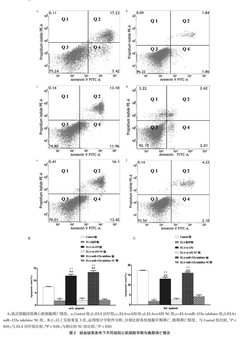

[摘要] 目的 觀察血管紧张素受体AT1相关的受体蛋白(APJ)的内源性配体ELABELA(ELA)对心肌细胞在缺血缺氧条件下凋亡的影响,并探讨miR-133a在其中的调控机制。 方法 将体外培养的心肌细胞分为Control组、ELA治疗组、ELA+siAPJ组、ELA+siAPJ NC组、ELA+miR-133a inhibitor组、ELA+ miR-133a inhibitor NC组,在缺血缺氧条件(无血清,1%体积分数O2)下培养24 h。免疫荧光鉴定心肌细胞标志物心肌α肌动蛋白(α-SA)和心肌肌钙蛋白T(cTnT)表达情况。流式细胞仪检测各组细胞凋亡情况。Western blot、qRT-PCR检测各组细胞APJ、miR-133a的表达情况。 结果 ①与Control组比较,ELA治疗组细胞早期凋亡率与晚期凋亡率显著减少(P < 0.01);②与对应的NC组细胞比较,ELA+siAPJ组、ELA+miR-133a inhibitor组细胞早期凋亡率与晚期凋亡率明显增加(P < 0.01);③与Control组比较,ELA治疗组细胞APJ的蛋白与mRNA表达量、miR-133a的mRNA表达量均明显升高(P < 0.01);④采用siRNAs阻断APJ的表达后,与ELA+siAPJ NC组比较,ELA+siAPJ组细胞APJ的蛋白与mRNA表达量、miR-133a的mRNA表达量均显著减少(P < 0.01)。采用siRNAs阻断miR-133a的表达后,ELA+miR-133a inhibitor组细胞APJ的蛋白与mRNA表达量与ELA+miR-133a inhibitor NC组比较,差异无统计学意义(P > 0.05),miR-133a的mRNA表达量显著减少(P < 0.01)。 结论 ELA能够抑制心肌细胞在缺血缺氧环境下的凋亡,此效应可能与其激活APJ之后上调miR-133a有关。
[关键词] ELABELA;血管紧张素受体AT1相关的受体蛋白;缺血缺氧;心肌细胞;细胞凋亡
[中图分类号] R331.31 [文献标识码] A [文章编号] 1673-7210(2019)05(c)-0012-06
[Abstract] Objective To investigate the effect of ELABELA (ELA), which is a ligand of angiotensin receptor AT1 associated endogenous protein (APJ), on cardiomyocytes apoptosis under ischemia-hypoxia condition and to explore the regulatory mechanism of miR-133a. Methods The cardiomyocytes were cultured for 24 h under ischemia-hypoxia conditions (without serum, 1% O2) and divided into control group, ELA treatment group, ELA+siAPJ group, ELA+siAPJ NC group, ELA+miR-133a inhibitor group and ELA+miR-133a inhibitor NC group. The expression of myocardial α-actin (α-SA) and cardiac troponin T (cTnT) were observed by immunofluorescence. Flow cytometry was used to detect apoptosis. Western blot and qRT-PCR were used to detect the expression of APJ and miR-133a. Results ①Compared with the control group, the rate of early and late apoptosis in ELA treatment group was significantly decreased (P < 0.01); ②compared with corresponding NC group, the early and late apoptosis rate of ELA+siAPJ group and ELA+miR-133a inhibitor group were significantly increased (P < 0.01); ③the expression of APJ and miR-133a in ELA treatment group was significantly higher than that in control group after 24 h culture (P < 0.01); ④after blocking the expression of APJ and miR-133a by siRNA, compared with that in the corresponding NC group, the protein and mRNA expression level of APJ and mRNA expression level of miR-133a were significantly decreased in ELA+siAPJ group (P < 0.01). The protein and mRNA expression level of APJ did not change significantly in ELA+miR-133a inhibitor group (P > 0.05), but the mRNA expression level of miR-133a was significantly decreased (P < 0.01). Conclusion ELA could inhibit the apoptosis of cardiomyocytes in ischemic and hypoxic environment. This effect might be related to up-regulation of miR-133a after activation of APJ.