吸入法建立急性一氧化碳中毒迟发型脑病大鼠模型
2019-07-18陈超王宝军项文平李月春郝喜娃庞江霞高浩然谢伟
陈超 王宝军 项文平 李月春 郝喜娃 庞江霞 高浩然 ?谢伟


[摘要] 目的 建立符合臨床特点的大鼠急性一氧化碳中毒迟发型脑病(DEACMP)模型。 方法 采用随机数字表法将认知功能筛查合格的雄性Wistar大鼠196只分为实验组和对照组,实验组164只,对照组32只。对照组吸入空气,实验组吸入一氧化碳(CO)气体,根据吸入CO浓度不同分为三组,分别为1000、3000、4000 ppm组,每组存活32只,连续吸入CO 40 min保持中毒浓度稳定;各组大鼠分别在中毒后7、14、21和28 d处死大鼠取材;采用分光光度计分析碳氧血红蛋白(COHb)水平;采用水迷宫训练评估各组大鼠中毒后认知功能的变化情况,采用HE染色方法观察大鼠海马细胞受损情况,明确建立可靠的大鼠DEACMP模型的方法。 结果 吸入CO后大鼠出现明显中毒表现,COHb水平与吸入CO浓度大小呈正相关(r = 0.748,P < 0.05),1000、3000、4000 ppm组及对照组大鼠死亡率分别为0%、40.74%、58.97%、0%,各组大鼠DEACMP发生率分别为6.25%、28.13%、31.25%、0%,在中毒后21、28 d大鼠DEACMP高发,选择死亡率低、发病率高、并结合大鼠饲养周期,明确大鼠吸入CO浓度3000 ppm,中毒后21 d可以建立DEACMP大鼠模型。 结论 大鼠吸入CO浓度3000 ppm 40 min,21 d后可以建立可靠DEACMP模型,为DEACMP的预防和治疗带来新的希望。
[关键词] 一氧化碳中毒;动物模型;迟发型脑病
[中图分类号] R74 [文献标识码] A [文章编号] 1673-7210(2019)05(c)-0004-04
[Abstract] Objective To establish a model of delayed encephalopathy acute carbon monoxide poisoning (DEACMP). Methods A total of 196 intelligence-qualified rats were divided into experimental group and control group by random number table method, with 164 rats in the experimental group and 32 rats in the control group. The experimental group used an acute static inhalation of carbon monoxide (CO) to establish a poisoned rat model. According to different concentrations of CO poisoning, they were randomly divided into 1000, 3000, 4000 ppm group, with 32 rats alive in each group. Each group continued to inhale for 40 minutes. Rats in each group were sacrificed at 7, 14, 21 and 28 d after poisoning. Spectrophotometer method was used to dynamically monitor the changes of carboxyhemoglobin (COHb) content in each group. Morris water maze test was used to evaluate cognitive function. HE staining was used to observe the cell damage in hippocampus. The best poisoning concentration and time for establishing a DEACMP rat model was established. Results After poisoned by CO, the rats showed typical symptoms of poisoning. There was a significant positive correlation between COHb content and CO concentration (r = 0.748, P < 0.05). The mortality rates of the 1000, 3000, 4000 ppm group and the control group were 0%, 40.74%, 58.97% and 0%, respectively. The incidence of DEACMP in each group was 0%, 6.25%, 28.13%, 31.25%, and 0%, and the 21 d and 28 d after being poisoned were high DEACMP time points. Based on the low mortality rate and high incidence rate, the CO poisoning concentration of 3000 ppm and the time of 21d were the best method to establish a DEACMP rat model. Conclusion A DEACMP rat model can be established with a static inhalation concentration of 3000 ppm CO for 40 minutes, and models can be successfully established 21 days after poisoning.
[Key words] Carbon monoxide poisoning; Animal model; Delayed encephalopathy
CO中毒是常见的窒息性中毒之一,广泛存在于人们生产生活环境中,当空气中CO浓度超过35 ppm即可出现中毒症状[1-3],中毒可引起多个脏器功能损害[4],一氧化碳中毒迟发型脑病(delayed encephalopathy after acute carbon monoxide poisoning,DEACMP)是其中较常见的并发症[5],发病率为0.2%~47.3%[6],其临床表现较重,以认知障碍为主,致残率较高,给患者生活带来严重影响[7]。目前对于DEACMP的发病机制及治疗虽然已有大量研究,但仍不能合理阐明,成功建立动物模型是进一步研究DEACMP的基础,将有助于阐明其发生过程,为临床预防与治疗提供可靠的实验依据。
1 材料与方法
1.1 实验动物
健康雄性的Wistar大鼠共208只,购自内蒙古大学动物研究培育中心,大鼠体重180~230 g,饲养于室温18~23℃且12 h明暗交替的室内,自由进食水。
1.2 主要仪器与试剂
动物CO中毒箱,自行设计,用钢化玻璃制成的体积为0.7 m×0.5 m×0.43 m的长方体,使用胶粘剂封闭中毒箱连接处,箱体分别设置进气口、出气口及CO浓度检测仪连接处;CO气体浓度检测仪,购自金盾安全防护有限公司及元特科技有限公司;Morris水迷宮及图像自动监视系统;Leica CM1950冰冻切片机;T6新悦-可见分光光度计;CO标准气体,购自佛山市科的气体有限公司。
1.3 动物训练
首先将大鼠放置在水迷宫平台上数秒熟悉环境,随后依次在4个象限进行训练,电脑自动记录大鼠运动轨迹和找到平台时间。训练5 d后选择逃避潜伏期<60 s,运动轨迹为直线式或趋向式的大鼠为合格,不合格者淘汰。
1.4 模型建立及分组情况
随机将196只训练合格的大鼠分为实验组及对照组,实验组164只,对照组32只,实验组给予急性静态吸入CO气体建立中毒模型[8-9],依据CO中毒浓度不同分为三组,即1000、3000及4000 ppm组,连续吸入CO 40 min保持中毒浓度稳定;对照组通入等量的空气,其他条件不变。因CO浓度不同大鼠死亡情况不同,为保证后期实验正常进行,中毒后每组各需要存活大鼠32只,随后将其分别随机分为四个时间点,即中毒后7、14、21和28 d处死大鼠取材,共计16组,每组各8只。
1.5 碳氧血红蛋白(carboxyhemoglobin,COHb)含量检测
分别在中毒后15、30、60、120和240 min,断尾取大鼠血,采用改良双波长定量法检测COHb含量,按照公式计算:COHb(%)=(2.44×A535/A578-2.68)×100%。
1.6 DEACMP模型建立成功的标准
各组大鼠分别于中毒后4个时间点行水迷宫定位航行实验,记录大鼠运动轨迹和逃避潜伏期,大鼠常规多聚甲醛心脏灌注固定后取脑行病理学观察。参考Ochi等[10]研究结果判断DEACMP模型是否成功:水迷宫结果提示大鼠认知功能发生障碍,并且HE染色发现大鼠脑内海马组织细胞受损。
1.7 统计学方法
采用SPSS 20.0对所得数据进行统计学分析,计量资料采用均数±标准差(x±s)表示,组间比较采用t检验,计数资料采用百分率表示,组间比较采用χ2检验。采用Pearson法进行单因素相关性分析,以P < 0.05为差异有统计学意义。
2 结果
2.1 大鼠CO中毒后表现
向中毒箱通入CO气体5~10 min后大鼠开始出现多动、表现活跃,15~20 min后出现瘫软无力、活动减少、不能站起、呼吸浅快,口周、耳部、肢端皮肤呈樱桃红色,3000 ppm及4000 ppm组大鼠中毒症状较重,部分发生抽搐、角弓反张、昏迷甚至死亡。解剖死亡大鼠发现多脏器充血伴局部点片状出血;脑组织高度充血,周围间质水肿,部分出现点状出血,与报道急性CO中毒表现相符。
2.2 中毒后大鼠COHb含量变化
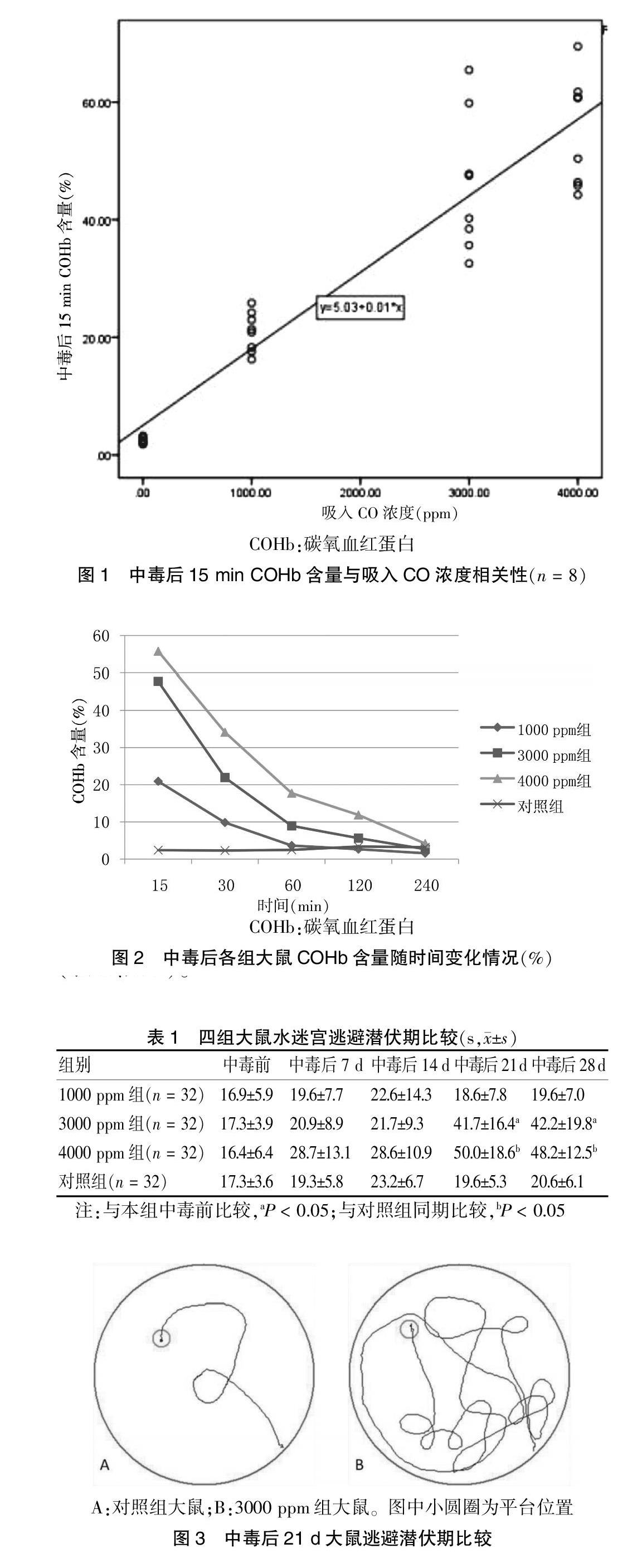
中毒箱中通入CO 15 min后测得各组大鼠COHb含量均升高,1000 ppm组COHb含量16.32%~22.92%,3000 ppm及4000 ppm组32.13%~75.65%,符合中、重度中毒标准,中毒后15 min测得COHb含量与吸入CO浓度大小呈正相关(r = 0.748,P < 0.05),见图1,COHb含量随时间变化情况见图2。
2.3 各组大鼠死亡情况比较
对照组及1000 ppm组大鼠在中毒过程中无死亡情况发生;3000 ppm组、4000 ppm组大鼠死亡多发生在中毒30~40 min。3000、4000 ppm组大鼠死亡率分别为为40.74%(22/54)、58.97%(46/78),对照组、1000 ppm组大鼠死亡率均为0%,实验组各组间死亡率比较,差异均有统计学意义(P < 0.05)。
2.4 DEACMP发生情况
2.4.1 水迷宫结果 实验结果显示,1000 ppm组大鼠各时间点逃避潜伏期与对照组比较,差异无统计学意义(P > 0.05);3000 ppm及4000 ppm组大鼠在中毒后21、28 d与对照组、1000 ppm组比较逃避潜伏期明显增加,差异有统计学意义(P < 0.05);与中毒前比较,3000 ppm及4000 ppm组大鼠在中毒后21、28 d逃避潜伏期明显增加,差异有统计学意义(P < 0.05)(表1、图3)。
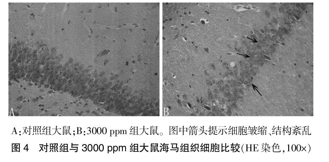
2.4.2 病理学结果 对照组大鼠海马组织细胞形态正常、细胞核、细胞膜结构完整清晰,3000 ppm组及4000 ppm组大鼠部分可见海马组织细胞排列稀疏、细胞萎缩、数量减少、大小不等、结构模糊不清,未发现明显坏死灶,见图4。
结合以上水迷宫与病理结果,判断各组大鼠DEACMP发生情况,对照组、1000 ppm组、3000 ppm组及4000 ppm组DEACMP的发生率分别为0(0/32)、6.25%(2/32)、28.13%(9/32)、31.25%(10/32);3000 ppm组与4000 ppm组DEACMP发生率比1000 ppm组明显升高,差异有统计学意义(P < 0.05);3000 ppm组与4000 ppm组之间DEACMP发病率比较,差异无统计学意义(P > 0.05);1000 ppm组大鼠在中毒后28 d有2只发生DEACMP,3000 ppm组在14 d有1只发病,在21、28 d均有4只发病,4000 ppm组在7、14 d分别有1只发病,在21、28 d分别有5只和3只发病,DEACMP在3000 ppm组与4000 ppm组中毒后21、28 d高发,差异有统计学意义(P < 0.05),相同浓度在21、28 d之间比较,差异无统计学意义(P > 0.05),结合大鼠饲养周期,选择死亡率低、发病率高的组别,明确大鼠吸入CO浓度3000 ppm、时间21 d可以建立可靠的DEACMP动物模型。
3 讨论
DEACMP患者常出现神经精神异常(智能、人格、行为等改变),病程较长,治疗困难,常遗留精神或认知障碍[11-12],这是目前临床工作中亟待解决的难题,深入研究DEACMP的基础是建立可靠的动物模型,本课题在Hara等[8-9]研究基础上进行了验证和改进,使实验更加安全、结果更加可靠。与腹腔注射CO比较[13],吸入法与日常中毒情况更接近,代表性更好;中毒时气体通过密封性良好的管道进入中毒箱,实验人员不易发生意外中毒,安全性较高;但吸入法存在随着中毒时间延长,中毒箱内氧气减少、二氧化碳潴留等情况,为了改善这些情况对实验结果的影响,本研究采用每次减少中毒大鼠只数,来增加实验的可靠性。
COHb含量高低是判断CO中毒严重程度的重要方法之一,相关研究[14-15]发现是否发生DEACMP与中毒的严重程度呈正相关。本研究发现DEACMP在3000 ppm组及4000 ppm组中发生率较高,且两组COHb含量可达中、重度中毒标准,说明中毒程度直接影响DEACMP的发生,该研究结果与相关研究[14-15]结果相符;中毒后15 min COHb含量与吸入CO浓度呈正相关,与Li等[15]研究结果一致,提示本研究建立的DEACMP模型可靠。
Morris水迷宫实验是反映认知功能变化较好的实验方法之一,其结果准确可信,是判断动物认知功能的重要方法[16-17]。实验前期首先通过水迷宫训练筛选出智力、运动功能正常的大鼠,淘汰了学习记忆不合格大鼠,避免了动物之间个体差异对实验结果的影响,使得实验结果更加可靠和准确。本研究水迷宫结果显示,3000 ppm组及4000 ppm组大鼠逃避潜伏期在中毒后21、28 d较对照组明显延长,差异有统计学意义(P < 0.05),说明在中毒后21、28 d两组大鼠认知功能发生改变。相关研究[18-20]发现CO中毒后脑内海马组织细胞广泛损害,病理学表现细胞结构不清、层数变薄、体积缩小等,本实验HE染色发现DEACMP大鼠海马组织细胞排列稀疏、细胞萎缩、数量减少、大小不等、结构模糊不清,说明中毒后大鼠海馬组织细胞存在损伤。根据病理学及水迷宫结果共同判定大鼠吸入CO浓度3000 ppm、时间21 d可以建立可靠的DEACMP模型,为DEACMP发病机制的研究提供了可靠基础,为DEACMP的预防与治疗带来新的希望。
[参考文献]
[1] Rose JJ,Wang L,Xu Q,et al. Carbon monoxide poisoning:pathogenesis,management,and future directions of therapy [J]. Am J Respir Crit Care Med,2017,195(5):596-606.
[2] Borger J. The danger of carbon monoxide poisoning associated with hookah use:an emergency physician′s perspective [J]. N C Med J,2017,78(6):424.
[3] McKenzie LB,Roberts KJ,Shields WC,et al. Distribution and Evaluation of a Carbon Monoxide Detector Intervention in Two Settings:Emergency Department and Urban Community [J]. J Environ Health,2017,79(9):24-30.
[4] Wang B,Xiang W,Xue H,et al. Efficacy of N-Butylphthalide and hyperbaric oxygen therapy on cognitive dysfunction in patients with delayed encephalopathy after acute carbon monoxide poisoning [J]. Med Sci Monit,2017, 23:1501-1506.
[5] Jorge A. Carbon monoxide poisoning [J]. Crit Care Clin,2012, 28:537-548.
[6] Goldstein M. Carbon monoxide poisoning [J]. J Emerg Nurs,2008,34:538-542.
[7] Xiang W,Xue H,Wang B,et al. Combined application of dexame-thasone and hyperbaric oxygen therapy yields better efficacy for patients with delayed encephalopathy after acute carbon monoxide poisoning [J]. Drug Des Devel Ther,2017,11:513-519.
[8] Hara S,Mizukami H,Kurosaki K,et al. Existence of a threshold for hydroxyl radical generation independent of hypoxia in rat striatum during carbon monoxide poisoning [J]. Arch Toxicol,2011,85:1091-1099.
[9] Hara S,Mizukami H,Kuriiwa F,et al. cAMP production mediated through P2Y(11)-like receptors in rat striatum due to severe,but not moderate,carbon monoxide poisoning [J]. Toxicology,2011,288:49-55.
[10] Ochi S,Abe M,Li C,et al. The nicotinic cholinergic system is affected in rats with delayed carbon monoxide encephalopathy [J]. Neurosci Lett,2014,569:33-37.
[11] Sun X,Xu H,Meng X,et al. Potential use of hyperoxygenated solution as a treatment strategy for carbon monoxide poisoning [J]. PLoS One,2013,8(12):e81779.
[12] Guan L,Zhang YL,Li ZY,et al. Salvianolic acids attenuate rat hippocampal injury after acute CO poisoning by improving blood flow properties [J]. Biomed Res Int,2015,2015:526483.
[13] Xiang WP,Xue H,Wang BJ,et al. Delayed encephalopathy of acute carbon monoxide intoxication in rats:potential mechanism and intervention of dexamethasone [J]. Pak J Pharm Sci,2014,27:2025-2028.
[14] Chen Q,Bai J,Li CR,et al. Changes of HbCO in the blood of rats with different CO concentration and inhalation time [J]. Fa Yi Xue Za Zhi,2016,32(6):410-412.
[15] Li Q,Bi MJ,Bi WK,et al. Edaravone attenuates brain damage in rats after acute CO poisoning through inhibiting apoptosis and oxidative stress [J]. Environ Toxicol,2016,31(3):372-379.
[16] Roderique JD,Josef CS,Newcomb AH,et al. Preclinical evaluation of injectable reduced hydroxocobalamin as an antidote to acute carbon monoxide poisoning [J]. J Trauma Acute Care Surg,2015,79:116-120.
[17] Zhang J,Wu H,Zhao Y,et al. Therapeutic effects of hydrogen sulfide in treating delayed encephalopathy after acute carbon monoxide poisoning [J]. Am J Ther,2016, 23(6):e1709-e1714.
[18] Li HM,Shi YL,Wen D,et al. A novel effective chemical hemin for the treatment of acute carbon monoxide poisoning in mice [J]. Exp Ther Med,2017,14(5):5186-5192.
[19] Xue L,Wang WL,Li Y,et al. Effects of hyperbaric oxygen on hippocampal neuronal apoptosis in rats with acute carbon monoxide poisoning [J]. Undersea Hyperb Med,2017,44(2):121-131.
[20] Watanabe S,Matsuo H,Kobayashi Y,et al. Transient degradation of myelin basic protein in the rat hippocampus following acute carbon monoxide poisoning [J]. Neurosci Res,2010,68(3):232-240.
(收稿日期:2018-01-24 本文編辑:金 虹)
