Effect of Xylazine Anesthesia on Glu and GABA Amino Acid Neurotransmitters in Rat
2019-04-12ZhangXintongLiLinaWangQiChangTianWenYajingYuanRuiDuXuemanTangSainanandGaoLi
Zhang Xin-tong, Li Li-na, Wang Qi, Chang Tian, Wen Ya-jing, Yuan Rui, Du Xue-man, Tang Sai-nan, and Gao Li
Heilongjiang Key Laboratory for Laboratory Animals and Comparative Medicine, College of Veterinary Medicine, Northeast Agriculture University,Harbin 150030, China
Abstract: Neurotransmitters of the central nervous system were the important way to study the mechanism of anesthesia. The effect of different doses of xylazine anesthetic on the glutamate (Glu) and γ-aminobutyric-acid (GABA) were investigated and the mechanism of xylazine anesthetic on the central nervous system were explored in this study. A total of 88 rats were randomly divided into three groups, including normal saline control group, group with low dose of xylazine and group with high dose of xylazine.Cerebrum, cerebellum, hippocampus, thalamus and brainstem were collected. The results showed that the concentration of Glu in the hippocampus, thalamus and brainstem decreasedfirst and then increased, but it increasedfirst and then decreased in the cerebrum and cerebellum during the period of anesthesia. The concentration of GABA in the cerebrum, thalamus, brainstem and hippocampus increased first and then decreased. The results showed that xylazine inhibited Glu and promoted GABA with different dose dependence. The results and methods could provide guides for the clinical use of xylazine.
Key words: brain, glutamate, γ-aminobutyric acid, rat, xylazine
Introduction
Xylazine (Rompun) is α2-adrenergic receptor agonist with the effects of central analgesia, sedation and muscle relaxation (Ehant et al., 2003; Vinje et al.,2002), which can directly inhibit the central nerve conduction block, the inhibition of central nervous system is related with the effect of autonomic nervous system (Chiara et al., 2016). The locus coeruleus has been showed to innervation directly from the pain pathway in the spinal cord and cranial nucleus, when the α2-receptor is excited on the locus coeruleus,noxious stimuli response from these sensors is delayed,so it is widely considered to be one of the major sites of action of xylazine in the central nervous system(Kitayama et al., 2002). It diffuses extensively and penetrates the blood brain barrier, due to the uncharged and lipophilic nature of the compound (Alkire et al.,1995). The sedative and analgesic effects of xylazine inhibit the transmission of neural impulses in the central nervous system (Mazzoli and Pessione, 2016).In recent years, the research in molecular and cellular mechanism of general anesthesia has causing a growing interest.
γ-aminobutyric acid (GABA) is a major inhibitory amino acid neurotransmitter and synthesized by Glutamate (Glu) catalyzed by glutamate decarboxylase(GAD). It is reported that GABA is one of the most important ways to explore the mechanism of anesthesia(Drabek et al., 2014). When the body is anesthetized,GABA will increase, which indicates that GABA plays an inhibitory role on the central nervous system(Sugiyama et al., 1992). It is found that the release of the GABA is also affected by the concentration of intracellular calcium, which shows a certain calcium dependent (Suria and Rasheed, 1994). In addition,some anaesthetic can also increase the concentration of GABA in brainstem, which indicates that GABA has been involved in central inhibition of anesthesia (Lasen and Langmoen, 1998).
Glutamate (Glu) releases into the synaptic cleft and acts on postsynaptic membrane receptor, then increases Na+permeability and causes excitatory postsynaptic potential (EPSP) (Musella et al., 2014). This study aimed to determine the effect of xylazine anesthesia on amino acid neurotransmitter and provided theoretical basis for further studies on the mechanism of xylazine anesthesia.
Materials and Methods
Animals and experimental groups
Male and female wistar rats, weighing (200±20) g,were provided by Northeast Agricultural University.At the first week, before the experiment, rats were fed in adaptive conditions. All the experiments were performed in accordance with the Ethical Committee for Animal. Eight rats were randomly divided into the control group (C), the rest was in the experimental group and divided into low dose of xylazine group(50 mg · kg-1) and high dose of xylazine group(70 mg · kg-1). Animals in the experimental group were divided into induction period (Ⅰ, collecting samples after injecting xylazine 8 min); anesthesia group (A, collecting samples when righting reflex disappeared); recoveryⅠperiod (R1, collecting samples when righting reflex recovered); recoveryⅡperiod (R2, collecting samples after righting reflex recovered for 3 h) and recovery Ⅲ period (R3, collecting samples after righting reflex recoverd for 5 h).
Sample collection
After intraperitoneal injection of saline solution 8 min for the control group, the rats were decapitated and brain tissues were collected immediately. Rats in experimental groups at corresponding time points were sacrificed and sampled. Cerebrum, cerebellum,thalamus, brainstem and hippocampus were quickly dispensed on ice, which were put into the freezing tube and stored in liquid nitrogen.
Determination of Glu content
Glu in the samples was determined via enzyme linked immunosorbent assay using the Glu kit (Nanjing Jiancheng Bioengineering Institute). Settedfive standard holes and added standard of different concentrations and 50 μL standard diluent in blank hole, 50 μL sample was added in the test holes. Then 50 μL solution A was added in each hole immediately, shaked gently and incubated at 37℃ for 1 h. Discarded the liquid in the hole, washed each hole with 350 μL of wash solution, soaked for 2 min and washed three times.Each hole received an addition of 100 μL solution B(compounded when it was in need) and incubated at 37℃ for 30 min. Dried and washed five times, each hole received an addition of 90 μL substrate solution,kept in dark place at 37℃. Then, 50 μL termination solution was added to each micropore to terminate.The OD value was measured with a microplate reader at wavelength of 450 nm immediately.
Determination of GABA concentration
GABA in the samples was determined via enzyme linked immunosorbent assay using the GABA kit(Nanjing Jiancheng Bioengineering Institute). Setted five standard holes and added standard of different concentrations and 50 μL standard diluent in blank hole, added 50 μL sample in the test sample holes.Then each hole received an addition of 50 μL solution A immediately, shaked gently and incubated at 37℃for 1 h. Discarded the liquid in the hole, washed each hole with 350 μL of wash solution, soaked for 1-2 min and washed three times. Each hole received an addition of 100 μL solution B (compound when it was in need) and incubated at 37℃ for 30 min. Dried and washed five times, each hole received an addition of 90 μL substrate solution, kept in dark place at 37℃(10-20 min). Then, 50 μL termination solution was added to each micropore to terminate. The OD value of each hole was measured with a microplate reader at wavelength of 450 nm immediately.
Statistical analysis
All the data were analyzed using GraphPad Prism 5.1(GraphPad Software Inc, USA). The data were analyzed by one-way ANOVA after normal distribution and homogeneity test of variance. All the test results were expressed as mean±standard deviation. When p<0.05,the values were considered statistically significant.
Results
Changes of behavior
After intraperitoneal injection of saline or xylazine,rats in the control group showed no abnormal changes,and hadflexible activities; most rats injected xylazine appeared hyperexcitability, restless and strong reaction in 4-6 min, then the rats became slow, depressed,reluctance to move, not sensitive to peripheral stimulation. The average disappearance time of righting reflex was (10.8±3.75) min, rats were found slow breathing,slow heartbeat, fatigue, drowsiness and urinary incontinence in this period; the average time for the recovery of righting reflex was (36.52±10.35) min.
Detection results of Glu concentration in different brain regions induced by high dose xylazine
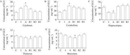
Glu in the cerebrum and cerebellum, which concentration of anesthesia group increased by 44.59% and 55.73% compared with that of the control group (p<0.05, Fig. 1A and B). In the thalamus and brainstem,the concentration of R1 group, decreased by 23.27% and 24.39% compared with that of the control group (p<0.05, Fig. 1D and E). The contents of Glu in the hippocampus of anesthesa group decreased by 51.58% compared with those of the control group (p<0.05, Fig. 1C).

Fig. 1 Results of Glu concentration in different brain regions induced by high dose xylazine
Detection results of GABA concentration in different brain regions induced by high dose xylazine
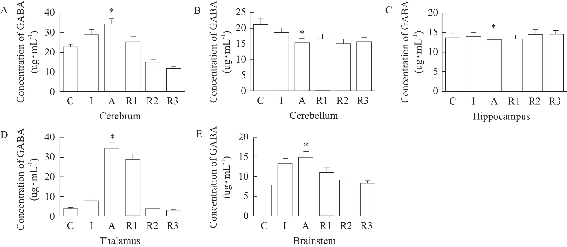
The concentration of GABA in anesthesia group,increased by 51.49% and 62.16% compared with that of the control group in the cerebrum and thalamus(p<0.05, Fig. 2A and D). The concentration in the cerebellum and hippocampus signified a trend of decreasingfirst and then picking up, the concentration of anesthesia group decreased by 32.43% and 4.75%compared with that of the control group (p<0.05, Fig. 2B and C). In brainstem, the results showed a tendency of decreasingfirst and then picking up, the lowest concentration of R1 group decreased by 24.39% compared with that of the control group (p<0.05, Fig. 2E).

Fig. 2 Results of GABA concentration in different brain regions induced by high dose xylazine
Detection results of Glu concentration in different brain regions induced by low dose xylazine
Glu in the cerebrum and cerebellum showed a tendency of picking upfirst and then decreasing. The level of Glu in anesthesia group increased by 14.68%and 42.96% compared with those of the control group in the cerebrum and cerebellum (p<0.05, Fig. 3A and B). In the thalamus and brainstem, the concentration showed a tendency of decreasing first and then picking up, the concentration in R1 group decreased by 10.99% and 13.30% compared with that of the control group (p<0.05, Fig. 3D and E).

Fig. 3 Results of Glu concentration in different brain regions induced by low dose xylazine
The concentration in hippocampus showed a tendency of descend, and decreased by 43.8% in anesthesia group compared with that of the control group (p<0.05, Fig. 3C).
Detection results of GABA concentration in different brain regions induced by low dose xylazine
The concentration of GABA in the cerebrum, thalamus and brainstem showed a tendency of picking up first and then decreasing. The highest concentration of anesthesia group increased by 29.46%, 24.08%,45.89% and 5.35%, respectively compared with that of the control group in the cerebrum, thalamus, brainstem and hippocampus (p<0.05, Fig. 4A, D, E and C).In the cerebellum, the change of GABA concentration was not obvious compared with each period (p>0.05,Fig. 4B).

Fig. 4 Results of GABA concentration in different brain regions induced by low dose xylazine
Discussion
The five brain regions of rats were the main effect sites of anesthetic drugs (Drabek et al., 2014).Under the action of anesthetics, changes of brain function accompanied by changes of neurotransmitter function. Studies have shown that the anesthetics drug could play a central role through a variety of neurotransmitters (Rudolph and Möhler, 2006). Glu was the most common free amino acid in the brain tissues and the main excitatory neurotransmitter in the central nervous system (Mazzoli and Pessione,2016). It has been found that the postsynaptic Glu receptor in sensitivity did not reduce, anesthetics could cause excitatory presynaptic Ca2+of postsynaptic Glu mediated influx, while reduced the release of Glu (Sugiyama et al., 1992). Glu related studies have shown that sevoflurane could reduce the content of Glu in brain, its action mechanism might be inhibiting the nerve cells by activating GABA A receptor and producing the corresponding effect of resistance to excitatory conduction, then indirectly inhibited the synthesis of Glu in pre-synaptic nerve cells, reduced the release amount of Glu in presynaptic membrane(Vinje et al., 2002). Nicol suggested that a certain dose of anesthetic drugs could enhance synaptic neurons or glial cells for Glu reuptake effects, it could be said that narcotic analgesics generally inhibited the release of Glu (Nicol et al., 1995). The concentration of Glu in thalamus, hippocampus and brainstem in xylazine anesthesia in rats was significantly decreased, and it was most obvious in hypothalamus and hippocampus(Figs. 1 and 3). The results were consistent with previous research conclusions of other anesthetic drugs on Glu. Literature indicated that enflurane anesthesia could inhibit the release of Glu. After anesthesia, contents of Glu in motor cortex and otherfive areas also had significantly decreased, especially in the hypothalamus and hippocampus areas (Li et al.,2015). Some people thought that inhaled anesthetics could reduce the release of presynaptic Glu, and enhance reuptake of Glu and block postsynaptic Glu receptors, thereby reduce excitatory synaptic transmission to produce the effect of general anesthesia(Lasen and Langmoen, 1998). Study had shown that sevoflurane anesthesia could inhibit the release of Glu.Therefore, the change of Glu concentration in thalamus and hippocampus might be induced by thiazide (Alkier et al., 1995).
GABA was an inhibitory neurotransmitter presented in the central nervous system, which had a role of strong nerve block. When presynaptic neurons received external signal stimulation, GABA could bind to the postsynaptic membrane GABA receptors,resulting in postsynaptic membrane hyperpolarization and inhibiting excitatory transmission. Related study showed that xylazine, ketamine and propofol could act on the cerebral cortex and lead to the content of GABA increases in the cerebral cortex (Suria and Rasheed,1994). From changes of GABA concentration in the high and low doses of xylazine groups in brain tissues, the trend of GABA showed a certain regularity with the changes of anesthesia behavior in rats (i.e.when rats went into anesthesia, the total content of GABA in brain area increased correspondingly, and when rats gradually recovered from anesthesia, the total contents of GABA decreased correspondingly),which was the most obvious in the hypothalamus and brainstem. The results showed that high and low doses of xylazine caused the concentration of GABA increased in brain tissue, which was positively correlated with the xylazine anesthesia. The xylazine mechanism anesthesia caused by GABA in central nervous system remained to be further researched.The experiment results showed that the contents of GABA in the cerebral cortex, cerebellum, hippocampus, the brain stem and thalamus decreased in turn. After intraperitoneal injection xylazine 50 mg · kg-1in rats, the concentration of GABA in cerebral cortex, brainstem, thalamus stage and hippocampus was significantly increased during the period of anesthesia compared with that of the control group, and the most obvious change appeared in thalamic tissues. The changes of GABA concentration in cerebellum were not significant. After intraperitoneal injection of xylazine at the dose of 70 mg · kg-1in rats, the concentration of GABA in the cerebellum and hippocampus decreased in the anesthesia phase. The concentration of GABA in the thalamus, cerebral cortex and brain stem still showed an increasing trend. The data showed that high dose of xylazine caused the concentration of GABA in the cerebellum and hippocampus decreasing. High and low doses of xylazine caused the difference of GABA concentration changes, and showed dose dependence increasing trend in thalamus, cerebral cortex and brainstem, which might be the reason that the high dose of xylazine caused the behavior of shortened induction period, prolonged anesthesia period and deepened anesthesia.
Conclusions
The concentrations of Glu in the hippocampus and thalamus of rats were significantly inhibited by 50 mg · kg-1and 70 mg · kg-1, so the hippocampal and thalamus might be the site of action for the changes of Glu caused by xylazine. At the same time, xylazine increased GABA concentration in the thalamus and cerebral cortex, so the cerebral cortex and thalamus might be the sites of the inhibitory amino acid neuro-transmitter effect of xylazine. The inhibition effect of xylazine on Glu was dose-dependent to different degrees, and the promotion effect on GABA concentration was dose-dependent to different degrees.
Xylazine anaesthesia had significant effects on the neurotransmitters secreted by neurons in vitro. Amino acid neurotransmitter Glu had a significant inhibitory effect, and GABA had a promoting effect. GABA could trigger membrane potential hyperpolarization,resulting in reduced excitatory conduction effect.Accordingly, it could be determined that GABA and Glu played an important role in anesthetic mechanism of xylazine.
杂志排行
Journal of Northeast Agricultural University(English Edition)的其它文章
- Effect of GmLEC1-A Expression on ABA Content at Germination Stage in Soybean (Glycine max)
- Proteomic Studies of Petal-specific Proteins in Soybean [Glycine Max (L.)Merr.] Florets
- Compatibility Screening of Plant Extracts Synergistic with Osthole
- Bioinformatics and Expression Pattern Analysis of Tomato nsLTP 2-like cDNA full-length Gene Clone
- Molecular Differentiation of Different Pathogenic Phenotypes of Infectious Bursal Disease Viruses by RT-PCR Combined with Restriction Fragment Length Polymorphism (RFLP) Assay
- Chlorophyll Content Retrieval of Rice Canopy with Multi-spectral Inversion Based on LS-SVR Algorithm
