Treatment of invasive fungal disease:A case report
2019-03-14XueFeiXiaoJiongXingWuYangChengXu
Xue-Fei Xiao, Jiong-Xing Wu, Yang-Cheng Xu
Abstract
Key words: Invasive fungal disease; Case report; Lymphadenectasis; Lymph node biopsy;Mycosis of lymph nodes; Amphotericin B
INTRODUCTION
Invasive fungal disease (IFD) is a common type of infection in daily clinical practice around the world.It is defined as fungus that invades body tissues, fluids, and blood,and its growth in these places causes inflammation reaction, leading to tissue damage and organ dysfunction.The incidence in patients with immunosuppression due to organ transplants, malignant tumors,etcis high (up to 20%-40%)[1].In recent years,with increasing numbers of immunosuppression in patients with diseases (e.g.,malignant tumors and acquired immune deficiency syndrome) and those who use immunosuppressive drugs, IFD incidence has increased dramatically, and the proportion is higher in patients with chronic diseases[2-6].Current estimates suggest that there are approximately 300 million life-threating fungal infections annually,resulting in 1.6 million deaths[7].Health impacts worldwide include high morbidity,an overall mortality of 30%–80%, and a multibillion dollar annual economic burden[8].
Lung is the most common target organ of fungal infection.Some specific fungi also have corresponding sensory organs.For example,Aspergillusoften diffuses in the brain, candida infection often appears in mucositis, and cryptococcal infection often involves the central nervous system[9].However, it is not common that the main manifestation is lymph node invasion.Unlike previously reported cases, we report a case of invasive mycosis with lymph node fungal infection as the predominant manifestation in a non-immunodeficient patient.
CASE PRESENTATION
Chief complaints
A 21-year-old man presented to the emergency room department with the chief complaints of repeated cough and abdominal pain associated with multiple lymph nodes enlargement.
History of present illness
The patient began to cough and expectorate 2 mo ago, but he refused treatment at that time.These symptoms continued to appear repeatedly.One month ago, he felt pain in his abdominal region with persistence of colic and paroxysmal exacerbation.There were many lymph nodes on the left side of his neck and groin, but there was no fever over the course of disease.His appetite was poor, and his weight decreased approximately 20 kg in 2 mo.
History of past illness
There were no significant comorbidities at admission.
Personal and family history
The patient was unmarried and childless, lived in a good environment.He denied smoking or drinking and had no personal or family history of other diseases.
Physical examination upon admission
Clinical examination revealed the presence of multiple swollen lymph nodes,especially on the left side of his neck and groin.The lymph nodes looked like peanuts with moderate hardness, and their borders were clear.There were no adhesions in the surrounding tissues, and an absence of tenderness.Lung auscultation revealed thick breathing sounds and dry and wet rales.
Laboratory examinations
Laboratory results including liver function, renal function, electrolytes, enzymology,and immunological tests, such as lymphocyte subsets, immunoglobulin, and immunoelectrophoresis, were normal.Blood culture, parasite detected, sputum acid fast staining, virology examination, rheumatoid factor tests, tuberculosis-antibody immunoglobulin G, tuberculosis-antibody immunoglobulin M tests, and human immunodeficiency virus (1+2) antibodies were negative.White cell count, neutrophil ratio, C-reactive protein, and erythrocyte sedimentation rate were elevated, and sputum culture showedKlebsiella pneumoniae.
Imaging examinations
The computed tomography showed there were many enlarged lymph nodes in the chest and abdominal cavity, with some distributed in the retroperitoneal space.We also found pulmonary atelectasis and infection in the left lung (Figure 1, Videos 1-3).
Other auxiliary examinations
In the first biopsy of the cervical lymph node, we found a few lymphocytes and multinucleated giant cells, with no tumor cells, and there tended to be lymph node granulomatous lesions (Figure 2).
In the second biopsy of the supraclavicular lymph node, we found lymph nodes with widespread degeneration and necrosis, and there were many spores and small quantities of hyphae in these tissues.There were many giant cell granulomas in the peripheral lymphoid tissues (Figure 3).
Bronchoscopy showed bilateral bronchial mucous that was uneven with hyperemia and edema.In addition, there were some small white ulcers.Blood samples as well as white glutinous secretions with filaments were seen in the airway.
FINAL DIAGNOSIS
Based on the imaging findings and the results of the secondary lymph node biopsy,the patient was finally diagnosed with mycosis of lymph nodes.
TREATMENT
After admission, he received regular antibiotic treatment and anti-tuberculosis treatment (Cefoperazone tazobactam 2 × 2 g/d,intravenous drip; Isoniazide 0.3 g;Rifampin 0.45 g; Pyrazinamide 3 × 0.5 g; Ethambutol 0.75 g/d, PO), but the treatment effect was not ideal.His temperature was raised gradually in the fifth day, and he started to present with respiratory failure (the oxygenation index less than 150 mmHg) and needed mechanical ventilation therapy.The general anti-infection and anti-tuberculosis treatment were invalid, so we stopped giving anti-tuberculosis drugs and switched to antifungal therapy using Caspofungin (50 mg/d, intravenous drip) for 7 d.The patient’s temperature, however, was still not under control.Therefore, we added Voriconazole (2 × 0.2 g/d, intravenous drip) to his treatment.Four days later, this change appeared to be invalid, and the patient’s temperature continued to rise.Then we conducted another lymph node biopsy (Figure 2), and at the same time, we began Amphotericin B (30 mg/d, intravenous drip) as the antifungal treatment and stopped using Caspofungin.As Amphotericin B was gradually added, Voriconazole was discontinued after 4 d of Amphotericin B.Figure 4 shows the timeline of drug intervention.
OUTCOME AND FOLLOW-UP
On the third day of Amphotericin B treatment, the patient’s temperature gradually returned to normal, and respiratory failure relieved.On the 15th day after admission,the patient was evacuated from the ventilator, and his condition tended to improve.He was then transferred out of the intensive care unit.After continued antifungal treatment for 1 mo in the respiratory department, he went back to the local hospital for further antifungal treatment for 2 mo and recovered.Figure 5 represents the timeline from the patient’s presentation to the final outcome.

Figure 1 Radiographic findings.
DISCUSSION
Clinical manifestations in fungal infection are various and lack of specificity, and they often appear in conjunction with other diseases and are easily concealed by the primary diseases.In general, the lung is the most common target organ in fungal infection.Some specific fungi also have corresponding target organs:Aspergillomycosis often spreads in the brain; mucosal inflammation is the most common manifestations in candidiasis; and cryptococcosis always involves central nervous system[9].Onychomycosis is considered to be one of the hallmarks of human immunodeficiency virus[10].However, swollen lymph nodes as the prominent manifestation are not common in fungal infections.
Many new antifungal drugs and dosage forms have been developed in recent years, but the incidence and mortality of IFD remains high[2,11-14].It has been reported that the mortality rates exceed 30% in patients diagnosed with IFD[15].In recent years,diagnostic testing has improved significantly, and the determination of some biomarkers, such as procalcitonin and presepsin, play an important role in the identification of fungal or bacterial infections[16-19].However, accurate diagnosis of IFD remains challenging.Fungal infections lack specific characteristic clinical manifestations and laboratory indicators, making early diagnosis difficult and the rate of missed diagnosis and misdiagnosis high[11].In this case, the patient was young and had no history of tumor or other immunodeficiency.The first lymph node biopsy indicated lymph node granulomatous lesions, where there is no specificity.Therefore,the implementation of empirical anti-bacterial and diagnostic anti-tuberculosis treatment was made.Obviously, there was no effect and the patient's condition gradually worsened, with onset of fever, shortness of breath, and the need for mechanical ventilation treatment.When conventional anti-infective treatment is ineffective or the disease advances progressively, the possibility of fungal infection should be taken into consideration.Antifungal treatment should be given appropriately, and lymph node biopsy should be performed again to find the pathogen.
Clarity and uniformity in defining these infections are important.At present,invasive fungal infection is mainly diagnosed by grading mode[1].The diagnostic basis is composed of four parts:Host (risk) factors, clinical evidence, mycological evidence,and histopathological evidence[1].The diagnostic level can be divided into three grades:Definite diagnosis, clinical diagnosis, and suspected diagnosis[1].Diagnostic criteria are shown in Tables 1-3[1].Infections caused byPneumocystis jirovecare not included.The criteria for definite diagnosis and clinical diagnosis (Tables 1 and 2)[1]include indirect tests, whereas the level of suspected diagnosis (Table 3)[1]include fungal etiology, although mycological evidence is lacking.These definitions have been adopted by most practice guidelines for IFD.The most commonly identified fungal species associated with IFD areCandidaspecies,Aspergillus,Cryptococcus, andPneumocystis[20].This case accorded with the grade of suspected diagnosis according to this standard.As there was no etiological basis, Caspofungin with relatively few side effects was given.In this case, Caspofungin was given first and then combined with Voriconazole.Voriconazole is the preferred antifungal drug for empirical antifungal therapy[21].Unfortunately, the patient's condition was not effectively controlled, and fever occurred (the body temperature rose to 40° C).At this point, lymph nodes biopsy was again carried out, revealing lymph node mycosis.The diagnosis of fungal infection was clear, but empirical antifungal therapy was ineffective.At this point,Amphotericin B was resolutely replaced for treatment, and the patient eventually recovered.However, due to technical limitations, we failed to clear the specific type of the fungal infection.Detection and characterization of drug resistancein vitrocould assist clinicians to select the best antifungal regimen[8].Evidence supports therapeutic drug monitoring to optimize clinical efficacy[22,23], and our future research efforts will focus on optimization this strategy.
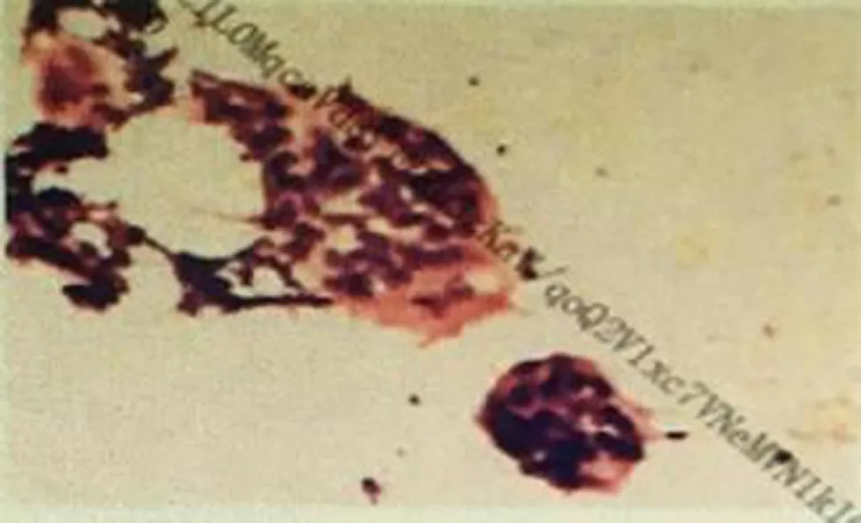
Figure 2 Biopsy of neck lymph node.
IFDs are characterized by insidious onset and lack of specificity of symptoms.Early neglect can cause delay of diagnosis and treatment, resulting in critical illness and life threatening complications.Therefore, effective antifungal therapy should be carried out once the definite diagnosis/clinical diagnosis is confirmed, and empirical antifungal therapy should also be carried out in the early stage for patients of suspected diagnosis with unclear pathogens.When empiric antifungal therapy is ineffective, it is important to change the antifungal drugs decisively.The patient eventually recovered and was discharged from the hospital, benefiting from early and timely empirical antifungal treatment, although ineffective, but winning the time and opportunity for the latter irrigation of changing antifungal drugs.
In summary, invasive mycosis is a common medical problem in the world.The positive rate of lymph node biopsy is not high.Once invasive fungal infection occurs,it is often accompanied by severe condition, long course, high medical cost, and poor prognosis.In addition, IFD has been shown to be a substantial financial burden to the health care system[24,25].Therefore, multi-stage and multi-site lymph node biopsies are the key to the diagnosis of the disease.Timely and effective antifungal treatment is essential for curing the disease.
CONCLUSION
The possibility of fungal infection should be considered when both empirical antiinfection and diagnostic anti-tuberculosis treatments are ineffective.The new antifungal drug was not the best treatment, and the empirical antifungal drugs do not necessarily work for every patient.Precise individualized treatment is needed.When routine antifungal therapy is invalid, it is appropriate to change the drug.When replacing antifungal drugs, it is necessary to consider the overlap and continuity of drugs.
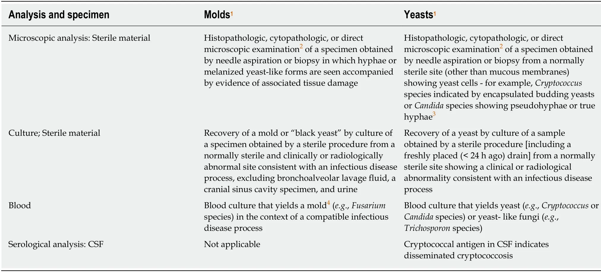
Table 1 Criteria for proven invasive fungal disease except for endemic mycoses

Table 2 Criteria for probable invasive fungal disease except for endemic mycoses

Probable IFD requires the presence of a host factor, a clinical criterion, and a mycological criterion.Cases that meet the criteria for a host factor and a clinical criterion but for which mycological criteria are absent are considered possible IFD.1Host factors are not synonymous with risk factors and are characteristics by which individuals predisposed to invasive fungal diseases can be recognized.They are intended primarily to apply to patients given treatment for malignant disease and to recipients of allogeneic hematopoietic stem cell and solidorgan transplants.These host factors are also applicable to patients who receive corticosteroids and other T cell suppressants as well as to patients with primary immunodeficiencies.2Must be consistent with the mycological findings, if any, and must be temporally related to current episode.3Every reasonable attempt should be made to exclude an alternative etiology.4The presence of signs and symptoms consistent with sepsis syndrome indicates acute disseminated disease, whereas their absence denotes chronic disseminated disease.5These tests are primarily applicable to aspergillosis and candidiasis and are not useful in diagnosing infections due to Cryptococcus species or Zygomycetes(e.g., Rhizopus, Mucor, or Absidia species).Detection of nucleic acid is not included, because there are as yet no validated or standardized Methods.TNF-α:Tumor necrosis factor-alpha; CT:Computed tomography; MRI:Magnetic resonance imaging; IFD:Invasive fungal disease.
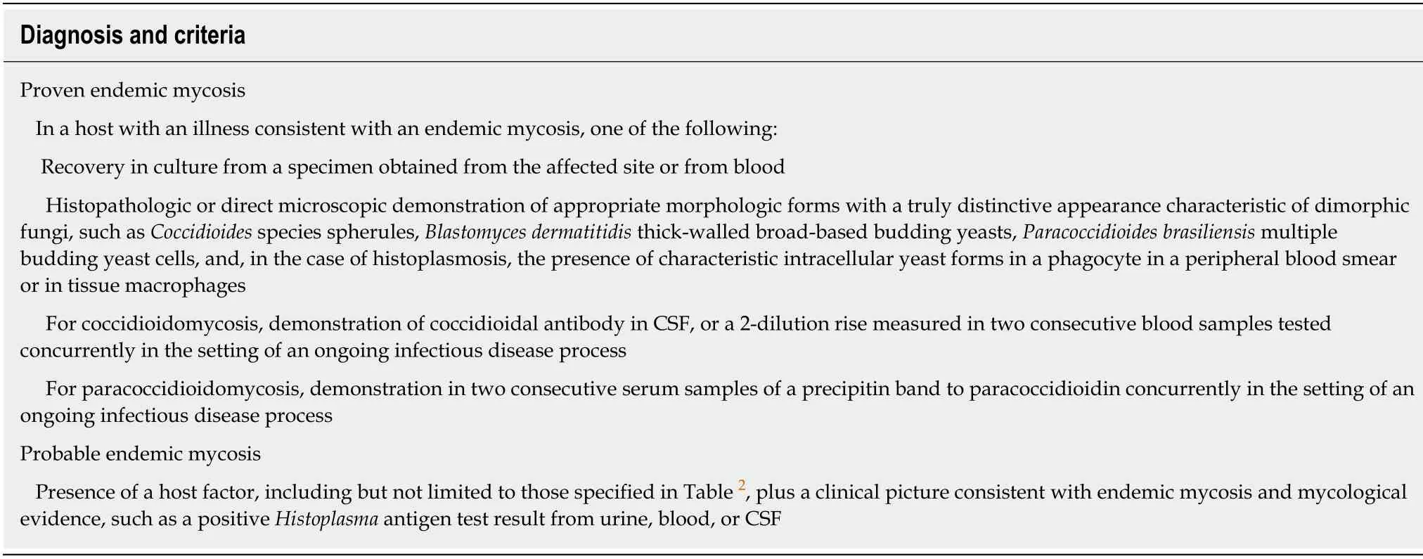
Table 3 Criteria for the diagnosis of endemic mycoses

Figure 3 Secondary biopsy of supraclavicular lymph node.
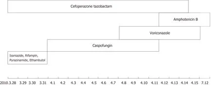
Figure 4 Timeline summarizing drug intervention.

Figure 5 Timeline summarizing patient’s information, clinical findings, diagnostic tests, diagnosis, pharmacological intervention, and follow up.
杂志排行
World Journal of Clinical Cases的其它文章
- Malignant syphilis accompanied with neurosyphilis in a malnourished patient:A case report
- Ex vivo revascularization of renal artery aneurysms in a patient with solitary kidney:A case report
- Pseudothrombus deposition accompanied with minimal change nephrotic syndrome and chronic kidney disease in a patient with Waldenström's macroglobulinemia:A case report
- Hepatocellular carcinoma successfully treated with ALPPS and apatinib:A case report
- Acute pancreatitis connected with hypercalcemia crisis in hyperparathyroidism:A case report
- Ulcerative colitis complicated with colonic necrosis, septic shock and venous thromboembolism:A case report
