Three-step coherent diffraction imaging systembased on parallel plates
2018-12-13LUOYongLITuoLIGuilinSHIYishi
LUO Yong, LI Tuo, LI Gui-lin, SHI Yi-shi*
(1.School of Optoelectronics,University of Chinese Academy of Sciences,Beijing 100049,China; 2.Academy of Opto-Electronics,Chinese Academy of Sciences,Beijing 100094,China; 3.School of Science,Xijing University,Xi′an 710123,China)
Abstract: In a traditional single beam multiple-intensity reconstruction(SBMIR) system, error is accumulated by multiple translational image sensors, which reduces the imaging effect and the effective resolution of the photoelectric imaging system. In this paper, a three-step coherent diffraction imaging system based on parallel plates is proposed. Three different diffraction planes are obtained by inserting or extracting two parallel plates and imaging and restoration reconstruction of complex amplitude objects are achieved. The numerical simulation and experiments show that the system overcomes the error accumulation problem of several translations in the SBMIR system, and one only needs to record three diffraction surfaces to avoid oversampling. The proposed optical system is easy to implement and has high repeatability.
Key words: the single-beam multiple-intensity;coherent diffraction imaging;parallel plates;complex amplitude
1 Introduction
引 言
Coherent Diffraction Imaging(CDI) is a lensless diffraction imaging technique[1-3]that has been rapidly developing with applications in adaptive X-ray imaging and related fields[4-8]. The CDI methods implemented in nowadays involve holography[9-10], wavefront detection reconstruction[11]and Gerchberg-Saxton′s(GS) algorithm for multiple diffraction information surfaces[12-13]. In general, single-step diffraction imaging is not suitable for complex amplitude recovery reconstruction because only one diffraction pattern is recorded. Therefore, an improved GS algorithm was produced[14-15], along with random binary pure phase modulation[16], rotational phase modulation[17], single-beam multi-intensity wavefront reconstruction(SBMIR) and other technical solutions[18-21]. However, among the many schemes, most of them are limited to the use of computers for numerical simulation. Also, specific optical imaging experiments for the schemes have not been implemented and they have no proposed specific experimental procedure. For this reason, further verification is needed to give plausibility to the methods and reproducibility of their experiments. Traditional SBMIR technology has been studied using numerical simulation analysis and specific experimentation. The phase recovery problem has also been solved. SBMIR methodology generally involves fixing a CCD camera on a precision stage and adopts a mechanical stepping mode. This easily causes the experimental image to have problems that result from shaking equipment, thereby reducing image resolution and quality. Furthermore, this technique usually requires that 10-20 diffractive faces be collected and recorded, which introduces the issue of oversampling defects. The above problems lead to difficulty repeating experiments and cause the technology to be less useful in real-world applications.
相干衍射成像(Coherent Diffraction Imaging,CDI),是一种无透镜衍射成像技术[1-3],从提出至今快速发展并应用于自适应成像、X射线成像等相关领域[4-8]。CDI的实现方法有基于全息术[9-10]、波前检测重建[11]和多个衍射信息面的Gerchberg-Saxton(GS)算法等[12-13]。通常,单步衍射成像因为只记录一幅衍射图像,无法适用于复振幅的恢复重建,所以基于此类相位恢复重建的问题,通常采用改进型的GS算法[14-15]、随机二元纯相位调制[16]、旋转相位调制[17]、单光束多强度波前重建(SBMIR)等技术方案[18-21]。然而,大部分方法都只局限于利用计算机进行数值模拟,具体的光学成像实验并未实现,也没有提出具体的实验方案。所以方法的实用性及实验的可重复性需要进一步验证。传统的SBMIR技术对数值模拟分析和具体实验都进行了研究,相位恢复问题得到了解决。但是,传统的SBMIR技术大多是将图像传感器CCD固定于精密平移台上,采用机械移动的步进方式,导致实验图像有抖动问题。从而降低了成像分辨率和质量,而且此技术通常需采集记录10~20个衍射面,有过采样的缺陷,上述问题及缺陷导致实验重复性较差,不利于技术方案的实际应用。
In order to solve and avoid the above issues, a three-step coherent diffraction imaging system based on parallel plates is proposed. The position of the CCD camera and the sample is fixed and two parallel plates are inserted in or extracted from the system. By doing so, three intensity information diffraction planes are quickly obtained and the sample pattern is eventually reconstructed by the recovery algorithm. The results of computer numerical simulation and actual optical experiments show that the system effectively avoids and solves the problem with shaking and the oversampling defects that exist in the traditional technical solutions. The proposed method is simple, quick to perform and repeatable while also producing images that are of significantly higher quality
为了解决和避免上述问题及缺陷,本文提出一种基于平行平晶的三步相干衍射成像系统,图像传感器CDD和样品的位置固定不变,采用依次在系统中插入或抽出2块平行平晶的方法,快速获得3幅强度信息衍射图,通过恢复算法最终重建样品图像。计算机数值模拟和实际的光学实验结果表明:该系统有效解决了传统技术方案的抖动问题与过采样的缺陷,最重要的是系统的成像效果显著提升,且具有实验可重复性高,操作简单快捷的特点。
2 Imaging System Structure and Method Principle Analysis
成像系统结构及方法原理分析
2.1 Traditional SBMIR Technology and System Implementation
传统SBMIR技术及系统实现
Before introducing the three-step coherent diffraction imaging system based on parallel plates, a brief discussion on the single-beam multi-strength reconstruction(SBMIR) technology scheme using a precision mobile platform will be presented. A typical SBMIR optical imaging system is shown in Fig.1(a). The CCD camera is fixed on a motorized precision stage where the rotation of a motor causes the precision stage to move along its track. The CCD camera records the diffraction intensity informationINof the object every time the precision stage moves by a distance of Δz. Anther scheme will be done by moving the position of the sample. Let the square of the intensity of the CCD acquisition recorded and the amplitude of the Fourier transform of the object be:
在介绍基于平行平晶的三步衍射成像系统之前,先简要讨论采用精密移动平台完成SBMIR技术方案。典型的SBMIR光学成像系统如图1(a)所示。图像传感器CCD固定在精密的机械移动平台上,通过电机转动使平移台沿着轨道方向移动,平移台每移动一段距离Δz,CCD就记录一次物体的衍射强度信息IN。另一种方案则是通过移动样品位置来实现。设CCD采集记录的强度信息与物体傅里叶变换的幅度成平方关系为:
IN=[F(ON)RZ+ΔZ]2, (1)
WhereFdenotes the Fourier transform operator,INis the object plane andRZ+ΔZis the diffraction distance. Typically, an SBMIR system requires that at least 3 diffraction intensity maps be recorded for wavefront reconstruction of the completed sample but often as many as 10-20 are used.
式中,F表示傅里叶变换算子,IN为物平面,RZ+ΔZ为衍射距离。通常情况下,典型的SBMIR系统需要采集记录的衍射强度图不少于3幅,一般为10~20幅,并以此完成的样品的波前重建工作。
2.2 Three-step Coherent Diffraction Imaging System Based on Parallel Plates
基于平行平晶的三步衍射成像系统
The proposed three-step coherent diffraction imaging system based on parallel plates is shown in Fig.1(b). The relative position of the object and the CCD camera is fixed. P1 and P2 represent two parallel flat crystal plates, which are illuminated by coherent light. The monochromatic plane wave in the system is vertically irradiated to the object plane after being collimated by a pinhole filter and a lens. It then reaches the CCD camera which records the surface after a diffraction of distancez. The system can complete the imaging processing in three steps:For the first step, after constructing and fixing the system device, the CCD camera is directly used to record the intensity informationI1of the first diffractive surface; In the second step, with the relative position of the CCD camera and the object unchanged, a parallel crystal plane P1 is inserted at any position between them. The CCD camera then collects and records the intensity informationI2of the second diffractive surface; In the third step, without disturbing the setup from the second step, another parallel flat crystal P2 is inserted in an arbitrary position between the object and the CCD camera, then the CCD camera is once again used to record the intensity informationI3of the third diffraction plane. After completing these three steps, the diffractive surface intensity information ofI1,I2,I3are each known. The positions of the object and CCD camera do not need to be moved or changed throughout the entire process and the completion time of the entire experiment is about 30 s. It does not involve the use of a precision stage, has no shaking in its system, no accuracy problems and has a simple experimental procedure that does not suffer from issues caused by oversampling.
研究提出的基于平行平晶的三步衍射成像系统如图1(b)所示,物体与图像传感器CDD的相对位置是固定不变的,P1和P2表示两块平行平晶,采用相干光照明,系统中的单色平面波,经针孔滤波器与透镜组合成的准直扩束系统后,垂直照射到物体平面,经过一段衍射距离z后到达图像传感器CDD记录面。系统经过3个步骤完成成像过程:第一步,搭建与固定好系统器件后,直接用CCD采集记录得到第一衍射面的强度信息I1;第二步,保持CCD与物体的位置不变,在它们之间的任意位置插入一块平行平晶P1,CCD采集记录后得到第二衍射面的强度信息I2;第三步,在第二步系统结构位置不变的基础上,在物体与CCD之间任意位置插入另一块平行平晶P2,CCD再次记录下第三幅衍射图的强度信息I3。最终,一共得到3个不同衍射面的强度信息I1,I2,I3。整个过程中,物体和CCD的位置是不需要移动及改变的,完成整个实验约需30 s。该系统不使用精度平移台,没有系统抖动、精度问题,也无过采样的复杂实验过程。

Fig.1 Structural comparison and principle analysis of proposed system 图1 系统结构比较及原理分析
The principle of the three-step coherent diffraction imaging system based on parallel plates is shown in Fig.1(c). The object is illuminated by a monochromatic coherent wave, and the object plane is diffracted by distancez0to meet a Fresnel plane, which is the first step of the system. The image then travels the distancez1to a second Fresnel plane, which is the second step of the system. Finally, it then continues to travel the distancez2to meet a third Fresnel plane, being the third step of the system. The above process is not completed by moving the CCD camera or the object through the precision stage, but instead by inserting parallel flat crystals between the CCD camera and the object, allowing the information to be reconstructed using the multiple intensities of the single beam. Of course, it should be pointed out that the above steps can be implemented in reverse, meaning that all the parallel flat crystals can be inserted first and then sequentially removed.
基于平行平晶的三步衍射成像系统的原理分析如图1(c)所示,用单色相干平面波照射物体,物体经过距离z0的衍射后得到第一衍射面,即前文所述的第一步;继续传播距离z1后得到第二衍射面,即前文所述的第二步;再继续传播距离z2后得到第三衍射面,即前文所述的第三步。以上过程不是通过精密平移台移动CCD或物体来实现的,而是采用在CCD与物体之间插入平行平晶得到单光束多强度的信息重建。当然,需要指出的是此系统上述步骤可以反向实施,即可先将所有的平行平晶插入,再每次抽取一块完成实验研究过程。
3 Key Algorithms and Numerical Simulation Analysis
关键算法及数值模拟分析
3.1 Key Algorithm Methodology
关键算法步骤
The algorithm of the three-step coherent diffraction imaging system based on parallel plates is based on the GS algorithm. The original diffraction plane is increased to three diffraction planes with different diffraction distances to recover and reconstruct the sample. It is through this method that the accuracy of the iterative algorithm and the convergence speed and recovery reconstruction effects are improved. An added benefit of the three-step coherent diffraction imaging system is that it has the ability to recover and reconstruct complex amplitude objects.
基于平行平晶的三步衍射成像系统的关键算法是以G-S算法为基础,由原来的一幅衍射图样增加为3幅不同衍射距离的衍射图样,以实现对样品的恢复重建,从而提高迭代算法的计算精确程度,及收敛速度和恢复重建效果。同时,三步衍射成像系统方法的一个最大优势在于可以对复振幅型的物体进行恢复重建。
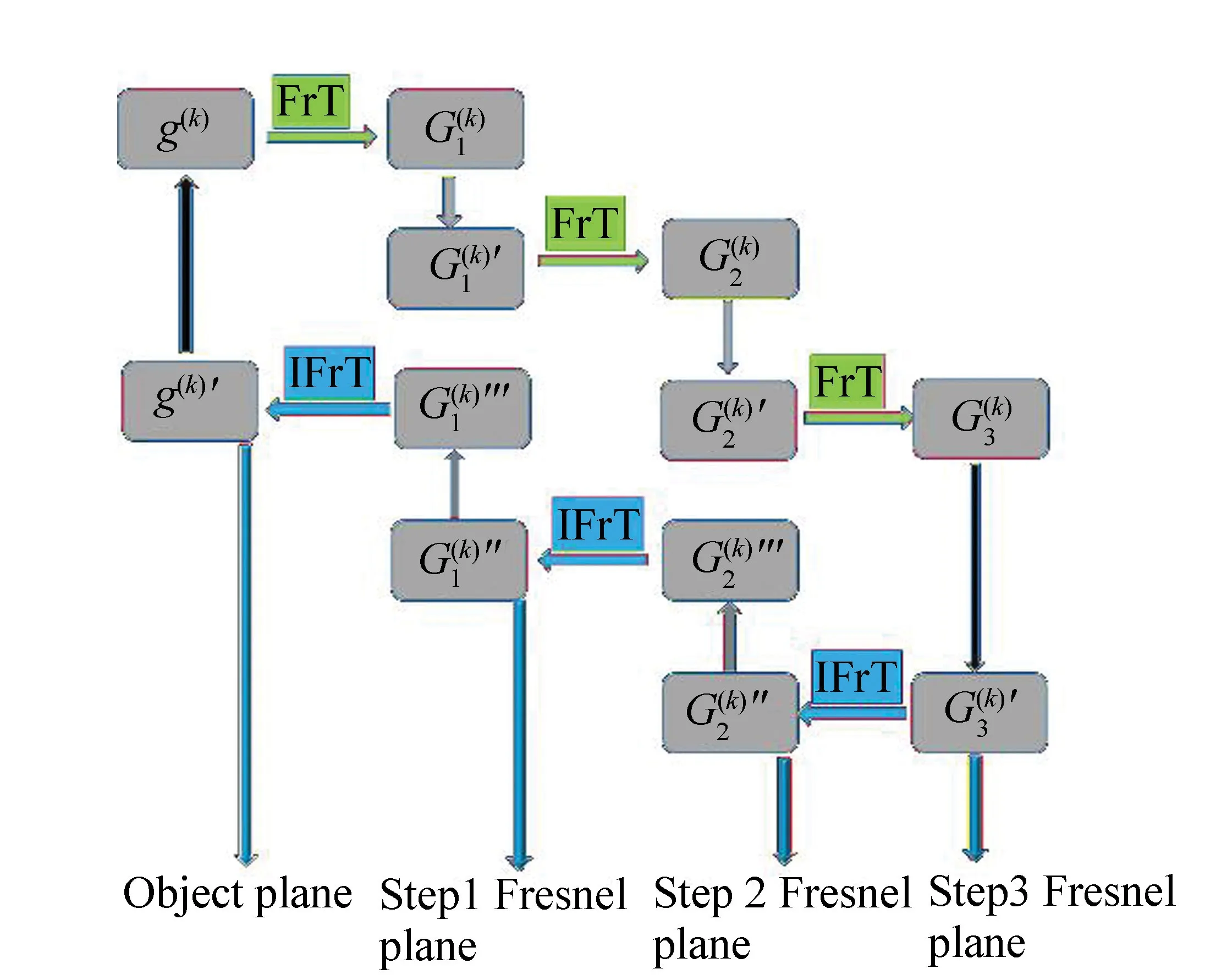
The algorithm key steps are shown in Fig.2, assuming

Fig.2 Block diagram of the key algorithm 图2 关键算法框图
关键算法步骤如图2所示,设
g(k)(x0,y0)=|g(k)(x0,y0)|·
exp[kφ0(x0,y0)] , (2)


(1)From the object plane positive to the first diffractive surface:
从物平面正向到第一衍射面:
(3)
(2)From the first diffractive surface positive to the second diffractive surface:
第一衍射面正向到第二衍射面:
(4)
(3)From the second diffractive surface positive to the third diffractive surface:
第二衍射面正向到第三衍射面:
(5)
(4)Reverse from the third diffractive surface to the second diffractive surface:
第三衍射面逆向到第二衍射面:
(6)
(5)Reverse from the second diffractive surface to the first diffractive surface:
第二衍射面逆向到第一衍射面:
(7)
(6)Reverse from the first diffractive surface to the object plane:
第一衍射面逆向到物平面:
(8)
When the sample is a pure amplitude type object, there is:
当样品为纯振幅型物体时,则有
g(k+1)(x0,y0)=|g(k)′(x0,y0)| , (9)
When the sample is a complex amplitude type object, there is:
样品为复振幅型物体时,则有
g(k+1)(x0,y0)=g(k)′(x0,y0) . (10)
3.2 Numerical Simulation Analysis
数值模拟分析
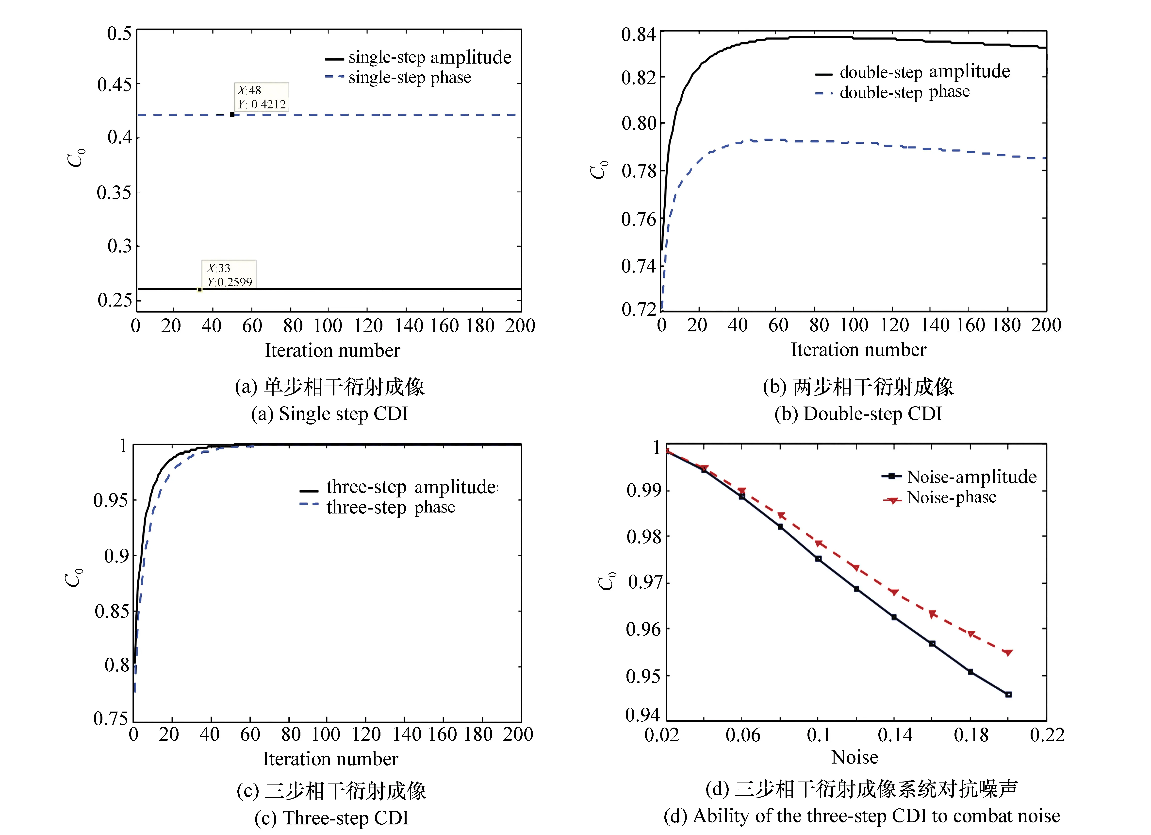
In order to further demonstrate the feasibility of the method using the three-step coherent diffraction imaging system, a numerical simulation analysis of the computer was first carried out, with the results shown in Fig.3. The single-step coherent diffraction image is calculated and analyzed is shown in Fig.3(a), the two-step diffraction imaging is shown in Fig.3(b) and the three-step diffraction imaging is shown in Fig.3(c). For convenience of comparison, the number of algorithm iterations is set to 200 times, the commonly used image evaluation function correlation coefficientCois used to judge the effect of restoration and reconstruction, and the range is generally [0,1]. The closer theCovalue is to 1, the closer the reconstruction is to the real object. If the value is smaller, the recovery quality is worse. Furthermore, the higher the deviation from the real object, the worse the imaging effect, affecting the iterative break and selection algorithm's number of iterations. For a pure amplitude type object, since there is no phase, the calculation is relatively simple and its detailed numerical simulation results are omitted. However, it should be pointed out that the convergence speed is very fast and theCovalue of the amplitude can quickly reach 1.
为了进一步论证三步衍射成像系统方法的可行性,首先进行了计算机数值模拟分析,结果如图3所示。分别计算了单步衍射成像(图3(a)),两步衍射成像(图3(b))、三步衍射成像(图3(c))。为方便比较,特将算法的迭代次数都设置为200次,采用常用图像评价函数相关系数Co来判断恢复重建效果,其取值范围一般为[0,1]。Co值越接近1说明恢复重建的物体越接近真实的物体。其值越小说明恢复质量越差,越偏离真实物体,成像效果越差,并以此来判断和选择算法的迭代停止条件。对于纯振幅型的物体而言,由于没有相位,所以较为简单。就不在给出其详细的数值模拟结果,但需要指出的是其收敛速度非常的快,且振幅的Co值能快速达到1。
In the process of computationally calculated numerical simulation, the sample pattern used is a grayscale image with a size of 256 pixel×256 pixel, the CCD camera′s pixel size is 6.45×10⁻⁶m/pixel, the laser′s wavelength is 632.8×10⁻⁹m. The sample is of the complex amplitude type, its phase distribution range is set to [-π,π], and the diffraction distances are set toz0=100 mm,z1=10 mm,z2=10 mm. The reconstruction of the complex amplitude of the sample is completed, and the correlationCocoefficient′s value is represented for the amplitude distribution and the phase distribution, respectively. In the simulated results, the black solid line is the value of the amplitude part correlation coefficient change, and the blue dotted line is the value of the phase part correlation coefficient change. In order to mark the value ofCoat a desired point in Fig.3 (a), the cursor included with the Matlab software package is used.
在进行计算机数值模拟分析过程中,使用的样品图样为灰度图,尺寸大小为256 pixel×256 pixel,图像传感器CCD的像素尺寸为6.45×10⁻⁶m/pixel,激光波长为632.8×10-9m,且样品为复振幅型,其相位分布范围设为[-π,π],将衍射距离设为z0=100 mm,z1=10 mm,z2=10 mm,完成对样品复振幅的恢复重建。对振幅分布与相位分布分别用相关系数Co值表示。在数值模拟结果中,其中实线为振幅部分相关系数变化值,蓝色点线为相位部分相关系数变化值。为了标注Co在某点的数值大小,在图3(a)中,使用了Matlab软件中自带的游标。
From the simulation results shown in Fig.3(a), under the same conditions, theCovalue range of the amplitude and phase fractions of the single-step diffraction imaging recovery reconstruction is less than 0.5. It is shown that only obtaining a single coherent diffraction intensity map is impossible to recover and reconstruct a complex amplitude object, which is why the method of single-step coherent diffraction imaging is not applicable to such objects. However, with the addition of a diffraction plane that has a distance ofz0+z1, the recovery and reconstruction effect resulting from two-step diffraction imaging is significantly improved, theCovalues of the amplitude and phase are higher than 0.5 and there is a tendency to converge. Nevertheless, late in the algorithm iteration, there is a slight decrease in morphology so a comprehensive evaluation shows that it cannot achieve the desired result. These results are shown in Fig.3(b). In contrast, the three-step coherent diffraction imaging process starts to converge when the algorithm passes 60 iterations and completely converges after about 70 iterations without any subsequent regression. Moreover, final convergenceCovalue of the recovery results, either the amplitude portion or the phase portion, reaches 1. These results show that the imaging quality of the system is continuously improved from one diffraction plane to three diffractions, that the algorithm completely converges to the third image, and that theCovalue reaches the optimal ideal value.

Fig.3 Numerical simulation analysis and comparison 图3 数值模拟分析及比较
图3(a)数值模拟结果表明,在相同的条件下,单步衍射成像恢复重建的振幅与相位部分的Co数值均低于0.5。说明仅有单幅衍射强度图是无法对复振幅型的物体进行恢复重建的,单步相干衍射成像方法不适用于复振幅型物体。然而,在增加一幅距离为z0+z1的衍射图后,两步衍射成像的恢复重建效果得到了明显提升,振幅及相位部分的Co值均高于0.5,且有收敛的趋势,但算法迭代到后面则出现了轻微的降低趋势。所以综合评价没有达到理想结果,结果如图3(b)。相比之下,三步相干衍射成像在算法迭代到60次时开始收敛且到70次左右完全收敛,后续没有任何下降的趋势。并且,无论是振幅部分还是相位部分,最终收敛的Co值均达到1。结果说明由一幅衍射图增加至三幅衍射的过程中,系统的成像质量不断的提升,且到第三幅时算法完全收敛,Co值也达到最佳的理想值。
In order to illustrate the robustness of the three-step coherent diffraction imaging system, a numerical simulation of the system′s ability to combat noise is added, as shown in Fig.3(d). Thex-value indicates that the system gradually increases the noise from 0, and the step size increases by 2%. When the noise increases to 20%, theCovalue of the amplitude and phase continues to exceed 0.94 with only minor fluctuations. Numerical simulation results show that the three-step diffraction imaging can effectively combat noise.
为了分析三步相干衍射成像系统方案的鲁棒性,对增加噪声系统进行了的数值模拟计算,结果如图3(d)所示。其中横坐标表示加入的噪声从0逐渐增加,增加步长为2%,当噪声增加至20%时,振幅与相位的Co值继续高于0.94,且变化幅度非常小。数值模拟结果表明,三步衍射成像对抗噪声能力良好。
4 Experimental results and analysis
实验结果及分析
4.1 Optical experiment results
光学实验结果
In order to demonstrate the feasibility of the three-step coherent diffraction imaging system based on parallel plates, the actual optical system imaging and recovery reconstruction work was carried out, and the corresponding SBMIR experiment based on precision translation stage was completed. These system structures are shown in Fig.1(a) and Fig.1(b). The experiment uses the following parameters: the laser is a coherent light source of a single plane with a wavelength ofλ=632.8 nm. The CCD camera′s pixel size is 6.45 μm/pixel, the number of intensities recorded by the experiment is 3 and the image size is 800 pixel×800 pixel. The above parameters are identical for both experiments. The difference between these experiments is that the traditional SBMIR system uses a precision translation stage where the moving CCD camera obtains diffractive surfaces at different distances. The accuracy of the translation stage is 0.01 μm and the maximum movement range is 15 mm. The three-step coherent diffraction imaging system based on parallel plates uses three parallel flat crystals, which are sequentially inserted into the system to obtain different diffractive surfaces. To an extent, the size of the parallel flat crystals does not affect the experimental operation so there is no outlined requirement, which is an advantage of this method. For more diffractive surfaces, the number of parallel flat crystals can be increased and the system will not need to be modified. Furthermore, there is no issue with shaking equipment or error caused by movement.
为了论证基于平行平晶的三步衍射成像系统的可行性,进行了实际的光学系统成像及恢复重建工作,并完成相对应的基于精密平移台的SBMIR实验,系统结构如图1(a)和图1(b)所示。实验参数设置如下:激光器为单色平面波的相干光源,波长λ=632.8 nm,图像传感器CCD像素尺寸为6.45 μm/pixel,实验所采集记录的强度信息图数量为3幅,采集图像尺寸为800 pixel×800 pixel,两个系统的实验参数完全相同。不同的是,传统的SBMIR采用的是精密平移台,移动CCD得到不同距离的衍射面,平移台的精度为0.01 μm,最大移动量程为15 mm。而基于平行平晶的三步衍射成像系统,使用的是三块平行平晶,依次插入系统中,以此得到不同的衍射面。在不影响实验操作的情况下,平行平晶的规格尺寸并没有严格要求,这也是此系统的一个优势,具有一定的自由度。如果需要更多衍射面,则增加平行平晶数量即可,不需要改动系统,故没有移动器件导致的系统抖动问题。
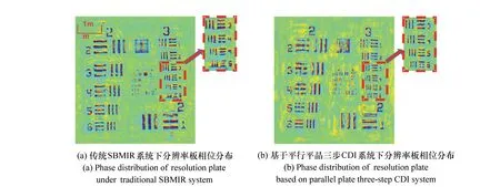
The experimental samples used were USAF 1951 resolution targets. The experimental results of recovery and reconstruction are shown in Fig.4. For the traditional SBMIR system, using a precision mobile platform requires that the CCD camera and sample be affixed to the platform, then, using movements of the platform, the CCD camera records the diffraction surface intensity information at different positions. In the experimental results given, in order to facilitate the comparison, the position of the sample to be tested and the CCD camera are fixed on the platform and the test was performed using moving steps of 3 mm. The translation stage is moved twice to create three steps and the CCD was allowed to record three experimental results. Finally, reconstruction was performed with the results shown in Fig.4(a).
实验样品均为USAF 1951分辨率板,恢复重建结果如图4所示。传统SBMIR系统使用了精密移动平台,其将CCD或待测样品固定于平台上,然后移动平台,CCD记录不同位置处的衍射面强度信息图。在给出的实验结果中,为了便于比较,待测样品位置固定不变,把CCD固定于平台上移动,移动步长为Δz=3 mm,分3个步骤移动2次平移台,CCD记录3次实验结果。最终恢复重建结果如图4(a)所示。

Fig.4 Comparison of phase distribution after restoration and reconstruction 图4 恢复重建后相位分布比较
In order to perform the three-step diffraction imaging system based on parallel plane, parallel flat crystals were inserted in three steps. The CCD camera recorded three experimental results which were eventually recovered and reconstructed. These results are shown in Fig.4(b).
基于平行平晶的三步衍射成像系统,使用平行平晶,分3个步骤两次插入平行平晶,CCD记录3次实验结果,最终进行恢复重建,结果如图4(b)所示。
Some areas in Figs.4(a) and (b) are highlighted in dotlines and then magnified. It can be clearly seen that the quality of (b) is better than that of (a), showing higher quality in experimental results given by the three-step coherent diffraction imaging system, when compared to the traditional SBMIR system method using a mobile platform.
图4(a)和(b)中,将虚线圈标出的部分放大相同倍数进行观察比较。通过对比可以明显看出图4(b)比图4(a)的质量好。实验结果表明:平行平晶的三步衍射成像系统比传统的使用移动平台的SBMIR系统实际成像效果好。
The experimental results also show that the phase distributions of some parts of the sample are reversed. The reason for this may be that the sample is tilted during the experiment, so that the parallel beam is inconsistent when it is irradiated onto the surface of the sample, causing errors in the reconstructed results.
同时实验结果显示,样品某些部位的相位分布存在翻转情况,原因可能是在实验过程中,样品有倾斜,使得平行光束照射到样品表面时并不一致,所以记录后重构结果有误差。
In order to further illustrate the three-step coherent diffraction imaging system based on parallel flat crystals and how a complex amplitude type object can be imaged and restored, the experiment of using a biological slice as a sample was performed with results shown in Fig.5. The experimental parameters used were the same as the preceding experiment with only the test sample being different.
为了进一步的说明基于平行平晶的三步衍射成像系统,能够对复振幅型物体成像并恢复重建,完成了以生物切片为样品的实验,如图5所示。所使用的实验参数与上述相同,只更换待测样品。

Fig.5 Experimental results of complex amplitude type samples 图5 复振幅型样品的实验结果
Fig.5(a) is a microscopic photograph of an original bio-slice sample. Fig.5(b) shows the reconstructed phase distribution under ordinary coherent diffraction imaging and Fig.5(c) is the amplitude distribution of the sample resulting from the parallel crystal plate system. The amplitude distribution after recovery is restored under a three-step diffraction imaging system. Fig.5(d) is the distribution of the phase portion. Fig.5(b) shows the recovery reconstruction results under ordinary(single-step) diffraction imaging technology. Since there is only one diffractive surface, the information that can be obtained is extremely limited. There is no effective constraint on the phase recovery of the sample so it is impossible to effectively restore and reconstruct the complex amplitude type object. From the results of Fig.5(c) and (d), it is clear that the three-step coherent diffraction imaging system based on parallel flat crystals can effectively image and recover reconstruction of complex amplitude objects.
在图5中,5(a)为原始生物切片样品的显微图,5(b)为普通相干衍射成像恢复重建后的相位分布,5(c)为基于平行平晶的三步衍射成像系统下恢复重建后的振幅分布,5(d)为相位部分的分布。图5(b)为普通(单步)衍射成像技术下的恢复重构结果。由于只有一个衍射面,所能够获得的信息极为有限,对样品的相位恢复没有有效的约束条件,所以无法对复振幅型的物体进行有效的恢复重建。图5(c)、5(d)的结果说明,基于平行平晶的三步衍射成像系统能够对复振幅型的物体进行有效的成像及恢复重建。
The experimental results show that the three-step coherent diffraction imaging system based on parallel plates in which flat crystals are simply inserted or extracted can decrease error resulting from system shaking and movement of a traditional SBMIR precision mobile platform. It can also effectively capture an image of amplitude and complex amplitude objects and restore them without oversampling. Furthermore, all of this is possible with high repeatability.
实验结果表明,基于平行平晶的三步衍射成像系统,采用插入或抽取平行平晶的方法,使系统避免了因机械移动而产生的系统抖动。能有效对振幅型,复振幅型的物体进行成像并恢复重建,不需要过采样,并且系统的重复性高。
A further benefit of the 3-step process is that it combats the problem where imaging systems often encounter field-of-view problems. Because the system's used sample size is generally much smaller than the size of the parallel flat crystals, and both are in the near-field range, the field of view of the system is unaffected by the flat crystals.
成像系统通常会涉及到视场问题,系统中由于使用的样品尺寸远小于平行平晶的尺寸,且都是在近场范围成像,因此系统的视场不会受到平晶的影响。
4.2 Error Discussion
误差讨论
The experimental results show that the imaging system has a certain amount of error and that the reconstructed sample has some ripples. In response to these problems, error analysis is performed from the following few aspects.
实验结果表明,成像系统有一定的误差,恢复重构的样品有一些波纹。针对这些问题,从以下几个方面进行误差分析。
Influence of optical component uniformity:The parallel crystal three-step coherent diffraction imaging system is influenced by the uniformity of the optical components of which it is comprised. In the process of manufacturing such optical components, the level and quality are not always ideal. One of the most important devices within this system is the parallel flat crystal. The light in the experiment was deviated after passing through the flat crystal, causing reconstruction results to be inaccurate and corrugated. Presumably, the lens of the system may also have this problem.
光学元件均匀性的影响,基于平行平晶的三步相干衍射成像系统,其中最重要的器件之一平行平晶,此类光学元件在加工制造过程中,工艺水平达不到理想要求,所以导致实验中衍射光束通过平晶后出现偏差,最终恢复重建结果有误差及波纹,当然系统中的透镜也可能有此问题。
The influence of the tilt of the optics in the imaging system:The need to insert or extract flat crystals in sequence in this operation cannot completely guarantee perfect alignment. Even the sample may be tilted. These tilts will cause the distance from the diffraction plane to the CCD camera to be inconsistent, which may cause ripples and errors in the experimental results.
光学器件的倾斜的影响,在成像系统中,需要依次插入或抽取平晶,在进行此操作时,无法完全保证绝对的平直而没有倾斜,同时样品也有可能出现倾斜,这些倾斜则会导致样品衍射到CCD记录面的距离不一致,因此可能会导致实验结果出现了波纹和误差。
External force vibration and air disturbance effects: In these optical experiments, all instruments are exposed to the environment, so external forces may cause the optical platform to vibrate. Such air disturbances in the system will affect the CCD recorded results which also has an impact on restoration and reconstruction.
外力振动及空气的扰动影响,进行光学实验,基所有的仪器都裸露在外,因此外力因素可能导致光学平台振动,系统周围的空气扰动,都会影响到CCD记录的结果。这同样对恢复重建的结果造成了一定的影响。
5 Conclusion
结 论
Based on the GS algorithm and the single-step diffraction lensless imaging method, a three-step coherent diffraction imaging system based on parallel flat crystals is proposed and compared with traditional SBMIR technology using a precision mobile platform. Using computerized numerical simulation and actual optical experimentation, it was demonstrated that a three-step coherent diffraction imaging system based on parallel flat crystals: is unlike the traditional SBMIR technology in that the required diffraction patterns are reduced from between 10 and 20 to merely 3; has no mechanical movement, or evidence of shaking in resulting imagery; does not suffer from reduced imaging resolution in photoelectric imaging systems resulting from movement; and is easily repeatable. As the system′s diffractive surfaces are added simply added to the system, the system succeeds in imaging and recovery reconstruction complex amplitude type objects and overcomes the hurdles that the single-step diffraction imaging cannot. At the same time, the proposed imaging system, being a lensless coherent diffraction imaging system, has no lens aberration problem. However, the parallel crystal plates used in the system, the required level of precision, as well as the tilt problem during plate insertion can have a negative impact on results. The three-step coherent diffraction imaging system based on parallel flat crystals proposed in this paper has a wide range of applications and a high value in the fields of diffraction imaging measurement, multi-wavelength imaging, biological microscopy, and optical information security.
在G-S算法和单步衍射无透镜成像方法的基础上,提出了基于平行平晶的三步衍射成像系统,并与使用精密移动平台的传统SBMIR技术进行比较,从计算机数值模拟和实际的光学实验进行论证。与传统SBMIR技术不同,基于平行平晶的三步衍射成像系统所需记录的衍射图由10~20幅,降低为3幅,更值得注意的是系统没有机械移动,没有图像抖动导致光电成像系统的成像分辨率降低的问题,也不需考虑移动的精度问题,且实验的重复性好;由单步衍射成像1个衍射面提升至3个衍射面,系统实现了对复振幅型物体的成像及恢复重建,有效克服了单步衍射成像对复振幅型物体无法恢复的缺陷。提出的成像系统,虽然是无透镜的相干衍射成像系统,没有透镜的像差问题,但系统中使用的平行平晶光学器件,其工艺水平及精度,还有插入过程中的倾斜问题对实验结果造成一定的影响。研究所提出的基于平行平晶的三步衍射成像系统,在衍射成像的测量,多波长成像,生物显微,光学信息安全等领域具有广泛的应用价值。
