The evolution of Epstein-Barr virus detection in nasopharyngeal carcinoma
2018-03-08YouQuanLiNiSannKhinMelvinChuaDivisionofRadiationOncologyNationalCancerCenterSingapore6960SingaporeOncologyAcademicClinicalProgramDukeNUSMedicalSchoolSingapore69547Singapore
You Quan Li, Ni Sann Khin, Melvin L.K. Chua,Division of Radiation Oncology, National Cancer Center, Singapore 6960, Singapore; Oncology Academic Clinical Program, Duke-NUS Medical School, Singapore 69547, Singapore
The detection of Epstein-Barr virus (EBV) in nasopharyngeal carcinoma (NPC) has evolved over the last 40 years, transitioning from simple serological tests of latent viral infection to extremely sensitive measurements of the circulating tumor virome. Compared to the former, cell-free(cf) EBV DNA quantification is considered superior for population-based screening, and possesses additional advantages of clinical prognostication and surveillance for subclinical recurrences. However, despite its broad utility, the clinical value of cf EBV DNA for prediction of treatment response remains uncertain, and is currently being investigated in prospective clinical trials. These lessons from EBV and NPC have since been tested in another emerging viral-associated head and neck cancer that is linked to the human papillomavirus (HPV).
EBV and nasopharyngeal carcinoma
The first association of EBV with tumorigenesis was reported by Woodliff through his observation in African endemic Burkitt’s lymphoma1. Since this seminal finding, associations of EBV with several other lymphoid and epithelial cancers have been reported, including NPC, as reported by Old and Clifford et al.2. Owing to this invariable etiological linkage(especially for NPC from endemic regions), serological markers of EBV infection, such as anti-IgA antibodies for early antigen (EA) and viral capsid antigen (VCA), have been investigated for screening of NPC. Following acute infection,EBV enters a phase of latency within B-lymphocytes of the host, and antibodies such as EBV-EA, -VCA, and -NA(nuclear antigen) persist at detectable levels throughout this phase. It is thus plausible that these serological markers potentially represent precursor signals of the eventual onset of NPC3; however, arguments against this notion relate to the ambiguous involvement of EBV during the process of NPC tumorigenesis. First, it is contentious if early or delayed exposure to the virus after birth determines the individual risk of developing NPC later in adult life4. Second, this conundrum is further compounded by the observation that EBV is not detected in pre-malignant lesions in high-risk individuals, thus suggesting that either 1) the downstream effects of EBV infection may be inconsequential in the irreversible malignant transformation of the epithelium or 2)EBV resides in other tissue types apart from the epithelium5.Of note, other molecular pathways such as p16 dysregulation have been implicated in the maintenance of the virome within a cell3. Third, EBV is especially resistant to transfecting epithelial cells, as opposed to lymphoid cells. For these reasons, the optimal screening strategy for NPC using serological markers of latent EBV infection remains elusive until today.
Historical studies using EBV serology
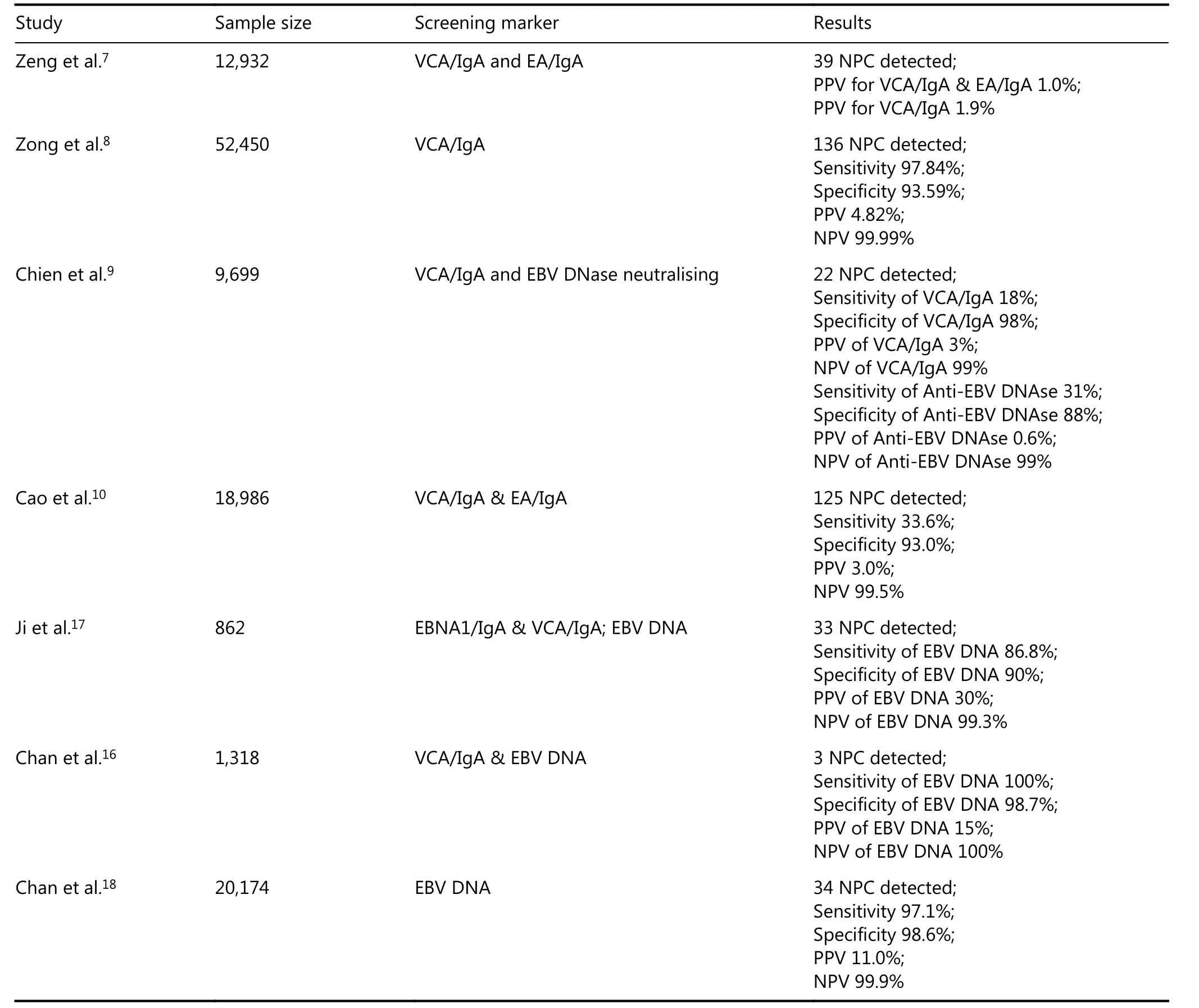
The focus of cancer screening has always been on early detection, coupled with the simplistic logic that inducing stage migration from advanced to early-stage disease will eventually improve population survival rates in the long term. This may hold true in NPC, since the prognosis of this disease is highly correlated with disease stage6. In this background, early screening studies in high-risk populations were first designed using EBV serological assays (Table 1). In these studies, high titers of IgA-VCA, IgA-EA, and EBV DNAse at baseline, with subsequent consecutive rises during follow-up, were predictive of an NPC diagnosis; however,false positive rates of 2%–18% were also reported using serological tests alone7-10. Expectedly, accuracy was enhancedwhen combinatorial markers were tested, compared to that observed using a single marker; the use of dual IgA-VCA and DNAse markers resulted in a higher rate of detection in a population of 9,699 Taiwan individuals (371 vs. 45 cases per 100,000 person-years)9. Similarly, Liu et al.11also showed an improved area under the curve (AUC) for NPC detection using dual marker selection as opposed to that of using a single marker in an independent Chinese cohort of 5,481 cases (AUC range of 0.87–0.97 vs. 0.77–0.95). While this may be true and heralds promise, a crucial issue that remains unresolved pertains to the uncertainty regarding the optimal combination of serological markers that would yield the highest accuracy for NPC screening. Owing to these limitations, coupled with the advent of circulating tumor EBV DNA testing, enthusiasm to implement EBV serological screening gradually waned over time.

Table 1 Summary of NPC screening studies.
The meteoric rise of circulating EBV DNA in NPC
Cell-free EBV DNA (cf EBV DNA) in the plasma was first reported as a biomarker for NPC by Lo and colleagues in 199912; briefly, these are short fragments of the EBV virome(<181 bp) that are supposedly released by cancer cells during apoptosis. Using real-time quantitative PCR targeted to the BamHI-W and EBNA-1 regions, Lo et al.12were able to demonstrate the presence of a circulating virome in a majority of NPC cases (55 of 57), but only in a few healthy controls (3 of 43). Importantly, this biomarker has clinical relevance; EBV DNA copy number was correlated to clinical disease stage, and in the post-treatment surveillance setting,persistent or detectable EBV DNA was predictive of an eventual tumor recurrence13. These findings were corroborated by Lin et al.14in a subsequent study. An interesting observation from these studies is the temporal sequence of cf EBV DNA detection relative to the onset of clinical disease (50–150 days in 6 cases), which suggests that cf EBV DNA is likely a surrogate for occult NPC tumor clones. This intuitively broadens the potential utility of EBV DNA in population-based screening, where the ultimate clinical goal is to detect early-stage disease.
cf EBV DNA was first combined with serum IgA-VCA antibody for screening, and the former method identified 75% of the false-positive cases detected by serology15. A subsequent moderate-sized population-based study of 1,318 volunteers in Hong Kong, SAR, China evaluated cf EBV DNA as a screening modality for NPC. Of the 69 individuals(5.2%) with a baseline positive test, only 3 early-stage NPC cases were identified by nasal endoscopy and MRI16. In a replicate study of 862 individuals from Southern China, the investigators reported a sensitivity of 86.8% (33 of 38 NPC cases) for NPC diagnosis using EBV DNA, but the sensitivity for early-stage disease was only 81% compared to 100% for cases with advanced disease17.
Against this background, the current study by Chan et al.18is seminal, since it represents the largest sample size considered until today, where 20,174 individuals were prospectively screened for NPC using cf EBV DNA from 2013 to 2016. Of the 309 cases with persistently elevated EBV DNA, 34 were eventually diagnosed with NPC (11.2%positive predictive value). The reported sensitivity and specificity of the assay were 97.1% and 98.6%, respectively.Crucially, 24 of the 34 (71%) cases diagnosed by screening presented with stage I and II disease, which compares favorably to an unscreened cohort in the same period [only 149 of 773 (19%) presented with stage I/II disease]; although not without potential selection bias, the early detection also corresponded to a superior progression-free survival [HR 0.10 (95% CI = 0.05–0.18)]. Overall, this study provides strong level IIA evidence to suggest implementing the cf EBV DNA assay as a method of NPC screening in high-risk individuals from endemic regions.
Nonetheless, to advocate caution, the short follow-up duration of the present study precludes an assessment of the long-term clinical impact, particularly for long-term survivorship. It is inconclusive if stage migration through screening will improve long-term overall survival, since survival rates of even advanced stages of NPC exceed 75%–80% with contemporary treatment of combination chemotherapy and intensity-modulated radiotherapy6. Next,the trajectory of tumorigenesis of NPC is unknown; it is possible that a subset of NPC cases directly progress to malignancy through a punctuated evolutionary process, and therefore screening will not alter the natural history of the disease in such cases. Third, the positive predictive value of the assay based on the current study is low at 11.0%, which is not unexpected given the gradual decline in NPC incidence even in the endemic parts of the world19. Should this declining trend continue, there would be a lesser need for NPC screening. Finally, the cost effectiveness of cf EBV DNA requires further investigation, especially when 593 individuals have to be screened to detect 1 NPC case; of note,the estimated cost burden has to include not only the cost of the assay but also that of subsequent investigations such as MRI and endoscopic examination, all of which are against life-years saved by screening.
Future of EBV DNA and cf tumor DNA technologies in head and neck cancers
Despite its multipurpose utility, cf EBV DNA remains, at best, a prognostic biomarker. Its role is limited for predicting therapeutic efficacy and influencing treatment recommendation, since it does not inform on the molecular vulnerabilities of the circulating occult tumor clones. The concept of using cf EBV DNA as a predictive biomarker is presently being tested in a prospective clinical trial (NRGHN001; ClinicalTrials.gov, NCT02135042); in this study,patients are stratified based on the presence or absence of this biomarker at the end of chemoradiotherapy, and patients harboring persistent cf EBV DNA copies will be referred to a randomized phase 2 study of adjuvant gemcitabine-paclitaxel compared to conventional cisplatin-5-fluorouracil. While the results of this clinical trial are awaited, Chan and colleagues20reported their findings of NPC-0502, which unexpectedly revealed no benefit of adjuvant gemcitabine-cisplatin over observation in patients with persistent cf EBV DNA afterchemoradiotherapy. Hence, a novel approach might be required. In this context, we now possess a catalog of mutational events occurring in NPC21-23. This opens the possibility of designing novel assays that can capture mutations of circulating tumor clones, which could then be exploited for designing novel paired drug-mutational targets studies in NPC.
Finally, it would be intuitive to transpose the findings observed in EBV-associated NPC onto another emerging virus-associated head and neck cancer - HPV oropharynx squamous cell carcinoma (HPV-OPSCC). However, several practical issues, including harmonization of the assay to measure cf HPV DNA, still need to be addressed prior to its clinical implementation. Nonetheless, few groups have reported an association between such a biomarker and advanced nodal status and overall TNM stage, with potential utility in clinical prognostication and monitoring of treatment response24,25.
Acknowledgements
This work was supported by the National Medical Research Council Singapore Transition Award (Grant No.NMRC/TA/0030/2014), and the Duke-NUS Oncology Academic Program Proton Therapy Research Program Fund.
Conflict of interest statement
No potential conflicts of interest are disclosed.
1.Epstein MA, Achong BG, Barr YM. Virus particles in cultured lymphoblasts from Burkitt's lymphoma. Lancet. 1964; 1: 702-3.
2.Ho HC, Ng MH, Kwan HC, Chau JC. Epstein-Barr-virus-specific IgA and IgG serum antibodies in nasopharyngeal carcinoma. Br J Cancer. 1976; 34: 655-60.
3.Tsao SW, Tsang CM, Lo KW. Epstein-Barr virus infection and nasopharyngeal carcinoma. Philos Trans R Soc Lond B Biol Sci.2017; 372(1732)
4.Poh SS, Chua ML, Wee JT. Carcinogenesis of nasopharyngeal carcinoma: an alternate hypothetical mechanism. Chin J Cancer.2016; 35: 9
5.Tsang CM, Deng W, Yip YL, Zeng MS, Lo KW, Tsao SW. Epstein-Barr virus infection and persistence in nasopharyngeal epithelial cells. Chin J Cancer. 2014; 33: 549-55.
6.Sun XM, Su SF, Chen CY, Han F, Zhao C, Xiao WW, et al. Longterm outcomes of intensity-modulated radiotherapy for 868 patients with nasopharyngeal carcinoma: an analysis of survival and treatment toxicities. Radiother Oncol. 2014; 110: 398-403.
7.Zeng Y, Jan MG, Zhang Q, Zhang LG, Li HY, Wu YC, et al.Serological mass survey for early detection of nasopharyngeal carcinoma in Wuzhou City, China. Int J Cancer. 1982; 29: 139-41.
8.Zong YS, Sham JS, Ng MH, Ou XT, Guo YQ, Zheng SA, et al.Immunoglobulin A against viral capsid antigen of Epstein-Barr virus and indirect mirror examination of the nasopharynx in the detection of asymptomatic nasopharyngeal carcinoma. Cancer.1992; 69: 3-7.
9.Chien YC, Chen JY, Liu MY, Yang HI, Hsu MM, Chen CJ, et al.Serologic markers of Epstein–Barr virus infection and nasopharyngeal carcinoma in taiwanese men. N Engl J Med. 2001;345: 1877-82.
10.Cao SM, Liu ZW, Jia WH, Huang QH, Liu Q, Guo X, et al.Fluctuations of Epstein-Barr virus serological antibodies and risk for nasopharyngeal carcinoma: a prospective screening study with a 20-year follow-up. PLoS One. 2011; 6: e19100
11.Liu Y, Huang QH, Liu WL, Liu Q, Jia WH, Chang E, et al.Establishment of VCA and EBNA1 IgA-based combination by enzyme-linked immunosorbent assay as preferred screening method for nasopharyngeal carcinoma: a two-stage design with a preliminary performance study and a mass screening in southern China. Int J Cancer. 2012; 131: 406-16.
12.Lo YM, Chan LY, Lo KW, Leung SF, Zhang J, Chan AT, et al.Quantitative analysis of cell-free Epstein-Barr virus DNA in plasma of patients with nasopharyngeal carcinoma. Cancer Res. 1999; 59:1188-91.
13.Chan KC. Plasma Epstein-Barr virus DNA as a biomarker for nasopharyngeal carcinoma. Chin J Cancer. 2014; 33: 598-603.
14.Lin JC, Wang WY, Chen KY, Wei YH, Liang WM, Jan JS, et al.Quantification of plasma Epstein-Barr virus DNA in patients with advanced nasopharyngeal carcinoma. N Engl J Med. 2004; 350:2461-70.
15.Leung SF, Tam JS, Chan ATC, Zee B, Chan LYS, Huang DP, et al.Improved accuracy of detection of nasopharyngeal carcinoma by combined application of circulating Epstein-Barr virus DNA and anti-Epstein-Barr viral capsid antigen IgA antibody. Clin Chem.2004; 50: 339-45.
16.Chan KCA, Hung ECW, Woo JKS, Chan PKS, Leung SF, Lai FPT,et al. Early detection of nasopharyngeal carcinoma by plasma Epstein-Barr virus DNA analysis in a surveillance program. Cancer.2013; 119: 1838-44.
17.Ji MF, Huang QH, Yu X, Liu ZW, Li XH, Zhang LF, et al.Evaluation of plasma Epstein-Barr virus DNA load to distinguish nasopharyngeal carcinoma patients from healthy high-risk populations in Southern China. Cancer. 2014; 120: 1353-60.
18.Chan KCA, Woo JKS, King A, Zee BCY, Lam WKJ, Chan SL, et al.Analysis of plasma Epstein-Barr virus DNA to screen for nasopharyngeal cancer. N Engl J Med. 2017; 377: 513-22.
19.Ferlay J, Soerjomataram I, Ervik M, Dikshit R, Eser S, Mathers C, et al. GLOBOCAN 2012 v1. 0, Cancer Incidence and Mortality Worldwide: IARC CancerBase No. 11 [Internet]. Lyon, France:International Agency for Research on Cancer 2013; Available from:http://globocan.iarc.fr.
20.Chan ATC, Hui EP, Ngan RKC, Tung SY, Cheng ACK, Ng WT, et al. A multicenter randomized controlled trial (RCT) of adjuvant chemotherapy (CT) in nasopharyngeal carcinoma (NPC) with residual plasma EBV DNA (EBV DNA) following primary radiotherapy (RT) or chemoradiation (CRT). J Clin Oncol. 2017;35: 6002
21.Lin DC, Meng X, Hazawa M, Nagata Y, Varela AM, Xu L, et al. The genomic landscape of nasopharyngeal carcinoma. Nat Gen. 2014;46: 866-71.
22.Park SM, Wong DJ, Ooi CC, Kurtz DM, Vermesh O, Aalipour A, et al. Molecular profiling of single circulating tumor cells from lung cancer patients. Proc Natl Acad Sci USA. 2016; 113: E8379-86.
23.Li YY, Chung GT, Lui VW, To KF, Ma BB, Chow C, et al. Exome and genome sequencing of nasopharynx cancer identifies NF-κB pathway activating mutations. Nat Commun. 2017; 8: 14121
24.Dahlstrom KR, Li GJ, Hussey CS, Vo JT, Wei QY, Zhao C, et al.Circulating human papillomavirus DNA as a marker for disease extent and recurrence among patients with oropharyngeal cancer.Cancer. 2015; 121: 3455-64.
25.Higginson DS, Scher ED, Yarusi B, Chan S, Mitrani L, Thompson C, et al. Use of human papillomavirus 16 (HPV16) cell free DNA for assessment of response to chemoradiation in HPV-Associated oropharyngeal cancer. Int J Radiat Oncol Biol Phys. 2015; 93:S78-9.
杂志排行
Cancer Biology & Medicine的其它文章
- Dual-specificity phosphatase 6 (DUSP6): a review of its molecular characteristics and clinical relevance in cancer
- Silencing of syndecan-binding protein enhances the inhibitory effect of tamoxifen and increases cellular sensitivity to estrogen
- EGFR tyrosine kinase inhibitor HS-10182 increases radiation sensitivity in non-small cell lung cancers with EGFR T790M mutation
- Calcium channel α2δ1 subunit as a novel biomarker for diagnosis of hepatocellular carcinoma
- Parkin protein expression and its impact on survival of patients with advanced colorectal cancer
- A new combined criterion to better predict malignant lesions in patients with pancreatic cystic neoplasms
