Nischarin-siRNA delivered by polyethyleniminealginate nanoparticles accelerates motor function recovery after spinal cord injury
2017-11-08YueminDingYuyingLiChuwangJaoJuangChenchenZhengShaohanJuangYangXuanXiaoyiSunXiongZhang
Yue-min Ding, Yu-ying Li, Chu wang, Jao Juang, Chen-chen Zheng, Shao-han Juang, Yang Xuan, Xiao-yi Sun, Xiong Zhang
1 Department of Clinical Medicine, School of Medicine, Zhejiang University City College, Hangzhou, Zhejiang Province, China
2 Department of Physiology, School of Medicine, Quzhou College of Technology, Quzhou, Zhejiang Province, China
3 Department of Pharmacy, Zhejiang University City College, Hangzhou, Zhejiang Province, China
4 Department of Basic Medicine, College of Medicine, Zhejiang University, Hangzhou, Zhejiang Province, China
How to cite this article: Ding YM, Li YY, Wang C, Huang H, Zheng CC, Huang SH, Xuan Y, Sun XY, Zhang X (2017) Nischarin-siRNA delivered by polyethylenimine-alginate nanoparticles accelerates motor function recovery aer spinal cord injury. Neural Regen Res 12(10):1687-1694.
Funding: is work was supported by the Natural Science Foundation of Zhejiang Province of China, No. LY15H250001 and LY14H090002;the National Natural Science Foundation of China, No. 81000535 and 81402872; the Medical Science and Technology Project Foundation of Zhejiang Province of China, No. 2014KYA166; and the Science and Technology Innovation Talents Development Plan Foundation for High School Students in Zhejiang Province of China, No. 2014R401186.
Nischarin-siRNA delivered by polyethyleniminealginate nanoparticles accelerates motor function recovery after spinal cord injury
Yue-min Ding1, Yu-ying Li2, Chu wang1, Jao Juang1, Chen-chen Zheng1, Shao-han Juang1, Yang Xuan1, Xiao-yi Sun3,*, Xiong Zhang4,*
1 Department of Clinical Medicine, School of Medicine, Zhejiang University City College, Hangzhou, Zhejiang Province, China
2 Department of Physiology, School of Medicine, Quzhou College of Technology, Quzhou, Zhejiang Province, China
3 Department of Pharmacy, Zhejiang University City College, Hangzhou, Zhejiang Province, China
4 Department of Basic Medicine, College of Medicine, Zhejiang University, Hangzhou, Zhejiang Province, China
How to cite this article: Ding YM, Li YY, Wang C, Huang H, Zheng CC, Huang SH, Xuan Y, Sun XY, Zhang X (2017) Nischarin-siRNA delivered by polyethylenimine-alginate nanoparticles accelerates motor function recovery aer spinal cord injury. Neural Regen Res 12(10):1687-1694.
A previous study by our group found that inhibition of nischarin promotes neurite outgrowth and neuronal regeneration in Neuro-2a cells and primary cortical neurons. In recent years, more and more studies have shown that nanomaterials have good prospects in treatment of spinal cord injury. We proposed that small interfering RNA targeting nischarin (Nis-siRNA) delivered by polyethyleneimine-alginate (PEIALG) nanoparticles promoted motor function recovery in rats with spinal cord injury. Direct microinjection of 5 μL PEI-ALG/Nis-siRNA into the spinal cord lesion area of spinal cord injury rats was performed. From day 7 aer surgery, Basso, Beattie and Bresnahan score was significantly higher in rats from the PEI-ALG/Nis-siRNA group compared with the spinal cord injury group and PEI-ALG/Control-siRNA group. On day 21 aer injection, hematoxylin-eosin staining showed that the necrotic area was reduced in the PEI-ALG/Nis-siRNA group. Immunohistochemistry and western blot assay results confirmed successful inhibition of nischarin expression and increased protein expression of growth-associated protein-43 in the PEI-ALG/Nis-siRNA group.ese findings suggest that a complex of PEI-ALG nanoparticles and Nis-siRNA effectively suppresses nischarin expression, induces expression of growth-associated protein-43, and accelerates motor function recovery aer spinal cord injury.
nerve regeneration; spinal cord injury; polyethylenimine; alginate; nanoparticles; nischarin; small interfering RNA; necrotic area;growth-associated protein-43; motor function; neural regeneration
Introduction
Nischarin is a novel protein discovered in 2000 by Alahari et al (2000). A previous study by our group found that nischarin inhibits neurite outgrowth and regeneration in neuronal cells (Ding et al., 2015). With RNA interference(RNAi) technology, small interfering RNAs (siRNA), small hairpin RNAs (shRNA), or microRNAs (miRNA) can act on target cells to knockdown expression of specific genes(Good et al., 2016; Koenig et al., 2017; Mohammadzadeh et al., 2017). Recently, PEI-ALG self-assembling peptide nanoparticles were shown to carry siRNAs and successfully mediate siRNA gene silencing in vitro (Sharma et al., 2009;Becker and McDonald, 2012; Blesch et al., 2012; Li et al.,2014). However, whether PEI-ALG nanoparticles can deliver nischarin-targeted siRNA (Nis-siRNA) to the spinal cord and promote motor function recovery aer SCI in animals is not known.
Using RNAi technology and novel nanoparticles in the present study, we aimed to prepare self-assembling nanocomplexes of PEI-ALG and Nis-siRNA for treating SCI, to preliminarily investigate novel methods for treatment of SCI.
Materials and Methods
Preparation of PEI-ALG nanoparticles
Branched PEI (25 kDa) and sodium alginate were purchased from Sigma-Aldrich (Merck-Millipore, Darmstadt, Germany). PEI-ALG nanoparticles were prepared according to a previously published method, but with certain modifications(Patnaik et al., 2006, 2010). Initially, 5 mg branched PEI (25 kDa) was dissolved in 500 mL distilled water. In a separate vessel, 1 mg alginate was dissolved in 100 mL distilled water under constant stirring at 90°C over 4 hours (Patnaik et al., 2010). Subsequently, the solutions were filtered through 0.22-μm membranes.e alginate solution was then slowly added to the PEI solution while stirring, and then vacuum concentrated in a rotary evaporator followed by lyophilizationon to a final concentration of 0.1 μg/μL.e mixture was incubated at room temperature for 30 minutes with continuous stirring.e final product of PEI-alginate nanoparticles was stored at 4°C until further use.
Preparation of PEI-ALG/siRNA complexes
A commercially available siRNA, containing a pool of three target-specific nischarin siRNA sequences (catalogue No.SC-108099; Santa Cruz Biotechnology, Santa Cruz, CA,USA), was used for preparation of PEI-ALG/siRNA complexes for gene silencing studies. Scrambled siRNA was used as a non-targeting control siRNA (Ctl-siRNA; catalogue No.SC-370007; Santa Cruz Biotechnology). To prepare PEIAlG/siRNA complexes, Nis-siRNA and Ctl-siRNA were diluted in RNase-free diethylpyrocarbonate-treated water to produce a 1 μg/μL solution. Next, 44 μL PEI-alginate nanoparticles (0.1 μg/μL) were mixed thoroughly with 6 μL siRNA solution.e ratio of polymer amine groups to siRNA phosphate groups in the complex was 10. Solutions were incubated at room temperature for 30 minutes to obtain PEIALG/Nis-siRNA or PEI-ALG/Ctl-siRNA complexes, which were subjected to size and zeta potential measurement using a Zetasizer Nano-ZS90 (Malvern Instruments, Malvern, UK)with the following settings: 14 measurements per sample;viscosity for water, 0.89 cP; temperature, 25°C.
Animals
A total of 45 adult female Sprague-Dawley rats weighing 200–250 g and aged 7–8 weeks old were obtained from the Experimental Animal Center of Zhejiang University of China (License No. SCXK (Zhe) 2014-0001). Rats were housed(four per cage) in a temperature (21 ± 1°C) and humidity (55± 5%) controlled environment under a 12-hour light/dark cycle with free access to standard food and water.
The study protocol was approved by the Animal Ethics Committee of Zhejiang University of China (ZJU2015-429-01).e experimental procedure followed the United States National Institutes of Health (NIH) Guide for the Care and Use of Laboratory Animals (NIH Publication No. 85-23, revised 1986).
Rat models of SCI
Rats were randomly divided into: SCI group, PEI-ALG group, PEI-ALG/control (Ctl)-siRNA group, and PEI-ALG/Nis-siRNA group (n = 10 in each group), as well as a sham group (n = 5).
Rats were anesthetized by intraperitoneal injection of sodium pentobarbital (50 mg/kg body weight; Sigma-Aldrich,St. Louis, MO, USA).e vertebral column was exposed and T10laminectomy was performed. Subsequently, the right half of the spinal cord was sectioned with a razor blade (Tachi et al., 2015). Rats in treatment groups received microinjections of 5 μL PEI-ALG, PEI-ALG/Ctl-siRNA, or PEI-ALG/Nis-siRNA into the central lesion site. Rats in the SCI group with no subsequent treatment were used as controls. Sham rats received laminectomy alone. After surgery, rats were treated with normal saline by subcutaneous injection into the fat layer of the back to replace lost fluids. To prevent infection, penicillin (100 mg/d) was administered twice daily for 3 days post-injury.
Right hindlimb movement was assessed by Basso, Beattie and Bresnahan (BBB) locomotor rating scale scores after the animals woke up from anesthesia. Rats that exhibited little or no hindlimb movement were considered successful models (n = 8 in each group), while those with incomplete loss of motor function with BBB scores > 8 were excluded(Umezawa et al., 2017). After assessing BBB locomotor rating scale, three rats in each group were sacrificed for western blot assays, with the remaining five used for morphological studies.
Motor function testing
Scoring according to BBB locomotor rating scale was performed by observers blinded to treatment status. To confirm successful hemisection, BBB scoring was performed on each animal prior to SCI and on the first day after SCI (Basso,2004). Subsequently, the scoring was repeated on days 3, 7,14, and 21 after SCI based on ipsilateral hindlimb performance.
Tissue processing
At the end of 21 days post-injury, five rats were randomly selected from each group and sacrificed with an overdose of sodium pentobarbital (100 mg/kg), then transcardially perfused with 0.1 M phosphate buffer, followed by ice-cold 4%paraformaldehyde (catalogue No. P6148, Sigma-Aldrich).Spinal cord tissue was harvested, fixed in 4% paraformaldehyde, and soaked overnight in a sequence of sucrose solutions (10%, 20%, and 30%). Tissue surrounding the lesion site was embedded in optimal cutting temperature compound (Sakura Finetek Japan Co., Ltd., Tokyo, Japan), fast frozen with dry ice, and stored at −80°C until processing.Spinal cord tissue blocks were cut into 30-μm serial longitudinal sections. Every fifth section was mounted on glass slides and stored at −20°C until analyzed.
Hematoxylin-eosin (HE) staining
For general histological examination, spinal cord sections (10 sections per rat) were stained using a HE staining kit (Beyotime Institute of Biotechnology, Inc., Haimen, China), according to the manufacturer’s instructions. In brief, sections were rinsed in distilled water and stained with hematoxylin for 5 minutes. Aer washing, sections were differentiated in 1% acid-alcohol for 30 seconds and incubated with eosin for 30 seconds. After dehydration with ethanol, sections were covered with xylene-based mounting medium and imaged using an Eclipse 50i microscope (Nikon Corporation, Tokyo,Japan). ImageJ software version 1.37 (NIH, Bethesda, MD,USA) was used to measure necrotic areas of spinal tissue in 3-mm segments. In each section, the necrotic area was determined and amount of each section summed per rat to produce total necrotic area per 3-mm segment.
Immunofluorescence staining
Spinal cord sections were subjected to immunostaining,as previously described (Ding et al., 2013). In brief, slices were dried at room temperature, rinsed with 0.01 M phosphate-buffered saline (PBS) for 30 minutes, and blocked in 10% normal goat serum (Gibco, New York, NY, USA) in PBS at room temperature for 2 hours to prevent nonspecific staining. Subsequently, sections were incubated with mouse anti-nischarin monoclonal antibody (1:500 dilution; catalogue No. 558262; BD Biosciences, San Jose, CA, USA) or rabbit anti-growth-associated protein (GAP)-43 polyclonal antibody (1:1,000 dilution; catalogue No. 8945; Cell Signaling Technology, Danvers, MA, USA) overnight at 4°C. Sections were then washed three times with PBS and incubated with fluorescein isothiocyanate (FITC)-labeled goat anti-mouse immunoglobulin (Ig)G (1:200 dilution; catalogue No. 115-095-003; Jackson ImmunoResearch Laboratories,West Grove, PA, USA) or Cy3-labeled goat anti-rabbit IgG(1:200 dilution; catalogue No. 111-165-003; Jackson ImmunoResearch Laboratories) at room temperature for 2 hours,followed by a PBS wash. Specimens were mounted onto gelatin-coated glass microscope slides, air-dried, mounted in glycerol-based mounting medium and observed under a confocal microscope (FV1000; Olympus, Tokyo, Japan).Relative intensity of FITC or Cy3 associated with nischarin or GAP-43 protein content, respectively, was measured using ImageJ soware version 1.37 (NIH).
western blot assay
Statistical analysis
Data are expressed as the mean ± SEM, and processed with SPSS 12.0 soware (SPSS, Chicago, IL, USA). Comparisons between groups were analyzed by two-way analysis of variance followed by Bonferroni’s multiple-comparison test or one-way analysis of variance followed by Scheffe’s post hoc test. P < 0.05 indicated statistically significant differences between values.
Results
Characterization of nanocomplexes and silencing efficiency of siRNA
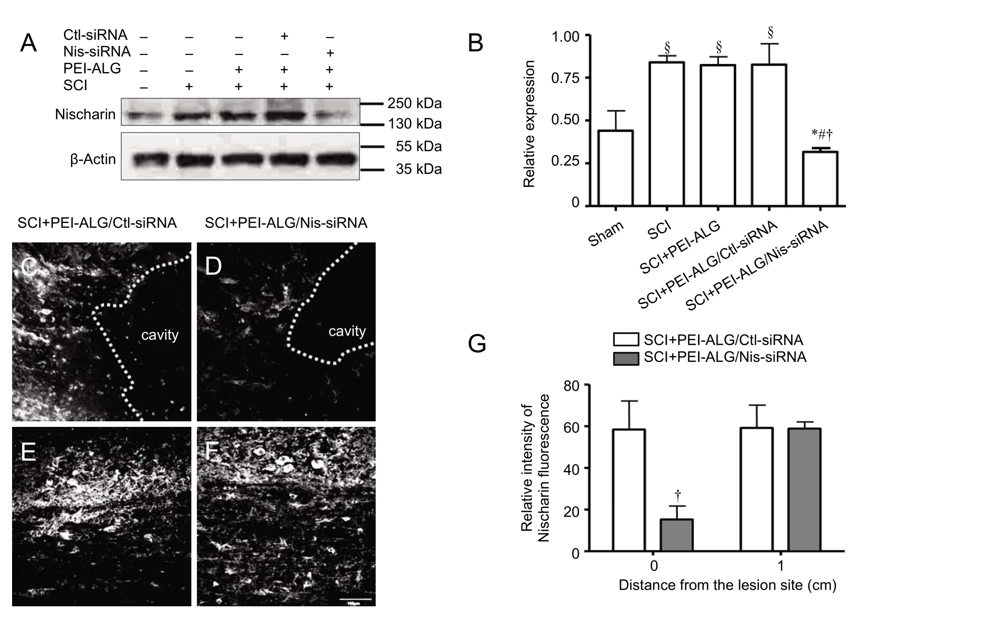
Figure 1 Nischarin protein expression in the injured spinal cord of rats.
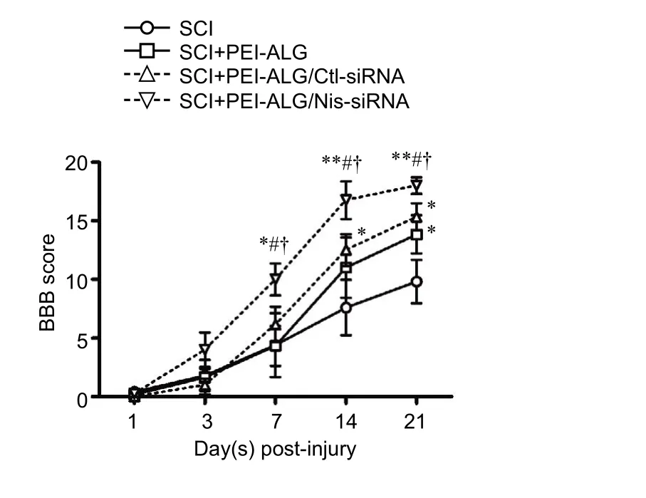
Figure 2 BBB score in rats with SCI at different time points.
Mean size and surface charge of PEI-ALG nanoparticles were 206.9 ± 36.3 nm and 10.1 ± 1.2 mV, respectively. After loading with Nis-siRNA, size and surface charge of the nanoparticles were 243.8 ± 43.6 nm and 8.9 ± 1.7 mV, respectively. Nanoparticles were injected into the injury site of spinal cord hemisection rat models. To measure gene silencing efficiency of Nis-siRNA delivered by nanocomplexes,nischarin expression levels in the injured region of the treated spinal cord were determined by western blot assay aer 3 weeks. Compared with the sham group, nischarin expression was upregulated in SCI groups (P < 0.05). Expression of nischarin protein in the PEI-ALG/Nis-siRNA group was significantly decreased compared with the SCI group (P < 0.05),PEI-ALG group (P < 0.05), and PEI-ALG/Ctl-siRNA group(P < 0.05). However, there was no difference among the SCI,PEI-ALG, and PEI-ALG/Ctl-siRNA groups (Figure 1AandB).ese findings were confirmed by immunofluorescence staining. Compared with the PEI-ALG/Ctl-siRNA group,nischarin expression in the PEI-ALG/Nis-siRNA group was significantly suppressed in the center of the lesion site (injection spot) (P < 0.05), but not the region 1 cm caudal to the injury epicenter (Figure 1C–G).
PEI-ALG/Nis-siRNA improved motor function of SCI rats

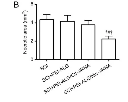
Figure 3 Hematoxylin-eosin staining of damaged areas in the spinal cord of rats on day 21 aer SCI.
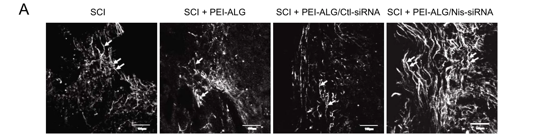
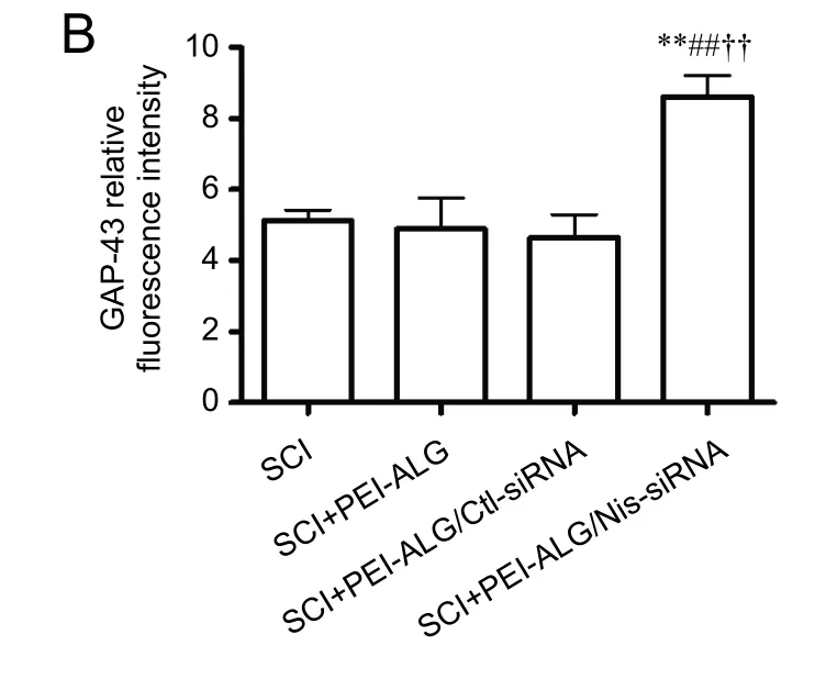
Figure 4 Effect of Nis-siRNA delivered by PEI-ALG nanoparticles on GAP-43 expression in the injured spinal cord of rats on day 21 aer SCI.
PEI-ALG/Nis-siRNA reduced necrotic area of injured spinal cord
To investigate the influence of PEI-ALG/Nis-siRNA on morphological changes and necrotic area in spinal cord tissue of rats in different groups, necrotic tissue in 3-mm cord segments were stained with HE and observed under light microscopy.e results showed that in the SCI group without treatment, spinal cord tissue structure was destroyed with massive infiltration of inflammatory cells and cavities. Similarly, in the PEI-ALG and PEI-ALG/Ctl-siRNA groups, inflammatory cell infiltration and cavities were also identified in spinal cord tissue. However, in the PEI-ALG/Nis-siRNA group, inflammatory cell infiltration at the injury site was not obvious with smaller cavities (Figure 3A). Quantification of necrotic area in different groups revealed that total necrotic area was reduced in the PEI-ALG/Nis-siRNA group compared with the SCI, PEI-ALG, and PEI-ALG/Ctl-siR-NA groups, (P < 0.05;Figure 3B). Consequently, PEI-ALG nanomaterial and Nis-siRNA may be used jointly for treating SCI, protecting injured cells, and reducing inflammatory cell infiltration and necrotic area.
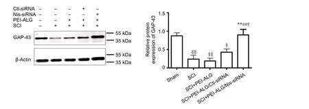
Figure 5 Effect of Nis-siRNA delivered by PEI-ALG nanoparticles on GAP-43 protein expression in the injured spinal cord of rats on day 21 aer SCI.
PEI-ALG/Nis-siRNA promoted GAP-43 protein expression in injured spinal cord
GAP-43 is a neuronal specific protein involved in outgrowth and regeneration of neuronal cells that is highly expressed during axonal regeneration. To clarify the mechanism through which PEI-ALG/Nis-siRNA promotes motor function recovery aer SCI, GAP-43 was detected by immunofluorescence staining on longitudinal spinal cord sections of rats from different groups. Compared with the other three groups, GAP-43 expression was increased around the injured area in the PEI-ALG/Nis-siRNA group, with a long filamentous, complete structure present. In contrast, it was less evident in the other three groups (Figure 4A). Quantification of immunofluorescence intensity revealed significantly higher GAP-43 protein expression in the PEI-ALG/Nis-siRNA group compared with the SCI, PEI-ALG, and PEI-ALG/Ctl-siRNA groups (P < 0.01;Figure 4B).
Western blot assay was further applied to detect GAP-43 protein content in injured spinal cord of rats.e results showed that relative expression of GAP-43 in the SCI, PEIALG, and PEI-ALG/Ctl-siRNA groups was significantly decreased compared with baseline expression in the sham group (P < 0.05). However, GAP-43 expression in the PEIALG/Nis-siRNA group was higher compared with the SCI group (P < 0.05), PEI-ALG group (P < 0.01), and PEI-ALG/Ctl-siRNA group (P < 0.05;Figure 5).
Discussion
Due to its high specificity, efficiency and other advantages,RNAi has become a potential treatment for SCI. For example, Sun et al. (2012) used Nogo-shRNA to effectively inhibit protein expression of Nogo and promote SCI repair in rats.
Qu et al. (2014) inhibited expression of the ephrin B3 gene,and consequently accelerated motor function recovery in SCI rats. Zukor et al. (2013) demonstrated that suppression of phosphatase and tensin homolog gene expression in mice promotes regeneration of corticospinal tract neurons. The novel protein nischarin, which acts on cytoskeletal actin through Rho-GTPase family members (Alahari et al., 2004;Reddig et al., 2005; Ding et al., 2008; Maziveyi and Alahari,2015), is highly expressed in the nervous system and inhibits neuronal cell migration (Ding et al., 2013). A recent study by our group found that neurite outgrowth is significantly promoted if nischarin expression is inhibited by siRNAs in vitro(Ding et al., 2015).us, we speculated that Nis-siRNA may stimulate axonal regeneration aer SCI in vivo.
Due to their good biodegradability, high flexibility, suitable mechanical properties, and absence of immunogenicity,nanoparticles have been studied in the field of biology and medicine, particularly in treatment of SCI, with promising results obtained (Kubinova and Sykova, 2010; Tyler et al.,2013; White-Schenk et al., 2015). For example, Kim et al.,(2009) used poly(lactic-co-glycolic acid)-based nanoparticles to deliver the drug methylprednisolone in vitro as well as in a SCI model in vivo.e study reported that local, sustained delivery of methylprednisolone via nanoparticles is significantly more effective in SCI treatment than systemic delivery. Tsai et al. (2006) produced a gel tunnel support with a complex of polyhydroxyethylmethacrylate and methyl methacrylate, filled it with methyl cellulose matrix and then implanted it into a rat with a completely transected T8spinal cord. Aer 8 weeks, regeneration of vestibular and red nucleus neurons was significantly increased.
Simultaneously, investigation of the combination of nanoparticles and RNAi technology for clinical treatment is being pursued (Acharya et al., 2017; Gentile et al., 2017).Recent research has focused on SCI treatment using nano-controlled release systems as siRNA carriers to achieve regulation of specific genes at the site of SCI (Li et al., 2016;Gao and Li, 2017). A previous study by our group has shown that ALG gel activates bone marrow mesenchymal stem cells and has unique advantages as a good carrier for stem cell transplantation in SCI treatment (Shi et al.,2011). Cationic PEI has been extensively studied in recent years for its high efficiency of gene delivery in vivo, thereby showing its therapeutic potential in humans (Lai, 2011;Patnaik and Gupta, 2013; Xun et al., 2014; Dabbaghi et al.,2017; Nakamura et al., 2017). Studies have revealed that PEI-ALG nanoparticles effectively deliver siRNAs, prevent enzymatic hydrolysis, and facilitate entry into cells for gene therapy (Patnaik et al., 2006; Patnaik et al., 2010). However,whether PEI-ALG nanoparticles can successfully deliver Nis-siRNA into cells in vivo to effectively interfere with target genes and promote rehabilitation of rats with SCI has remained elusive.
In the present study, a rat hemisection model of SCI was established. PEI-ALG nanoparticles were injected into the injured area, and hind limb motor function and morphological changes of spinal cord tissue in rats from different groups examined. As expected, nischarin expression in the center of lesion sites was significantly decreased at 3 weeks after PEIALG/Nis-siRNA injection. Efficient delivery distance of PEIALG nanoparticles was limited to the injection site, since nischarin expression was not different from the Ctl-siRNA group in the region 1 cm distal to the lesion site. Compared with the SCI, PEI-ALG, and PEI-ALG/Ctl-siRNA groups, BBB score in rats of the PEI-ALG/Nis-siRNA group began to increase from day 7 following SCI, which indicates that suppression of nischarin expression by Nis-siRNA effectively accelerated motor function recovery of SCI rats. HE staining showed less inflammatory cell infiltration and smaller cavities around the SCI site in the PEI-ALG/Nis-siRNA group. Immunofluorescence staining indicated that PEI-ALG/Nis-siRNA treatment increased GAP-43 expression in the injured spinal cord, which was confirmed by western blot assay. GAP-43 is highly expressed in neuronal growth cones, and GAP-43 upregulation is a useful marker of neurite growth or axonal regeneration(Ondarza et al., 2003; Novotna et al., 2011).us, suppression of nischarin expression may promote axonal regeneration by upregulating GAP-43 protein expression, although the specific mechanisms require further study.
Of note, the present study also found higher BBB scores in the PEI-ALG and PEI-ALG/Ctl-siRNA groups than those in the SCI group, particularly on day 21. Based on a previous study by our group, the beneficial effect of PEI-ALG and PEI-ALG/Ctl-siRNA is likely due to the positive impact of ALG hydrogel (Shi et al., 2011). Furthermore, the in vivo therapeutic effect of ALG was recently reported in SCI rat models (Grulova et al., 2015; Tamosaityte et al., 2015).
In summary, PEI-ALG/Nis-siRNA nanoparticles efficiently inhibit in vivo expression of nischarin protein, upregulate expression of GAP-43, and have a beneficial role in motor function recovery aer SCI.
Acknowledgments: We thank the staff from Department of Basic Medicine, College of Medicine, Zhejiang University, China, for their assistance in the preparation of PEI-ALG/siRNA complexes.
Author contributions:YMD designed the study and wrote this paper.YYL, CW, HH, CCZ, SHH and YX were responsible for the experimental conditions. XYS and XZ were responsible for study design, obtained funding, and provided technical support. All authors approved the final version of the paper.
Conflicts of interest:None declared.
Research ethics:e study protocol was approved by the Animal Ethics Committee of the Zhejiang University (ZJU2015-429-01). The experimental procedure followed the United States National Institutes of Health Guide for the Care and Use of Laboratory Animals (NIH Publication No.85-23, revised 1986).
Data sharing statement:Datasets analyzed during the current study are available from the corresponding author on reasonable request.
Plagiarism check:Checked twice by ienticate.
Peer review: Externally peer reviewed.
Open access statement:is is an open access article distributed under the terms of the Creative Commons Attribution-NonCommercial-ShareAlike 3.0 License, which allows others to remix, tweak, and build upon the work non-commercially, as long as the author is credited and the new creations are licensed under identical terms.
Acharya R, Saha S, Ray S, Hazra S, Mitra MK, Chakraborty J (2017)siRNA-nanoparticle conjugate in gene silencing: a future cure to deadly diseases? Mater Sci Eng C Mater Biol Appl 76:1378-1400.
Ahuja CS, Fehlings M (2016) Concise review: bridging the gap: novel neuroregenerative and neuroprotective strategies in spinal cord injury. Stem Cells Transl Med 5:914-924.
Alahari SK, Lee JW, Juliano RL (2000) Nischarin, a novel protein that interacts with the integrin alpha5 subunit and inhibits cell migration. J Cell Biol 151:1141-1154.
Alahari SK, Reddig PJ, Juliano RL (2004) The integrin-binding protein Nischarin regulates cell migration by inhibiting PAK. EMBO J 23:2777-2788.
Baptiste DC, Tighe A, Fehlings MG (2009) Spinal cord injury and neural repair: focus on neuroregenerative approaches for spinal cord injury. Exp Opin Investig Drugs 18:663-673.
Basso DM (2004) Behavioral testing aer spinal cord injury: congruities, complexities, and controversies. J Neurotrauma 21:395-404.
Becker D, McDonald JW, 3rd (2012) Approaches to repairing the damaged spinal cord: overview. Handb Clin Neurol 109:445-461.
Bin S, Zhou N, Pan J, Pan F, Wu XF, Zhou ZH (2017) Nano-carrier mediated co-delivery of methyl prednisolone and minocycline for improved post-traumatic spinal cord injury conditions in rats. Drug Dev Ind Pharm 43:1033-1041.
Blesch A, Tuszynski MH (2009) Spinal cord injury: plasticity, regeneration and the challenge of translational drug development. Trends Neurosci 32:41-47.
Blesch A, Fischer I, Tuszynski MH (2012) Gene therapy, neurotrophic factors and spinal cord regeneration. Handb Clin Neurol 109:563-574.
Dabbaghi M, Kazemi Oskuee R, Hashemi K, Ahami Goli A (2017)Evaluating polyethyleneimine/DNA nanoparticles-mediated damage to cellular organelles using endoplasmic reticulum stress profile. Artif Cells Nanomed Biotechnol doi: 10.1080/21691401.2017.1304406.Ding Y, Milosavljevic T, Alahari SK (2008) Nischarin inhibits LIM kinase to regulate cofilin phosphorylation and cell invasion. Mol Cell Biol 28:3742-3756.
Ding Y, Li Y, Lu L, Zhang R, Zeng L, Wang L, Zhang X (2015) Inhibition of nischarin expression promotes neurite outgrowth through regulation of pak activity. PLoS One 10:e0144948.
Ding Y, Zhang R, Zhang K, Lv X, Chen Y, Li A, Wang L, Zhang X, Xia Q (2013) Nischarin is differentially expressed in rat brain and regulates neuronal migration. PLoS One 8:e54563.
Gao SJ, Liu Y, Wang HJ, Ban DX, Cheng SZ, Ning GZ, Wang LL,Chang J, Feng SQ (2017) New approach to treating spinal cord injury using PEG-TAT-modified, cyclosporine-A-loaded PLGA/polymeric liposomes. J Drug Target 25:75-82.
Gao W, Li J (2017) Targeted siRNA delivery reduces nitric oxide mediated cell death aer spinal cord injury. J Nanobiotechnol 15:38.
Gentile E, Oba T, Lin J, Shao R, Meng F, Cao X, Lin HY, Mourad M,Pataer A, Baladandayuthapani V, Cai D, Roth JA, Ji L (2017) Cationic liquid crystalline nanoparticles for the delivery of synthetic RNAibased therapeutics. Oncotarget doi: 10.18632/oncotarget.18421.
Good ME, Begandt D, DeLalio LJ, Johnstone SR, Isakson BE (2016)Small interfering RNA-mediated connexin gene knockdown in vascular endothelial and smooth muscle cells. Methods Mol Biol 1437:71-82.
Grulova I, Slovinska L, Blasko J, Devaux S, Wisztorski M, Salzet M,Fournier I, Kryukov O, Cohen S, Cizkova D (2015) Delivery of alginate scaffold releasing two trophic factors for spinal cord injury repair. Sci Rep 5:13702.
Gunther MI, Weidner N, Muller R, Blesch A (2015) Cell-seeded alginate hydrogel scaffolds promote directed linear axonal regeneration in the injured rat spinal cord. Acta Biomater 27:140-150.
Kim YH, Ha KY, Kim SI (2017) Spinal cord injury and related clinical trials. Clin Orthop Surg 9:1-9.
Kim YT, Caldwell JM, Bellamkonda RV (2009) Nanoparticle-mediated local delivery of Methylprednisolone aer spinal cord injury. Biomaterials 30:2582-2590.
Kubinova S, Sykova E (2010) Nanotechnology for treatment of stroke and spinal cord injury. Nanomedicine (Lond) 5:99-108.
Lai WF (2011) In vivo nucleic acid delivery with PEI and its derivatives:current status and perspectives. Expert Rev Med Devices 8:173-185.
Li J, Liu Y, Xu H, Fu Q (2016) Nanoparticle-delivered IRF5 siRNA facilitates M1 to M2 transition, reduces demyelination and neurofilament loss, and promotes functional recovery after spinal cord injury in mice. Inflammation 39:1704-1717.
Li T, Wang GD, Tan YZ, Wang HJ (2014) Inhibition of lymphangiogenesis of endothelial progenitor cells with VEGFR-3 siRNA delivered with PEI-alginate nanoparticles. Int J Biol Sci 10:160-170.
Maziveyi M, Alahari SK (2015) Breast cancer tumor suppressors: a special emphasis on novel protein nischarin. Cancer Res 75:4252-4259.
Mohammadzadeh R, Saeid Harouyan M, Ale Taha SM (2017) Silencing of bach1 gene by small interfering RNA-mediation regulates invasive and expression level of miR-203, miR-145, matrix metalloproteinase-9, and CXCR4 receptor in MDA-MB-468 breast cancer cells.Tumour Biol doi:10.1177/1010428317695925.
Nakamura Y, Sato H, Nobori T, Matsumoto H, Toyama S, Shuno T,Kishimura A, Mori T, Katayama Y (2017) Modification of ligands for serum albumin on polyethyleneimine to stabilize polyplexes in gene delivery. J Biomater Sci Polym Ed doi:10.1080/09205063.2017.13287 30.
Nejati-Koshki K, Mortazavi Y, Pilehvar-Soltanahmadi Y, Sheoran S,Zarghami N (2017) An update on application of nanotechnology and stem cells in spinal cord injury regeneration. Biomed Pharmacother 90:85-92.
Novotna I, Slovinska L, Vanicky I, Cizek M, Radonak J, Cizkova D(2011) IT delivery of ChABC modulates NG2 and promotes GAP-43 axonal regrowth after spinal cord injury. Cell Mol Neurobiol 31:1129-1139.
Ondarza AB, Ye Z, Hulsebosch CE (2003) Direct evidence of primary afferent sprouting in distant segments following spinal cord injury in the rat: colocalization of GAP-43 and CGRP. Exp Neurol 184:373-380.
Patnaik S, Gupta KC (2013) Novel polyethylenimine-derived nanoparticles for in vivo gene delivery. Expert Opin Drug Deliv 10:215-228.
Patnaik S, Arif M, Pathak A, Singh N, Gupta KC (2010) PEI-alginate nanocomposites: efficient non-viral vectors for nucleic acids. Int J Pharm 385:194-202.
Patnaik S, Aggarwal A, Nimesh S, Goel A, Ganguli M, Saini N, Singh Y, Gupta KC (2006) PEI-alginate nanocomposites as efficient in vitro gene transfection agents. J Control Release 114:398-409.
Qu Y, Zhao J, Wang Y, Gao Z (2014) Silencing ephrinB3 improves functional recovery following spinal cord injury. Mol Med Rep 9:1761-1766.
Reddig PJ, Xu D, Juliano RL (2005) Regulation of p21-activated kinase-independent Rac1 signal transduction by nischarin. J Biol Chem 280:30994-31002.
Shahriari D, Koffler J, Lynam DA, Tuszynski MH, Sakamoto JS (2016)Characterizing the degradation of alginate hydrogel for use in multilumen scaffolds for spinal cord repair. J Biomed Mater Res A 104:611-619.
Sharma HS, Ali S, Tian ZR, Patnaik R, Patnaik S, Lek P, Sharma A,Lundstedt T (2009) Nano-drug delivery and neuroprotection in spinal cord injury. J Nanosci Nanotechnol 9:5014-5037.
Shi CY, Ruan LQ, Feng YH, Fang JL, Song CJ, Yuan ZG, Ding YM (2011)Marrow mesenchymal stem cell transplantation with sodium alginate gel for repair of spinal cord injury in mice. Zhejiang Da Xue Xue Bao Yi Xue Ban 40:354-359.
Song Z, Wang Z, Shen J, Xu S, Hu Z (2017) Nerve growth factor delivery by ultrasound-mediated nanobubble destruction as a treatment for acute spinal cord injury in rats. Int J Nanomed 12:1717-1729.
Sun HH, Gao F, Liu B, Yu HT, Kong N, Liu GM (2012) Inhibition of Nogo expression to promote repair after spinal cord injury. Chin Med J (Engl) 125:4044-4048.
Tachi Y, Okuda T, Kawahara N, Kato N, Ishigaki Y, Matsumoto T (2015)Expression of hyaluronidase-4 in a rat spinal cord hemisection model. Asian Spine J 9:7-13.
Tamosaityte S, Galli R, Uckermann O, Sitoci-Ficici KH, Later R, Beiermeister R, Doberenz F, Gelinsky M, Leipnitz E, Schackert G, Koch E,Sablinskas V, Steiner G, Kirsch M (2015) Biochemical monitoring of spinal cord injury by FT-IR spectroscopy-effects of therapeutic alginate implant in rat models. PLoS One 10:e0142660.
Tsai EC, Dalton PD, Shoichet MS, Tator CH (2006) Matrix inclusion within synthetic hydrogel guidance channels improves specific supraspinal and local axonal regeneration after complete spinal cord transection. Biomaterials 27:519-533.
Tyler JY, Xu XM, Cheng JX (2013) Nanomedicine for treating spinal cord injury. Nanoscale 5:8821-8836.
Umezawa H, Naito Y, Tanaka K, Yoshioka K, Suzuki K, Sudo T, Hagihara M, Hatano M, Tatsumi K, Kasuya Y (2017) Genetic and pharmacological inhibition of p38alpha improves locomotor recovery aer spinal cord injury. Front Pharmacol 8:72.
White-Schenk D, Shi R, Leary JF (2015) Nanomedicine strategies for treatment of secondary spinal cord injury. Int J Nanomed 10:923-938.
Xun MM, Liu YH, Guo Q, Zhang J, Zhang QF, Wu WX, Yu XQ (2014)Low molecular weight PEI-appended polyesters as non-viral gene delivery vectors. Eur J Med Chem 78C:118-125.
Zukor K, Belin S, Wang C, Keelan N, Wang X, He Z (2013) Short hairpin RNA against PTEN enhances regenerative growth of corticospinal tract axons aer spinal cord injury. J Neurosci 33:15350-15361.
Copyedited by James R, Norman C, Wang J, Li CH, Qiu Y, Song LP, Zhao M
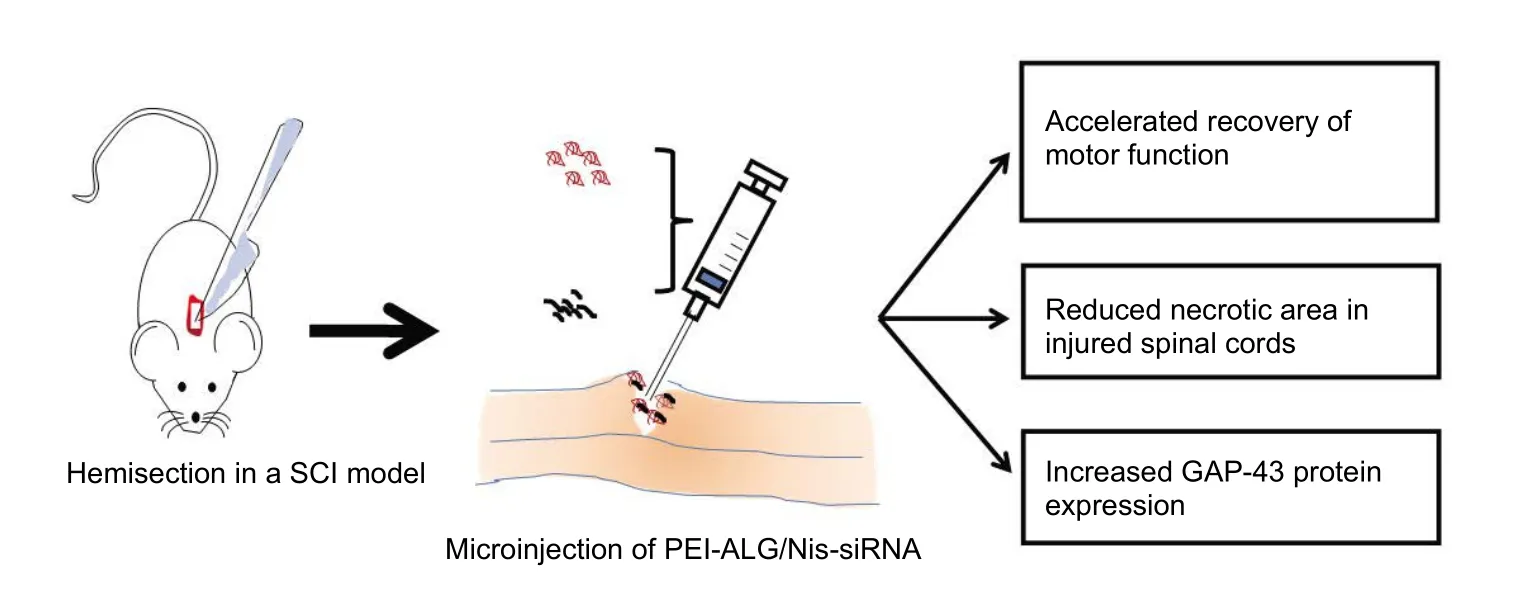
Graphical Abstract
Polyethyleneimine-alginate (PEI-ALG) nanoparticles carried nischarin-siRNA (Nis-siRNA) accelerates motor function recovery of spinal cord injury (SCI) rats by increasing the protein expression of growthassociated protein-43 (GAP-43)
*Correspondence to:
Xiong Zhang, Ph.D. or
Xiao-yi Sun, Ph.D.,xiongzhang@zju.edu.cn or sunxiaoyi@zucc.edu.cn.
orcid:
0000-0003-2235-569X
(Xiong Zhang)
0000-0002-6864-1715
(Xiao-yi Sun)
10.4103/1673-5374.217348
Accepted: 2017-08-20
杂志排行
中国神经再生研究(英文版)的其它文章
- Matrix bound vesicles and miRNA cargoes are bioactive factors within extracellular matrix bioscaffolds
- Diffusion tensor tractography studies on mechanisms of recovery of injured fornix
- Using 3D bioprinting to produce mini-brain
- Beta secretase activity in peripheral nerve regeneration
- Embracing oligodendrocyte diversity in the context of perinatal injury
- On the road towards the global analysis of human synapses