肺癌人源性肿瘤组织异种移植模型的组织学变化及其p63、napsin A和TTF-1的表达差异
2017-09-27黄海荣胡文娟许龙祥恽时锋
方 天,黄海荣,程 熙,胡文娟,刘 彪,许龙祥,董 敏,恽时锋*
(1.南京军区南京总医院比较医学科,南京 210002; 2.南京军区南京总医院心胸外科,南京 210002)
研究报告
肺癌人源性肿瘤组织异种移植模型的组织学变化及其p63、napsin A和TTF-1的表达差异
方 天1,黄海荣2,程 熙2,胡文娟1,刘 彪1,许龙祥1,董 敏1,恽时锋1*
(1.南京军区南京总医院比较医学科,南京 210002; 2.南京军区南京总医院心胸外科,南京 210002)
目的建立肺癌人源性肿瘤组织异种移植(patient-derived tumor xenograft,PDX)裸鼠模型,检测原代肺癌组织标本和第三代(F3)PDX模型组织标本内组织形态结构的变化和p63、napsin A和TTF-1的表达差异性。方法取临床肺癌新鲜手术切除标本12例,裸鼠皮下移植建立PDX模型,HE染色比较原代患者肿瘤组织与移植瘤组织结构和细胞形态变化与否;免疫组化的方法检测原代组织和F3代组织标本内p63、napsin A和TTF-1的表达水平的差异性。显微镜观察。结果成功建立传至第三代的PDX模型5例,4例肺鳞癌,1例肺腺癌。PDX模型和患者原代肿瘤具有相同的组织学特点和排列结构;p63在肺鳞癌患者及其PDX模型癌细胞中表达阳性率为84.3%和96%,两者之间差异无显著性(P> 0.05),而在肺腺癌中呈阴性表达。Napsin A在肺腺癌患者及其PDX F3癌细胞中阳性表达率分别为66%和72.4%,两者之间差异无显著性(P> 0.05)。TTF-1在肺腺癌患者及其PDX F3癌细胞中阳性表达率分别为91%和85%,两者之间差异也无显著性(P> 0.05)。Napsin A和TTF-1在肺鳞癌中均呈阴性表达。结论本实验室所建立的肺癌PDX模型基本保留了部分原代肿瘤的组织学特性,为临床前药效的个性化筛选评估以及生物标志物的鉴定提供了有效的研发资源。
PDX模型;肺癌;p63;Napsin A;TTF-1
将临床的新鲜肿瘤组织样品移植到免疫缺陷小鼠体内所建立的人源性肿瘤组织异种移植(patient-derived tumor xenograft,PDX)模型,能够较好地保留原发肿瘤的微环境和基本特质,能够基本复制患者肿瘤的异质性,保持人体原发肿瘤分子学、基因学和病理学的复杂性,为肿瘤研究提供了一个较细胞系接种模型更具优势的体内模型[1, 2]。但是,经过裸小鼠异种移植稳定传至第三代的PDX模型病理特性与人原发肺癌病理改变是否具有相同的组织学特点和排列结构,TTF-1、p63和napsin A的表达是否会出现差异,能否保留好原代肿瘤组织所特有的性质和微环境,能否更好地代表不同患者个体仍需要做进一步研究。本研究旨在比较原代肺癌组织与稳定传代至第三代的PDX肺癌模型中肿瘤组织的组织学结构的一致性,并鉴别TTF-1、p63、napsin A三种蛋白在原代肺癌组织和PDX肺癌模型中的表达情况。
1 材料和方法
1.1实验动物、肿瘤标本和主要试剂
SPF级BALB/c nu/nu雄性裸鼠50只,6~8周龄,规格为:体重20~25 g,购自北京维通利华实验动物技术有限公司【SCXK (京) 2012-0001】,饲养在南京军区南京总医院比较医学科SPF级屏障系统中心实验室【SYXK (军) 2012-0014】。所有肺癌肿瘤标本来自南京军区南京总医院心胸外科,标本获取前已通过伦理审核,并得到患者及家属同意。2016年3月~2016年11月,本院经组织病理证实,肺癌患者12例,男9例,女3例,年龄51~78岁,平均年龄62.6岁。其中,肺鳞癌8例,肺腺癌4例。主要试剂中,鼠抗人TTF-1、p63蛋白单克隆抗体及EnVision二步法试剂盒均为丹麦DAKO公司产品,napsin A多克隆抗体是福州迈新公司生产的即用型试剂盒。动物伦理审查批号:2016DWLS-0710。
1.2人肺癌PDX模型的建立及传代方法
取病人新鲜肿瘤组织标本(F0),直径10 mm,取材后立即置于新鲜培养基内冷藏保存。将肿瘤在培养基内分割成1 mm × 1 mm × 1 mm大小的肿瘤组织块,将组织块移植到5只BALB/c nu/nu裸鼠的皮下(F1)。移植部位分别为左、右腋下和背部。整个操作过程在超净工作台中完成。剩下的肿瘤组织,一半置于4%甲醛中固定;另一半液氮快速冷冻后转入-80℃冰箱内保存。
当皮下生长的肿瘤生长到300~500 mm3左右时,完整剥离肿瘤,在培养基内分割成1 mm × 1 mm × 1 mm大小的肿瘤组织块,将组织块移植到5只小鼠的皮下(F2)。整个操作过程在超净工作台中完成。传代达到F3取肿瘤组织块,取部分肿瘤组织用4%多聚甲醛固定,进行HE染色、免疫组化染色并与患者肿瘤组织比较。
1.3HE和免疫组化分析
将原代肿瘤组织即患者肿瘤组织和裸鼠体内传至F3代的肿瘤组织用4%多聚甲醛固定后,石蜡包埋,组织切片并进行苏木精-伊红(hematoxylin-eosin,HE)染色,观察肿瘤组织病理学变化。TTF-1、p63和napsin A蛋白检测采用免疫组织化学EnVision二步法,应用福州迈新公司生产的即用型试剂盒及即用型抗TTF-1、p63、napsin A抗体。所有组织标本经体积分数4%甲醛溶液固定,常规脱水,石蜡包埋,4 μm厚连续切片,常规脱蜡水化。严格按免疫组织化学试剂盒说明书进行操作。
1.4TTF-1、p63和napsinA阳性率判定
本研究中所选三种标志物TTF-1、p63、napsin A免疫组化染色(immunohistochemistry,IHC)的判断方法和标准是:光镜下每张切片选取随机的10个视野计数300个癌细胞,napsin A蛋白阳性细胞指在肿瘤细胞的细胞浆染色;TTF-1和p63蛋白阳性细胞则指肿瘤细胞细胞核染色,而细胞质及细胞膜未见染色。肿瘤组织中不超过10%的癌细胞染色为阴性,超过10%的癌细胞染色为阳性;当标本细胞几乎全部表达阴性但局灶区域呈强阳性表达时记为局灶阳性,本研究中局灶阳性记为阳性范畴[3]。
1.5统计学方法


表1 TTF-1、p63和napsin A蛋白在肺鳞癌和肺腺癌及其PDX模型中的表达阳性率/%
2 结果
经过10个多月的传代培养,共获得成功传至第三代的PDX模型5例,其中肺鳞癌4例,肺腺癌1例。每代平均历时3个半月。
2.1HE结果
如图1所示,PDX模型和从患者体内取得的原代肿瘤具有相同的组织学特点和排列结构,较好地保持了肿瘤细胞原有的排列结构和肿瘤细胞的异型性。
2.2免疫组织化学结果
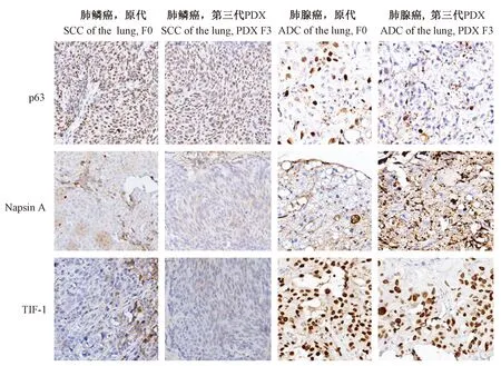
p63蛋白在肺鳞癌患者癌细胞和其第三代PDX模型癌细胞胞核中均呈强阳性表达;而在肺腺癌患者癌细胞及其PDX模型癌细胞中则极少数表达,少于5%,即呈阴性表达。Napsin A蛋白在肺鳞癌患者癌细胞和其第三代PDX模型癌细胞中呈阴性表达,而在肺泡上皮上呈阳性表达;napsin A蛋白在肺腺癌患者癌细胞和其第三代PDX模型癌细胞胞质中均呈强阳性表达。TTF-1蛋白在肺鳞癌患者癌细胞和其第三代PDX模型癌细胞中均呈阴性表达;而在肺腺癌患者癌细胞和其第三代PDX模型癌细胞胞核中均呈强阳性表达。实验结果见图2。
2.3p63、napsinA和TTF-1蛋白表达情况比较分析
p63在肺鳞癌患者癌细胞中表达阳性率为84.3%,明显高于肺腺癌患者及其第三代PDX模型(PDX F3)癌细胞中阳性表达率(4.8%,8.3%),差异有显著性(P< 0.01);与其相对应的PDX F3中癌细胞阳性率(96%)相比,差异无显著性(P> 0.05)。Napsin A在肺鳞癌癌细胞及其PDX F3癌细胞均呈阴性表达,两者之间无差异;明显低于肺腺癌患者及其PDX F3癌细胞中阳性表达率(66%,72.4%),差异有显著性(P< 0.01);而napsin A在肺腺癌患者及其PDX F3癌细胞两者之间表达阳性率差异无显著性(P> 0.05)。TTF-1在肺鳞癌癌细胞及其PDX F3癌细胞均呈阴性表达,两者之间差异无显著性(P> 0.05),而在肺鳞癌及其PDX F3肿瘤组织中肺泡上皮细胞均有少量表达;明显低于肺腺癌患者及其PDX F3癌细胞中阳性表达率(91%,85%),差异有显著性(P< 0.01);而TTF-1在肺腺癌患者及其PDX F3癌细胞两者之间表达阳性率差异无显著性(P> 0.05)。见表1。

注:A(×200)、B(×400):肺鳞癌患者原代肿瘤;C( ×200)、D(×400):肺鳞癌第三代PDX模型移植瘤;E( ×200)、F(×400):肺腺癌患者原代肿瘤;G( ×200)、H(×400):肺腺癌第三代PDX模型移植瘤。图1 患者原代肿瘤及其PDX模型肿瘤组织HE染色图Note. A (×200), B (×400): Primary tumor of squamous cell carcinoma (SCC) of the lung;C (×200), D (×400): F3 generation of PDX model of SCC of the lung;E (×200), F (×400): Primary tumor of adenocarcinoma (ADC) of the lung;G (×200), H (×400): F3 generation of PDX model of ADC of the lung.Fig.1 Histological architecture of the primary tumor and the F3 PDX model tissues. HE staining

图2 p63、napsin A和TTF-1分别在肺鳞癌和肺腺癌原代肿瘤组织及其PDX模型肿瘤组织中的表达(×200 )Fig.2 Expression of p63, napsin A and TTF-1 in the tumor tissues of SCC and ADC of the lung and the F3 PDX models
3 讨论
人源性肿瘤组织异种移植(patient-derived tumor xenograft,PDX)模型是指直接将新鲜外科手术肿瘤组织、活检组织或者原代细胞通过异位(通常指皮下或肾包膜)[4]或原位种植到免疫缺陷小鼠体内[5],依靠小鼠提供的环境生长的一种异种移植模型。PDX模型已被证实是一个有特殊意义的在很多肿瘤类型临床前药物测试的研究工具。虽然PDX模型保留了原代患者肿瘤的组织病理学、基因组结构以及药敏反应,PDX肿瘤的克隆型分布与患者原始肿瘤相比会有所变化。而且研究发现,PDX模型中更具侵袭性的肿瘤克隆很可能成为主导,根据基因组和转录组分析结果表明PDX模型的确显示了较细胞系接种模型更具转移性和复发性[6]。本研究结果证明,经过连续三代PDX模型稳定移植传代之后,肺鳞癌和肺腺癌PDX模型肿瘤组织病理特性仍然与人原发肺癌组织具有相同的细胞异型性、组织学特点以及紧密的排列结构。TTF-1、p63和napsin A蛋白在肺癌及其PDX模型中表达虽然有一些变化,但差异无显著性(P>0.05)。本研究在一定程度上证明本实验室所建立的肺癌PDX模型基本保留患者肿瘤的组织学特性,遗传学特征以及肿瘤特异性,但具体原代患者肿瘤的微环境,包括细胞外基质,免疫细胞以及肿瘤微环境因子有没有差异或者差异是否显著则需要进一步的深入研究。
本实验选择p63、napsinA和TTF-1三种蛋白作为肺鳞癌和肺腺癌的诊断依据是因为TTF-1/napsin A对肺腺癌有较高敏感性和特异性,而p63蛋白则对肺鳞癌的敏感性和特异性较高。TTF-1是对肺和甲状腺组织有特异性的标记物,在肺癌中有高表达[7-9]在非小细胞肺癌中阳性表达强度可作为一个独立的预后指标,在肺小细胞癌中有较高的阳性率,且在一部分原发性肺腺癌具有较高敏感性和特异性[10-13]。p63作为肿瘤抑制基因p53家族的新成员,在肿瘤的发生发展中起着重要作用[14, 15],其对鳞癌的敏感性和特异性均很高,且p63蛋白的表达与肺癌的分化程度呈正相关,而与病人临床分期呈负相关,可能对病人预后有提示作用[16, 17]。Napsin A可在异常增生的II型肺泡上皮和原发性肺腺癌和极少数大细胞癌中表达,而在其他类型原发性肺癌,如鱗状细胞癌和小细胞癌中均不表达[18, 19],在原发性肺腺癌的发生、发展中发挥了重要作用[20]。p63、napsin A和TTF-1三种蛋白联合检测可提高肺癌的诊断正确率,但结果仍有假阳性和假阴性表达存在,因此无论在肺癌的病理诊断还是在原代肺癌及其PDX模型研究鉴定中,都应该综合细胞形态学等各方面因素进行统计分析。
[1] Tentler JJ, Tan AC, Weekes CD, et al. Patient-derived tumour xenografts as models for oncology drug development [J]. Nat Rev Clin Oncol, 2012, 9(6): 338-350.
[2] Burgenske DM, Monsma DJ, Dylewski D, et al. Establishment of genetically diverse patient-derived xenografts of colorectal cancer [J]. Am J Cancer Res, 2014, 4(6): 824-837.
[3] Mukhopadhyay S, Katzenstein AL. Subclassification of non-small cell lung carcinomas lacking morphologic differentiation on biopsy specimens: utility of an immunohistochemical panel containing TTF-1, napsin A, p63, and CK5/6 [J]. Am J Surg Pathol, 2011, 35(1): 15-25.
[4] Dong X, Guan J, English JC, et al. Patient-derived first generation xenografts of non-small cell lung cancers: promising tools for predicting drug responses for personalized chemotherapy [J]. Clin Cancer Res, 2010, 16(5): 1442-1451.
[5] Cho YB, Hong HK, Choi YL, et al. Colorectal cancer patient-derived xenografted tumors maintain characteristic features of the original tumors [J]. J Surg Res, 2014, 187(2): 502-509.
[6] Kopetz S, Lemos R, Powis G. The promise of patient-derived xenografts: the best laid plans of mice and men [J]. Clin Cancer Res, 2012, 18(19): 5160-5162.
[7] Kargi A, Gurel D, Tuna B. The diagnostic value of TTF-1, CK5/6, and p63 immunostaining in classification of lung carcinomas [J]. Appl Immunohistochem Mol Morphol, 2007, 15(4): 415-420.
[8] Tan D, Li Q, Deeb G, et al. Thyroid transcription factor-1 expression prevalence and its clinical implications in non-small cell lung cancer: a high-throughput tissue microarray and immunohistochemistry study [J]. Hum Pathol, 2003, 34(6): 597-604.
[9] Stenhouse G, Fyfe N, King G, et al. Thyroid transcription factor 1 in pulmonary adenocarcinoma [J]. J Clin Pathol, 2004, 57: 383-387.
[10] Ueno T, Linder S, Elmberger G. Aspartic proteinase napsin is a useful marker for diagnosis of primary lung adenocarcinoma [J]. Br J Cancer, 2003, 88(8): 1229-1233.
[11] Ueno T, Elniberger G, Weaver TE, et al. The aspartic protease napsin A suppresses tumor or growth independent of its catalytic activity [J]. Lab Invest, 2008, 88(3): 256-263.
[12] Ahmad MF, Raman B, Ramakrishna T, et al. Effect of phosphorylation on αB-crystallin: Differences in stability, subunit exchange and chaperone activity of homo and mixed oligomers of αB-crystallin and its phosphorylation-mimicking mutant [J]. J Mol Biol, 2008, 375(4): 1040-1051.
[13] Andley UP, Song Z, Wawrousek EF, et al. differential protetive activity of αA- and αB-crystallin in lens epithelial cells [J]. J Biol Chem, 2000, 275(47): 36823-36831.
[14] Uramoto H, Sugio K, Oyama T, et al. Expression of the p53 family in lung cancer [J]. Anticancer Res, 2006, 26(3A): 1785-1790.
[15] Au NH, Gown AM, Cheang M, et al. p63 expression in lung carcinoma: a tissue microarray study of 408 cases [J]. Appl Immunohistochem Mol Morphol, 2004, 12(3): 240-247.
[16] Barbieri CE, Tang LJ, Brown KA, et al. Loss of p63 leads to increased cell migration and up-regulation of genes involved in invasion and metastasis [J]. Cancer Res, 2006, 66(15): 7589-7597.
[17] Massion PP, Taflan PM, Jamshedur Rahman SM, et al. Significance of p63 amplification and overexpression in lung cancer development and prognosis [J]. Cancer Res, 2003, 63(21): 7113-7121.
[18] Chuman Y, Bergman A, Ueno T, et al. Napsin A, a member of the aspartic protease family, is abundantly expressed in normal lung and kidney tissue and is expressed in lung adenocarcinomas [J]. FEBS Lett, 1999, 462(l-2): 129-134.
[19] Hirano T, Auer G, Maeda M, et al. Human tissue distribution of TA02, which is homologous with a new type of aspartic proteinase, napsin A [J]. Jpn J Cancer Res, 2000, 91(10): 1015-1021.
Histologicalchangesandexpressionlevelsofp63,napsinAandTTF-1inlungcancersandtheirpatient-derivedtumorxenograftmodelsinnudemice
FANG Tian1, HUANG Hai-rong2, CHENG Xi2, HU Wen-juan1, LIU Biao1, XU Long-xiang1, DONG Min1, YUN Shi-feng1 *
(1.Department of Comparative Medicine; 2.Department of Cardiothoracic Surgery; Nanjing General Hospital of Nanjing Military Region, Nanjing 210002, China)
ObjectiveTo establish patient-derived tumor xenograft (PDX) models of lung cancer in nude mice, and test the changes of histomorphological architecture and the expression levels of p63, napsin A and TTF-1 in the primary lung cancer tissues and the tissues of third-generation (F3) PDX models.MethodsTissue pieces of surgical specimens from 12 lung cancer patients(8 squamous cell carcinomas and 4 adenocarcinomas)were taken and subcutaneously engrafted into nude mice to establish PDX models. Samples of the primary lung cancer tissues from patients and its PDX model in mice after the F3 generation were examined by pathology using HE staining. The changes of expression levels of p63, napsin A and TTF-1 were detected by immunohistochemistry.ResultsA total of 5 cases of the F3 PDX models were established successfully, including 4 cases of squamous cell carcinoma (SCC) and one case of adenocarcinoma (ADC). The tissue samples of the PDX model showed the same histological features of the primary tumor tissues. The p63 protein was positively expressed in 84.3% of the SCC tissues and 96% of the PDX models, showing a non-significant difference (P> 0.05), while negatively expressed in the ADC tissues. The positive rate of napsin A protein expression was 66% of the ADC tissues and 72.4% of the F3 PDX models, without a significant difference between them (P> 0.05). The expressing rate of TTF-1 protein was 91% of the ADC tissues and 85% of the F3 PDX models, without a significant difference (P> 0.05) as well. In addition, both napsin A and TTF-1 were negatively expressed in the SCC tissues.ConclusionsThe PDX models of lung cancers we have established retained some of the key features of the primary cancers, such as histological architecture, genomic signature, cellular heterogeneity and drug responsiveness, providing an effective resource for the research and development of personalized screening and evaluating of the drug efficacy in clinical trials and the identification of biomarkers for drug responsiveness.
Patient-derived tumor xenograft model, PDX model; Lung cancers; p63; Napsin A; TTF-1
R-33
A
1671-7856(2017) 09-0060-05
10.3969.j.issn.1671-7856. 2017.09.011
2017-03-10
方天(1984-),女,硕士,技师。E-mail: Fangtianlove@126.com
恽时锋(1965-),男,博士,主任技师,教授,研究生导师,研究方向:医学实验动物学。E-mail: yunshifeng1@163.com
