OCT观察不同剂量阿托伐他汀对ACS患者脂质斑块纤维帽的影响
2017-09-20王世奇胡烨文贺甫威叶红华
王世奇 胡烨文 贺甫威 叶红华
OCT观察不同剂量阿托伐他汀对ACS患者脂质斑块纤维帽的影响
王世奇 胡烨文 贺甫威 叶红华
目的 通过光学相干断层扫描技术(OCT)观察不同剂量阿托伐他汀对急性冠状动脉综合征(ACS)患者冠状动脉易损脂质斑块纤维帽及脂质核心角度的影响。方法 选取接受经皮冠状动脉介入(PCI)的ACS患者24例,根据他汀剂量不同分为常规剂量组12例和强化剂量组12例,PCI术后分别接受常规阿托伐他汀20、40~80mg治疗。手术即刻及术后第9个月行OCT检查,记录脂质斑块纤维帽的部位及数量,比较斑块最薄纤维帽厚度和脂质核心角度。结果 常规剂量组易损斑块纤维帽厚度随访与基线期分别为(175.42±36.02)、(50.41±6.58)μm(P<0.01),强化剂量组分别为(233.33±88.35)、(49.12±7.33)μm(P<0.01);两组随访期易损斑块纤维帽厚度差异无统计学意义(P>0.05);常规剂量组TCFA脂质核心角度随访与基线期分别为(72.9±29.3)°、(127.6±50.8)°(P<0.01),强化剂量组分别为(74.6±32.9)°、(132.6±51.3)°(P<0.01),两组患者基线与随访时脂质核心角度差异均无统计学意义(均P>0.05)。结论 阿托伐他汀可使随访期TCFA厚度增大,脂质核心角度减小,采用OCT可重点观察TCFA厚度。
急性冠状动脉综合征 光学相干断层显像 易损斑块纤维帽 脂质核心角度
急性冠状动脉综合征(ACS)是心血管病患者发生 心脏事件的主要原因,其主要病理基础是动脉粥样硬化斑块发生破裂,且在此基础上继发血栓形成[1-2]。临床上将容易发生破裂,继发血栓形成的斑块称为易损斑块,其病理学特征包括活动性炎症、薄的纤维帽和大的脂质核心、内皮剥脱伴表面血小板聚集、斑块有裂隙或损伤、表面钙化斑、黄色有光泽的斑块、斑块内出血和正性重构等[3-4]。而薄纤维帽脂质斑块(TCFA)即斑块纤维帽<65μm,脂质核心超过斑块面积40%以及巨噬细胞浸润的斑块,占易损斑块的75%以上。将纤维帽薄、脂质核心角度大、易破裂的不稳定斑块变成纤维帽厚、脂质核心角度小、不易破裂的稳定性斑块是预防再次发生ACS最有效方法[5-7]。光学相干断层扫描技术(OCT)利用近红外光探测组织微米级结构的技术,类似血管内超声成像原理,区别在于用远红外光波代替声波。其最大的优势是分辨率高,轴向分辨率为10μm,侧向分辨率为20μm,约高出血管内超声(IVUS)10倍[8-10],目前已广泛用于在体冠状动脉粥样硬化斑块性质的检测及参数的测量。本研究应用OCT对ACS患者脂质斑块纤维帽、脂质核心角度进行检测,分析常规剂量他汀及强化剂量他汀对斑块纤维帽、脂质核心角度的影响,现报道如下。
1 对象和方法
1.1 对象 选择2011年2月至2014年4月本院心内科收治的确诊为ACS患者24例,其中男16例,女8例,年龄32~76岁,平均59.8岁。根据他汀类药物治疗剂量不同分为常规剂量组12例;ST段抬高型心肌梗死(STEMI)8例,非ST段抬高型心肌梗死(NSTEMI)2例,不稳定心绞痛(UAP)2例。强化剂量组12例;STEMI 7例,NSTEMI 1例,UAP 4例。两组患者的性别、年龄等一般资料比较差异均无统计学意义(均P>0.05),详见表1。

表1 两组患者一般资料比较[例(%)]
1.2 方法
1.2.1 治疗方法 常规剂量组患者均口服阿托伐他汀(美国辉瑞公司,商品名:立普妥,20mg/片)20mg;强化剂量组中1例口服阿托伐他汀80mg,3例口服60mg,8例口服40mg(1例患者口服80mg阿托伐他汀4周后因肝酶升高改为服用40mg,1例患者口服60mg阿托伐他汀8周后因肝酶升高改为40mg)。
1.2.2 检测方法 两组患者术前均口服阿司匹林(德国拜耳公司,商品名:拜阿司匹林,100mg/片)及氯吡格雷(法国赛诺菲公司,商品名:波立维,75mg/片)各300mg,PCI后对脂质斑块纤维帽进行OCT检查,记录TCFA的部位及数量并测量破裂或非破裂斑块最薄纤维帽厚度并确定目标斑块,术后口服相应剂量的阿托伐他汀,治疗9~12个月对目标斑块复查OCT,观察纤维帽厚度和脂质核心角度的变化。
1.2.3 观察指标 比较两组患者目标脂质斑块纤维帽厚度、脂质核心角度,并且将住院时的数据(基线期)与治疗后随访的数据(随访期)进行对比,随访期为治疗9~12个月。比较两组患者主要不良心血管事件(MACE,包括全因死亡、非致死性心肌梗死、靶血管血运重建、不稳定心绞痛)发生率。
1.3 统计学处理 采用SPSS 20.0统计软件,计量资料以表示,组间比较采用t检验;计数资料以百分率表示,组间比较采用χ2检验。P<0.05为差异有统计学意义。
2 结果
2.1 两组患者目标脂质斑块纤维帽厚度、脂质核心角度比较 见表2。

表2 两组患者目标脂质斑块纤维帽厚度、脂质核心角度比较
由表2可见,两组患者基线期脂质斑块纤维帽厚度差异无统计学意义(P>0.05),随访期强化组患者纤维帽厚度较常规组增大(P<0.05),两组随访期纤维帽厚度较基线增厚(均P<0.01)。两组患者基线期及随访期脂质核心角度差异均无统计学意义(均P>0.05),两组随访期目标脂质核心角度均较基线期减小(均P<0.01)。
2.2 两组并发症比较 两组患者在随访期间均未发生MACE(包括全因死亡、非致死性心肌梗死、靶血管血运重建、不稳定心绞痛);2例患者服用80mg阿托伐他汀时出现肝酶升高,分别为187U/L和263U/L,将剂量调整至40mg,1个月后复查恢复至正常水平,所有患者均未出现乏力、肌肉酸痛、肌酶升高等不良反应。
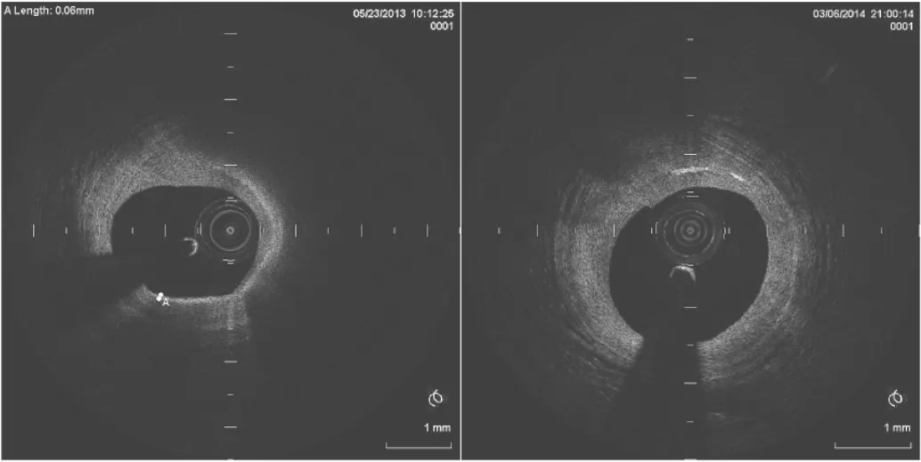
2.3 两组患者随访期目标斑块性质 常规组中3例患者随访期目标斑块由脂质斑块转变为纤维斑块,强化组中5例患者转化为纤维斑块。OCT下纤维帽光学特征见图1-2。

图1 OCT下纤维帽光学低亮度特征(为异质、模糊的边缘伴信号低反射和高衰减)

图2 OCT下纤维帽光学高亮度特征(纤维斑块特点为均质、高反射性和低衰减性光学信号)
3 讨论
Ozaki等[11-13]研究表明早期、强化剂量阿托伐他汀能显著减少ACS患者心血管事件,其机制主要包括减少斑块中脂质成分,增加斑块表面纤维帽厚度,改善内皮舒张功能,上调一氧化氮合成酶的活性,减少炎症反应等。Puri研究[14-16]显示中等剂量(20mg)阿托伐他汀可逆转冠状动脉粥样斑块,Ambrose研究[17-19]显示强化剂量(80mg)阿托伐他汀较常规剂量(40mg)普伐他汀更能阻断动脉粥样硬化斑块进展,上述两项研究中主要应用虚拟组织学血管内超声(VH-IVUS)来评估斑块体积及斑块成分,但因其分辨率相对较低,不能准确测量斑块纤维帽厚度和脂质核心角度。本研究应用OCT直接测量常规剂量及强化剂量阿托伐他汀干预后TCFA纤维帽厚度及脂质斑块角度的变化,探讨ACS患者他汀类药物的治疗策略。
本研究结果显示,ACS患者无论常规剂量组和强化剂量他汀组,其随访期TCFA的纤维帽均明显增厚,脂质成分明显减少,巨噬细胞浸润减少,且强化剂量他汀组斑块纤维帽较常规剂量组增厚更为明显,而两组患者脂质斑块脂质核心角度减少差异无统计学意义。孙丽娜等[20-24]的研究显示5mg阿托伐他汀较20mg阿托伐他汀口服12个月能显著增加冠状动脉粥样硬化斑块纤维帽厚度、减少脂质核心和巨噬细胞浸润,本研究进一步加大了阿托伐他汀剂量,结果显示20mg阿托伐他汀组,40~80mg阿托伐他汀组患者TCFA纤维帽增厚更加明显,但两组患者在随访期间均未出现心血管事件,提示常规剂量和强化剂量阿托伐他汀均能有效减少ACS患者MACE,但强化治疗使TCFA纤维帽较常规治疗明显增厚是否能带来进一步的临床获益可能需要更长时间的随访。研究期间两组患者服用阿托伐他汀后均未出现肌酸肌痛乏力、肌酶升高等不良反应,2例强化组患者肝酶出现一过性增高,减少阿托伐他汀剂量后恢复至正常水平,显示了阿托伐他汀良好的安全性。
本资料的局限性在于研究入选病例数较少,需进一步扩大样本量或进行多中心随机双盲对照研究;随访时间较短,易损斑块纤维帽增厚程度是否有更多临床获益需延长随访时间;强化组患者使用的阿托伐他汀剂量为40~80mg,平均剂量为48.3mg,由于病例数较少,未进行更细的亚组分析。
[1]KomukaiK,Kub o T,Kitabata H,etal.Effectofatrovastatin therap y on fib rous cap thickness in coronary atherosclerotic p laq ue as assessed by op ticalcoherence tomography[J].Am Coll Card iol,2014,64:2207-2217.
[2]NaghaviM,Libby P,Falk E,etal.From vulnerab le p laque to vulnerable p atient:a call for new d efinitions and risk assessment strateg ies:Part I[J].Circulation,2003,108:1664-1672.
[3]Virmani R,Burke A P,Farb A,et al.Patholog y of the vulnerab le p laq ue[J].Am CollCard iol,2006,47:C13-18.
[4]Davies M J.Anatomic features in victims of sudden coronary d eath.Coronary artery p athology[J].Circulation,1992,85:19-24.
[5]van d er WalAC,Becker AE,van d er Loos C M,etal.Site ofintimal rup ture or erosion of thrombosed coronary atherosclerotic p laq ues is characterized b y an inflammatory process irrespective ofthe d ominantp laq ue morp hology[J].Circulation,1994,89:36-44.
[6]Kramer M C,Rittersma S Z,d e Winter R J,et al.Relationship of thromb us healing to und erlying p laq ue morp holog y in sud den coronary d eath[J].Am CollCard iol,2010,55:122-132.
[7]Jang I K,Bouma B E,Kang D H,et al.Visualization of coronary atherosclerotic p laq ues in patients using optical coherence tomograp hy:comp arison with intravascular ultrasound[J].Am Coll Cardiol,2002,39:604-609.
[8]Yab ushita H,Bouma B E,Houser S L,et al.Characterization of human atherosclerosis b y op ticalcoherence tomog rap hy[J].Circulation,2002,106:1640-1645.
[9]Tearney G J,Regar E,Akasaka T,etal.Consensus standards for acquisition,measurement,and reporting of intravascular optical coherence tomography studies:a report from the International Working Group for Intravascular OpticalCoherence Tomography Stand ardization and Valid ation[J].J Am Coll Cardiol,2012,59: 1058-1072.
[10]Kub o T,ImanishiT,Takarada S,etal.Assessment ofculprit lesion morp hology in acute myocardialinfarction:ab ility of op tical coherence tomog rap hy comp ared with intravascular ultrasound and coronary ang ioscop y[J].J Am CollCard iol,2007,50:933-939.
[11]OzakiY,Okumura M,IsmailT F,etal.Coronary CT ang iograp hic characteristics ofculpritlesions in acute coronary synd romes not related to p laque rup ture as d efined b y op tical coherence tomog rap hy and ang ioscop y[J].Eur Heart J,2011,32:2814-2823.
[12]Yonetsu T,Kakuta T,Lee T,et al.In vivo critical fibrous cap thickness forrupture-p rone coronary plaq ues assessed b y opticalcoherence tomog raphy[J].EurHeartJ,2011,32:1251-1259. [13]Fujii K,Kob ayashi Y,Mintz G S,et al.Intravascular ultrasound assessment of ulcerated rup tured p laques:a comparison of culp rit and nonculp rit lesions of p atients with acute coronary synd romes and lesions in patients without acute coronary synd romes[J].Circulation,2003,108:2473-2478.
[14]PuriR,Nissen S E,Ballantyne C M,etal.Factors underlying reg ression ofcoronary atheroma with potentstatin therap y[J].Eur HeartJ,2013,34:1818-1825.
[15]Falk E,Shah P K,Fuster V.Coronary plaque disruption[J].Circulation,1995,92:657-671.
[16]RioufolG,Finet G,Ginon I,etal.Multip le atherosclerotic plaque rupture in acute coronary syndrome:a three-vessel intravascularultrasound study[J].Circulation,2002,106:804-808.
[17]Ambrose J A,Tannenbaum M A,Alexopoulos D,et al.Angiograp hic p rog ression of coronary artery disease and the developmentofmyocard ialinfarction[J].J Am CollCardiol,1988,12: 56-62.
[18]Little WC,Constantinescu M,Ap p leg ate R J,etal.Can coronary ang iog rap hy p redict the site of a sub seq uent myocardial infarction in patients with mild-to-mod erate coronary artery disease?[J].Circulation,1988,78:1157-1166.
[19]van der Wal A C,Becker A E.Atherosclerotic plaq ue rup tured pathologic basis of plaq ue stab ility and instability[J].Card iovasc Res,1999,41:334-344.
[20]孙丽娜,王宁夫,李虹,等.急性冠状动脉综合征患者短期大剂量他汀治疗的有效性及安全性研究[J].浙江医学,2014,36(15):1291-1293, 1320.
[21]俞章平,余晗俏,钟忆周,等.依折麦布联合阿托伐他汀对急性冠状动脉综合征患者血脂指标及血管内皮功能的影响[J].中国生化药物杂志,2014,36(6):110-112.
[22]韩战营,何冉,卢长青,等.依折麦布联合阿托伐他汀对急性冠状动脉综合征患者血脂及血管内皮功能的影响[J].中国动脉硬化杂志, 2013,21(12):1114-1118.
[23]李芝峰,殷跃辉.不同剂量阿托伐他汀治疗老年人急性冠状动脉综合征临床疗效观察[J].中华老年医学杂志,2012,31(12):1048-1050.
[24]葛彦彦,马晓静,鹿克风,等.阿托伐他汀强化治疗对ACS患者PCI效果的改善作用及其机制[J].山东医药,2016,56(37):44-46.
Effect of different doses of atorvastatin on vulnerable plaque,fibrous cap and lipid core angle of coronary arteries in patients with acute coronary syndrome
WANG Shiqi,HU Yewen,HE Fuwei,et al.Heart Center of Ningbo First Municipal Hospital,Ningbo 315000,China
Acute coronary synd rome Op ticalcoherence tomog rap hy Vulnerab le p laq ue fib er cap Lip id core ang le
20mg atorvastatin after PCIand those in hig h d ose g roup received 40-80mg atorvastatin.Patients und erwentopticalcoherence tomography(OCT)examination before and 9 months after PCI,the number and location of thin-cap fib roatheroma(TCFA)were record ed and the fibrous cap thickness and plaq ue lipid core point were measured. Results The vulnerab le p laque fib rous cap thickness were increased after PCI compared with b aseline b oth in routine dose group(175.42±36.02 vs 50.41±6.58μm,P<0.05)and hig h d ose g roup(233.33±88.35 vs 49.12±7.33μm,P<0.05),and there was sig nificantd ifference between two group s d uring the follow-up period(P<0.047).The TCFA lip id core ang le atfollow-up was decreased comp ared to b aseline b oth in routine d ose group (72.9±29.3°vs 127.6±50.8°,P<0.05)and in hig h d ose g roup(74.6± 32.9°vs 132.6± 51.3°,P<0.05),and there was no sig nificant d ifference b etween two g roup s at b aseline and follow-up. Conclusion Comp ared with the b aseline,the thickness of TCFA in ACS p atients with routine d ose or hig h d ose atorvastatin are increased,and the lip id core ang le are d ecreased sig nificantly.
2016-09-27)
(本文编辑:马雯娜)
10.12056/j.issn.1006-2785.2017.39.17.2016-1521
宁波市科技计划项目(2011C51003)
315000宁波市第一医院心脏中心
叶红华,E-mail:coolwind19820209@126.com
【 Abstract】 Objective To investig ate the effects of d ifferent d oses of atorvastatin on vulnerab le p laq ue,fib rous cap and lip id core ang le of coronary arteries in p atients with acute coronary synd rome(ACS). Methods Twenty four ACS p atients und erg oing p ercutaneous coronary intervention(PCI)from 2011 to 2014 in our hosp italwere rand omly d ivid ed into two g roup s with 12 in each.Patients in routine d ose g roup
