Derlin-1在宫颈癌组织的表达及其相关性研究*
2017-07-24于春丽
于春丽
(泰山医学院附属泰山医院,山东 泰安 271000)
*
Derlin-1在宫颈癌组织的表达及其相关性研究*
于春丽
(泰山医学院附属泰山医院,山东 泰安 271000)
目的 观察Derlin-1在宫颈癌组织中的表达水平,探讨Derlin-1表达与宫颈癌的相关性。 方法 回顾性分析2014年10月—2016年10月期间我院收治的宫颈癌患者30例,另收集同期我院子宫肌瘤行全子宫切除术患者作为观察组,采用免疫组化SABC法检测其Derlin-1水平,分析Derlin水平与年龄、分期、癌组织分化程度及病理类型的关系。结果 正常宫颈组织中Derlin-1的表达为率为3.2%(2/61),宫颈癌组织中Derlin-1的表达率为51.7% (31/60),宫颈癌组织中Derlin-1的阳性表达水平显著高于正常宫颈组织(χ2=4.96,P<0.001);且随着FIGO分期的上升,Derlin-1的表达水平也显著上升,IB1+B2期患者Derlin-1阳性表达率为92.3%,显著高于IIA1+A2期患者Derlin-1的阳性表达率(58.8%)(χ2=8.107,P<0.05);结论 Derlin-1的阳性表达与宫颈癌的发病有关,且随着FIGO分期的上升,Derlin-1的表达水平也显著上升,与年龄,分化程度及病理类型均无关。
Derlin-1;宫颈癌组织;相关性研究;FIGO分期
宫颈癌是全世界女性高发的第三大恶性肿瘤,我国宫颈癌的发病率仅次于乳腺癌,每年新增的宫颈癌患者约14万,占全球宫颈癌年发病率的三分之一,近几年来有资料显示,宫颈癌患者在发达国家的患病率及死亡率显著下降,但在发展中国家,其患病率及死亡率仍然保持在较高水平,且发病群体逐渐呈现年轻化的趋势[1-3],因此,在传统放、化疗基础上,寻找新的靶点显得非常迫切。Derlin-1是存在于內质网上的跨膜蛋白,有文献表明,其异常表达与多种肿瘤的发病有关[4-11],因此,本研究旨在探讨Derlin-1在宫颈癌组织中的表达及其相关性。
1 资料与方法
1.1 一般资料
选择我院2015年12月至2016年12月我院接收的宫颈癌患者30例,其中对照组男性16例,女性15例,年龄17~64岁,平均年龄(40±7.6)岁;按病理类型分,其中鳞癌17例,腺癌 13例;根据2015年FIGO分期标准:IB1期7例,IB2期6例,ⅡA1 8例,ⅡA2 9例;按癌细胞的分化程度分:其中高度分化4例,中度分化12例,低度分化14例;因子宫肌瘤行全子宫切除术患者的正常宫颈组织30 例作为对照。两组患者年龄及其他一般资料无统计学差异(P>0.05)。
1.2 纳入标准
所有患者均确诊为宫颈癌,所有入选患者均无重大脏器疾病,无其他肿瘤史,且所有患者均未采用放化疗治疗, 本研究经我院医学伦理委员会批准,所有患者均签署知情同意书。
1.2 方法
1.2.1 样本收集方法 采用宫颈活检标本或宫颈手术切除的标本,甲醛固定,将每个组织蜡块作 4 μm 连续切片 3 张,1 张用作 HE 染色,以供病理科医生明确诊断,1 张用于Derlin-1免疫组织化学检测,1 张备用。
1.2.2 Derlin-1检测方法 采用免疫组化SABC法检测其Derlin-1水平。
1.2.3 检测过程 (1)组织切片:将“1.2.1”中制备的组织蜡块裱于载有APES的玻片上,于60 ℃ 烤3 h,室温保存待用。(2)脱蜡:二甲苯I 10 min—二甲苯II 10 min—100%酒精 I 10 min—100%酒精 II 5 min—85%酒精3 min—75%酒精 3 min—蒸馏水清洗3 次,每次3 min。(3)阻断内源性过氧化物酶活性:将标本置于3%H2O2中,室温孵育10 min,蒸馏水冲洗3 次,每次5 min。(4)抗原修复:将标本置于0.01 M 枸橼酸盐缓冲液中煮沸,爆炒2 min,冷却至室温,然后PBS冲洗3 次,每次5 min。(5)BSA封闭:每张切片滴加5%BSA,室温保湿盒内孵育20 min,甩去多余液体,不冲洗。(6)滴加一抗:每张切片滴加适量一抗,4 ℃过夜,过夜后在室温下复温半小时,PBS冲洗3次,每次2 min。(7)滴加二抗滴加生物素化山羊抗兔Ig G,室温保湿盒内孵育20 min,PBS冲洗3次,每次2 min。(8)SABC孵育:滴加SABC,室温保湿盒内孵育20 min,PBS冲洗4次,每次5 min。(9)DAB 显色:按照说明配置 DAB 显色试剂,每张切片滴加适量,室温下显色,镜下控制反应时间,然后蒸馏水冲洗干净。(10) 复染,分化苏木素复染 2 min,蒸馏水冲洗。盐酸酒精分色。(11) 脱水:透明75% 酒精 5 min—85%酒精5 min—100%酒精 I 5 min—100%酒精II 5 min—二甲苯20 min。(12)封片:滴加中性树胶,盖玻片封片。(13) 阅片:显微镜下观察实验结果。
1.3 统计学分析
2 结 果
2.1 Derlin-1在宫颈癌组织与正常宫颈组织中的表达情况比较
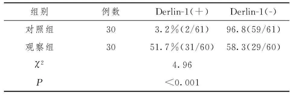
实验检测了Derlin-1在宫颈癌组织及正常宫颈组织的表达情况,如表1所示,正常宫颈组织中Derlin-1的表达为率为3.2%(2/61),宫颈癌组织中Derlin-1的表达为51.7% (31/60),宫颈癌组织中Derlin-1的阳性表达水平显著高于正常宫颈组织(χ2=4.96,P<0.001)。

表1 Derlin-1在宫颈癌组织与正常 宫颈组织中的表达情况比较(%)

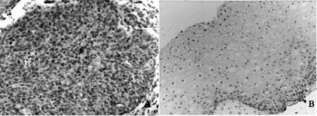
图1 Derlin-1在宫颈癌组织与正常宫颈组织中的表达情况图(×400)
A Derlin-1在正常宫颈组织中的表达情况;B Derlin-1在宫颈癌组织中的表达情况
2.2 宫颈癌组织中Derlin-1的表达与年龄的相关性
如表2所示, 55岁≥的患者有7人,其中Derlin-1阳性表达率为57.1%, <55岁的患者共23人,其中,Derlin-1阳性表达率为47.8%,二者相比,无统计学意义(χ2=0.096,P>0.05)。
2.3 宫颈癌组织中Derlin-1的表达与分期的相关性
如表3所示,随着FIGO分期的上升,Derlin-1的表达水平也显著上升,IB1+B2期患者Derlin-1阳性表达率为92.3%,显著高于IIA1+A2期患者Derlin-1的阳性表达率(58.8%)(χ2=8.107,P<0.05)。
2.4 宫颈癌组织中Derlin-1的表达与分化程度的相关性
如表4 所示,高、中低分化程度的宫颈癌患者组织中,Derlin-1的阳性表达率分别为42.9%、44.4%及57.1%,三者的阳性表达无统计学差异。
2.5 宫颈癌组织中Derlin-1的表达与病理类型的相关性 如表5所示,病理类型为鳞癌的患者Derlin-1阳性表达率为52.9%,腺癌的患者阳性表达率为46.1%,二者的阳性表达率无统计学差异。

表2 宫颈癌组织中Derlin-1的表达与年龄的相关性(%)

表3 宫颈癌组织中Derlin-1的表达与分期的相关性(%)

表4 宫颈癌组织中Derlin-1 的表达与分化程度的相关性(%)

表5 宫颈癌组织中Derlin-1 的表达与病理类型的相关性(%)
3 讨 论
宫颈癌是全世界女性高发的第三大恶性肿瘤,我国宫颈癌的发病率仅次于乳腺癌,每年新增的宫颈癌患者约14万,占全球宫颈癌年发病率的三分之一,近几年来有资料显示,宫颈癌患者在发达国家的患病率及死亡率显著下降,但在发展中国家,其患病率及死亡率仍然保持在较高水平,且发病群体逐渐呈现年轻化的趋势[1-3],Derlin-1 是存在于内质网膜上的跨膜蛋白,与内质网降解密切相关,参与错误折叠蛋白的逆向转运,其表达异常与肿瘤等多种疾病有关。Derlin-1 作为候选的癌基因,与多种恶性肿瘤的发病有关,因此,本实验通过探讨Derlin-1在宫颈癌组织中的表达与年龄、分期、分化程度及病理类型的相关性,旨在为抗宫颈癌新药作用靶点的研究提供参考。研究结果显示,正常宫颈组织中Derlin-1的表达为率为3.2%(2/61),宫颈癌组织中Derlin-1的表达为51.7% (31/60),宫颈癌组织中Derlin-1的阳性表达水平显著高于正常宫颈组织(χ2=4.96,P<0.001),提示Derlin-1的阳性表达与宫颈癌的发病有关;且随着FIGO分期的上升,Derlin-1的表达水平也显著上升,IB1+B2期患者Derlin-1阳性表达率为92.3%,显著高于IIA1+A2期患者Derlin-1的阳性表达率(58.8%)(χ2=8.107,P<0.05),提示Derlin-1的表达水平与宫颈癌的发病进程有关,此外Derlin-1的表达与年龄,分化程度及病理类型均无关。
综上所述,Derlin-1的阳性表达与宫颈癌的发病及发展均明显相关,可以作为治疗及新抗癌药物研究的靶点。
[1] Stevenson B R,Siliciano J D,Mooseker M S,et al.Identification of ZO-1:a high molecular weight polypeptide associated with the tight junction(zonula occludens) in a variety of epithelia.J Cell Biol 1986,103(3):755.
[2] Yon emura S,Itoh M,Nagafuchi A,et al.Cell-to-cell adherens junction formation and actin filament organization: similarities and differences between non-polarized fibroblasts and polarized epithelial cells[J].J Cell Sci.1995,108:127
[3] Schulze A,Standera S,Buerger E,et al.The ubiquitin-domain protein HERP forms a complex with components of the endoplasmic reticulum associated degradation pathway[J].Mol Biol, 2005, 354(5):1021-1027.
[4] Schaheen B,Dang H,Fares H.Derlin-dependent accumulation of integral membrane proteins at cell surfaces[J].J Cell Sci, 2009,122(Pt 13):2228-2239.
[5] Katiyar S,Joshi S,Lennarz WJ.The retrotranslocation protein Derlin-1 binds peptide: N-glycanase to the endoplasmic reticulum[J].Mol Biol Cell, 2005,16(10): 4584-4594.
[6] Greenblatt EJ,Olzmann JA,Kopito RR.Derlin- 1 is a rhomboid pseudo-protease required for the dislocation of mutant α-1 antitrypsin from the endoplasmic reticulum[J].Nat Struct Mol Biol,2011,18(10):1147-1152.
[7] Ran Y,Hu H,Hu D,et al.Derlin-1 is overexpressed on the tumor cell surface and enables antibody-mediated tumor targeting therapy[J].Clinical Cancer Research,2008,14(20):6538-6545.
[8] Bioulac-Sage P,Laumonier H,Laurent C,et al.Benign and malignant vascular tumors of the liver in adults[J].Semin Liver Dis,2008,28(3):302-314.
[9] Hu D,Ran YL,Zhong X,et al.Overexpressed Derlin-1 inhibits ER expansion in the endothelial cells derived from human hepatic cavernous hemangioma[J].J Biochem Mol Biol,2006,39(6): 677-685.
[10] Shah R,Rosso K,Nathanson SD.Pathogenesis,prevention,diagnosis and treatment of breast cancer[J].World J Clin Oncol,2014,5(3):283-298.
Expression of Derlin-1 in cervical cancer and its correlation
YU Chun-li
(Taian Central Hospital,Taian 271000,China)
Objective:To investigate the expression of Derlin-1 in cervical cancer and explore the correlation between the expression of Derlin-1 and cervical cancer. Methods Thirty patients with cervical cancer admitted to our hospital between October 2014 and October 2016 were retrospectively analyzed. The patients who underwent total hysterectomy were collected and Derlin-1 level were analyzed by immunohistochemical SABC method. The relationship between the level of Derlin and age, stage, histological differentiation and histological type were analyzed. Results The expression rate of Derlin-1 was 3.2% (2/61) in normal cervical tissue and 51.7% (31/60) in cervical cancer. The positive expression of Derlin-1 in cervical cancer tissues (χ2=4.96,P<0.001). The expression level of Derlin-1 was also significantly increased with the increase of FIGO stage, the positive expression rate of Derlin-1 was 92.3% in IB1+B2 stage, which was significantly higher than that in normal cervical tissue The positive expression rate of Derlin-1 was higher in stage IIA1+A2 patients(58.8%) (χ2=8.107,P<0.05). Conclusion The positive expression of Derlin-1 is related to the pathogenesis of cervical cancer. With the increase of FIGO staging, The expression level of Derlin-1 was also significantly increased, but not with age, differentiation and histological type.
Derlin-1; cervical cancer; correlation; FIGO staging
于春丽(1970—),女,山东泰安人,副主任医师,硕士,主要从事临床妇产科工作。
R711
A
1004-7115(2017)08-0859-03
10.3969/j.issn.1004-7115.2017.08.007
2017-05-20)
