老年股骨转子间骨折闭合复位内固定失败的影响因素分析
2017-07-01金浙凯
金浙凯
老年股骨转子间骨折闭合复位内固定失败的影响因素分析
金浙凯
目的 探讨闭合复位内固定术治疗老年股骨转子间骨折患者手术失败的相关因素,为临床骨科治疗提供参考。方法 2013年5月至2016年6月老年股骨转子间骨折患者100例,所有患者均予以闭合复位内固定术治疗,分析导致其手术失败的相关因素。结果 内固定失败患者7例,进一步分析其失败的影响因素,患者的骨折类型、Singh指数、并发基础病、TAD>25mm及Cleveland分区均是内固定失败的风险因素(P<0.05)。结论 应用闭合复位内固定术治疗老年股骨转子间骨折患者,其患者的骨折类型、Singh指数、并发基础病、TAD>25mm及Cleveland分区是内固定失败的风险因素。
闭合复位内固定术 股骨转子间骨折 内固定失败 影响因素
股骨转子间骨折常发生在老年骨质疏松患者,由于保守治疗的并发症和病死率均较高,通常主张进行手术治疗。目前常用的手术方法有髓内固定和髓外固定术。随着髓内固定手术的发展和技术设备的逐渐成熟,闭合复位髓内固定术已经成为治疗的主要方法,尤其是针对不稳定骨折的治疗[1]。本文探讨闭合复位内固定术治疗老年股骨转子间骨折患者手术失败的相关因素,为临床骨科治疗提供参考。
1 临床资料
1.1 一般资料 2013年5月至2016年6月本院老年股骨转子间骨折患者100例。男65例,女35例;年龄62~79岁,平均年龄(66.12±6.76)岁。骨折类型:Ⅰ型25例、Ⅱ型20例、Ⅲ型26例、Ⅳ型29例。
1.2 方法 闭合复位内固定术:硬脊膜外麻醉成功后,患者仰卧位,用C型臂X线机透视下骨折复位,做一个侧切口约4cm,外侧股骨转子在10cm以上,导针插入,扩髓后将股骨近端防旋螺钉(PFNA)置入,并固定螺旋叶片和锁定螺钉,冲洗后缝合[2]。
1.3 观察指标[3]统计所有手术患者的失败例数,并分析导致其失败的影响因素。
1.4 统计学方法 采用SPSS17.0统计软件。计数资料用χ2检验,P<0.05为差异有统计学意义。
2 结果
2.1 内固定失败的单因素分析 通过统计所有患者手术情况发现,其中内固定失败患者共7例。进一步分析其失败的临床影响因素发现,Ⅰ~Ⅲ级的Singh指数、并发基础病、尖顶距(TAD)>25mm且使用激素药物患者的内固定失败率显著高于Ⅳ~Ⅵ级的Singh指数、无基础病、TAD<25mm,且未使用激素药物的患者,差异有统计学意义(P<0.05),见表1。

表1 内固定失败的单因素分析[n(%)]
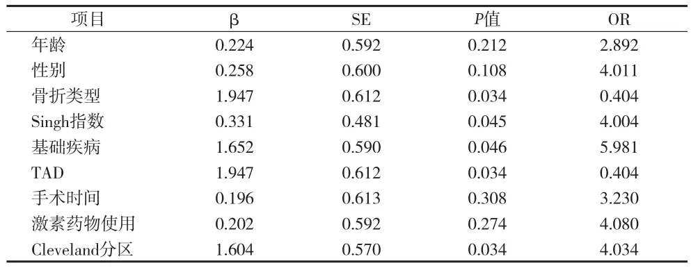
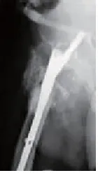
2.2 Logistic分析内固定失败的影响因素 通过Logistic分析,患者骨折类型、Singh指数、并发基础病、TAD>25mm及Cleveland分区是内固定失败的风险因素(P<0.05)。见表2、图1。

表2 多因素分析内固定失败的影响因素

图1 股骨粗隆骨折术后内固定松动

图2 TAD>25mm致术后2个月内固定松动
3 讨论
股骨转子间骨折系指股骨颈基底至小转子水平以上部位所发生的骨折,是老年人最常见的骨折之一,其发生率与股骨颈骨折相仿[4]。骨折多与骨质疏松有关,女性多于男性。由于转子部血液循环丰富,骨折后极少不愈合,故其预后远较股骨颈骨折为佳。闭合复位内固定术的特点是内固定坚强,有较高的稳定性和较强的把持力,抗剪切能力强,术后能早期进行功能锻炼[5-6]。同时手术创伤小、出血少、易于耐受,在不稳定性骨折尤其是高龄患者治疗上更具优势。闭合复位对骨折断端血供干扰小,有利于骨折愈合。老年人的骨质密度在冬季由于季节性变化更为减低,骨小梁变得极为脆弱[7]。闭合复位内固定术是一种融合微创技术的新型内固定手术方式。该技术适用于大部分股骨近端骨折,尤其是老年骨质疏松患者[8]。术后早期即可离床活动,避免长时间卧床可能出现的术后并发症。该手术具有切口小、闭合复位、骨折固定牢固、手术时间短、并发症少、术后恢复快等优点,成为适宜于高龄患者的手术新路径[9-10]。
本资料中,内固定失败原因:(1)Ⅰ、Ⅱ型骨折的总体损伤情况相对较轻,而Ⅲ、Ⅳ型骨折为转子间的粉碎性骨折,多数患者有不同程度的骨移位,成为术后骨骼不稳定的内在诱因[11-12]。(2)基础疾病,如糖尿病等是影响骨质疏松的重要因素,可降低患者的骨量,易造成固定后螺钉脱落或移位且增加术后并发症及感染的发生[13]。(3)TAD<25mm,可给予螺钉最佳的支撑,而>25mm时,则表明置入位置深度不足,增加失败的几率[14]。(4)螺钉固定于股骨头颈的中央或偏下后方时,其固定效果较理想,主要原因为头颈区骨质较厚,该区靠近股骨距,相较于其他位置,对骨折块的把持度最强[15]。
综上所述,应用闭合复位内固定术治疗老年股骨转子间骨折患者,其患者的骨折类型、Singh指数、并发基础病、TAD>25mm及Cleveland分区是内固定失败的风险因素。
[1] 项东, 吕建华, 彭亮, 等. 应用股骨近端髓内钉治疗老年人股骨转子间骨折(附105例报告). 浙江临床医学, 2014, 16(2): 259-260.
[2] 尚松, 蒋赞利. 股骨转子间骨折内固定失败的相关危险因素分析. 东南大学学报(医学版), 2013, 32(3): 324-326.
[3] 童培建, 吴寒松, 赵鹏, 等. 股骨转子间骨折内固定失败的风险评估. 中华骨科杂志, 2012, 32(7): 654-658.
[4] Malyszko J. Mechanism of endothelial dysfunction in chronic kidney disease. Clin Chim Acta,2010,411(19/20):1412-1420.
[5] 赵建峰,谢垒,方志祥,等.闭合复位PFNA内固定治疗老年股骨粗隆间骨折.浙江临床医学,2014(12):1961-1963.
[6] Orlandi RR, Kenndy DW. Revision endoscopic frontal sinus surgery.Otolaryngol.Clin North Am. 2011,34(1):77-90.
[7] Koreas G B, editor. Combine traditional Chinese and Western medicine clinical results. Rev Endocr Metab Disord,2013:73.
[8] Kew J, Rees GL, Close D. Multiplanar reconstructed computed tomography images improves depiction and understanding of the anatomy of the frontal sinus and recess. Am J Rhinol, 2013, 16(2):19-23.
[9] Shelbourne KD, Brueckmann RR. Rush-pin fiXation of supracondylar and intercondylar fractures of the femur. J Bone Joint Surg Am, 2014, 64(2):161-169.
[10] Stammberger HR, Kenney DW. Paranasal sinuses: Anatomic terminology and nomenclature. Ann oto rhinol laryngol, 2013, 167(suppl): 7-16.
[11] W.S.B.Lee,K.-F.The agger nasi cell: the key to understanding the anatomy of the frontal recess. Otolaryngol Head Neck Surg, 2014, 12(9): 497-507.
[12] Choi Bi, Lee HJ, Han JK, et al. Detection of hypervascular nodular hepatocellur carcinomas: value of triphasic helical CT compared with iodized oil CT. AJR, 2013,157(2):219-224
[13] Khan MA, Combs CS, Brunt EM, et al. Positron emission tomography scanning in the evaluation of hepatocellular carcinoma. Ann Nucl Med, 2012,14(2):121-126
[14] Tabit CE, Chung WB, Hamburg NM, et al. Endothelial dysfunction in diabetes mellitus: molecular mechanisms and clinical implications. Rev Endocr Metab Disord,2014,11(1):61-74.
[15] Endemann DH, Schiffrin EL. Endothelial dysfunction. J Am Soc Nephrol, 2015,15(8):1983-1992.
Objective To explore the clinical course of treatment of senile intertrochanteric fractures in patients with closed reduction and internal fi xation,analysis of related factors leading to operation failure,lay the foundation for the clinical treatment of Department of orthopedics. Methods 100 cases of femoral intertrochanteric fractures were treated in our hospital from May 2013 to June 2009. All patients were treated with closed reduction and internal fi xation,and the related factors leading to the failure of the operation were analyzed. Results Through the statistics of all patients,7 cases were found in patients with internal fi xation failure. Further analysis of the clinical factors affected the failure of that class 4~6 Singh index,TAD,25mm,with basic diseases and the use of hormone drugs in patients with internal fi xation failure rate was signif i cantly higher than the 1~3 Singh index,basic diseases,TAD was less than 25,and did not use hormone drugs in patients with signif i cant difference(P<0.05). And the patient's fracture type,Singh index,concurrent basic disease,TAD>25mm and Cleveland partition could be used as the risk factors of internal fi xation failure,the difference was signif i cant(P<0.05). Conclusion The clinical treatment of intertrochanteric fractures in the elderly patients,the application of closed reduction and internal fi xation,the patients with fracture type,Singh index,concurrent disease,TAD>25mm based and Cleveland partition can be used as internal fi xation failure risk factors.
Closed reduction and internal fi xation Intertrochanteric fractures Internal fi xation failure Inf l uencing factors
321017 浙江省金华市中医医院
