人体体液多肽组质谱分析方法优化
2017-03-01高璐,谷岩,陈峰
高 璐,谷 岩,陈 峰
人体体液多肽组质谱分析方法优化
高 璐,谷 岩,陈 峰
人体体液中多肽组成分近年来获得越来越多的关注。 不同体液作为不同器官、细胞在生理或病理状态下的分泌产物,对其成分特定改变的监测具有无创性、早期、敏感特异地预测人体健康状态、疾病变化、治疗疗效、药物毒性等体内变化方面的潜力。 而体液成分多而复杂,体液系统易受干扰,富集、提取分离出相对分子质量整体较低的多肽组成分并进行特定多肽成分的准确计量非常重要而具有挑战性。目前对人体体液多肽组成分的研究多采用基质辅助激光解吸电离飞行时间质谱法。 对此方法的实验偏差进行分析、提出改进方法,可以提高人体体液多肽组成分分析的准确性。应用基质辅助激光解吸电离飞行时间质谱法对人体体液多肽组成分进行质谱分析是人体健康状况临床检查的新思路,其中特定多肽组成分有潜力成为监测人体相关部位健康状况的生物标志物。
体液;多肽组;基质辅助激光解吸电离飞行时间质谱法;偏差;优化
人体体液多肽组成分的准确分析,特定多肽成分的准确计量很有希望帮助体液多肽组成分监测成为人体内病理等许多变化的新方法。应用基质辅助激光解吸电离飞行时间质谱(matrix-assisted laser desorption/ionization time-of-flight mass spectrometry,MALDI-TOF MS) 法对人体体液中多肽组成分进行质谱分析是人体相关部位健康状况临床检查的新思路[1-4,7-13],体液中特定多肽组成分有潜力成为监测人体健康状况的生物标志物[10-32],也必将成为今后的研究热点之一。 现将人体体液多肽组成分 MALDITOF MS 法分析实验偏差及操作优化方面的研究进行综述。
1 MALDI-TOF MS 法实验偏差
偏差主要来源于样本外部及样本内部2个方面。 样本外部偏差因素主要由样本蛋白/多肽提取,样本保存、离心分离、冻融,样本收集时机,基质液种类选择、准备,实验仪器性能等方面的偏差导致产生;而样本内部偏差因素主要是样本受血液及白细胞干扰,样本来源个体的年龄及性别差异等方面的偏差导致产生。
1.1 外部偏差
1.1.1 样本蛋白 /多肽提取 3 名健康个体[1],上午 10 时(餐后至少 2 h)取等量唾液入唾液收集管,每毫升唾液中加入 10 μL 苯甲基磺酰氟 (0.1 mol/ L) 、1 μL 胃蛋白酶抑制剂(1 mmol/L) 、20 μL 蛋白酶抑制因子混合物(P2714),均购自美国 Sigma 公司;之后样本分为离心组与非离心组 2 组,每组各300 μL 初始量,各组内再等分试样分别以有机溶剂沉淀法、硫酸铵沉淀法、酸沉淀法进行样本沉淀;部分条件处理组加以超滤步骤,之后的试样等分、固相萃取、MALDI板测定位点、质谱采集步骤各组样本实验条件均相同。质谱分析显示对于蛋白组成分的获取:①离心组以酸沉淀提取较未以酸沉淀提取组蛋白获取量高,乙腈、胍、硫酸铵处理组蛋白获取率较低;②所有提取方法组中非离心组均比离心组蛋白获取量高,尤其是乙醇组与丙酮组。 三氟乙酸组与乙腈/超滤组中离心组与非离心组蛋白获取量相似。 对于多肽组成分的获取,除乙腈组、乙醇组、丙酮组外,其他所有组中离心前进行提取步骤者多肽组成分获取量高,其中碳酸氢铵/超滤组结果最佳。
1.1.2 样本冻融周期、保存条件 获取条件相同的脑脊液样本[2]进行 3 组实验,第 1 组:实验样本分别于 4、21 ℃ 分别培养 0、1、3、6、24、48、72 h;第 2 组:样本收集后分别于-80、-196 ℃条件下保存 4 周;第3 组:样本保存过程中分别经冻融周期 1、2、3 次。之后的试样等分、固相萃取、MALDI板测定位点、质谱采集步骤各组样本实验条件均相同。质谱分析显示,4 ℃ 培养样本可稳定保存 72 h 无降解;而 21 ℃培养样本 24 h 后即出现多肽组成分差异(信噪比改变),48 h 后差异更加明显。 -80、-196 ℃ 条件下样本保存,质谱信号强度结果未观察到差异有统计学意义。样本经冻融周期3次较1次相关信号少量改变,可分辨质谱信号少量减少。
1.1.3 样本收集时机 样本(尿液)[3]分别来自一次晨尿、二次晨尿,之后的样本保存、离心分离、试样等分、固相萃取、MALDI板测定位点、质谱采集步骤各组样本实验条件均相同。 质谱分析显示,一次晨尿与二次晨尿样本质谱信号存在显著不一致,存在部分质荷比信号强度差异有统计学意义。唾液样本的质谱分析实验相似,样本收集时机的不同可能导致样本成分的显著改变,进而增加实验结果的偏差。
1.1.4 基质液的种类及准备方法 实验样本为标准样本(多肽/蛋白混合物)及条件相同的人血清样本[4],各分 4 组实验,分别使用 α-氰基-4-羟基肉桂酸( α-cyano-4-hydroxycinnamic acid,CHCA) 、芥子酸(sinapinic acid, SA) 、 龙胆 酸 (2,5-dihydroxybenzoic acid,DHB)、2,5-二羟基苯乙酮(2,5-dihydroxyacetophenone,DHAP)4 种基质,均为德国 Merck 公司产品;每组内每种基质分别采用晾干后点样法(drieddroplet, DD)[5]、 样 本 /洗 样 (sample/wash method,SM)[6]2 种基质准备方法。 之后的试样等分、固相萃取、MALDI板测定位点、质谱采集步骤各组样本实验条件均相同。 质谱分析显示:①DD 法于 4 种基质中都获得更多同质结晶型,质谱结果更优。 ②SM 法实验结果整体较 DD 法差,可重复性、再现性、分辨率、信噪比均较低,背景干扰较大,检测能力较低。 ③从整体实验结果数据分析来看,CHCA 作为基质实验结果最差。 就可重复性和再现性而言,SA基质结合 DD 法(SA/DD)实验结果最稳定。 ④DHB/ DD法实验结果良好。 此结果与之前研究结果不符,原因可能在于评价标准是背景干扰的大小而非峰值数量的多少。⑤从整体实验结果数据分析来看,DHAP/DD 法实验结果最优(样本为人血清时),其使用的限制条件在于准备步骤较复杂。
1.1.5 实验仪器性能 对实验仪器性能的分析主要在于仪器的质量准确性及批内、批间重复性[2]。 利用相对峰强存在 10 种特征信号的脑脊液样本,分别采用疏水作用色谱 C8 磁珠( 德国 Autoflex Bruker 公司) ,弱阳离子交换磁珠(MB-weak cation exchange,MB-WCX,德国 Autoflex Bruker 公司) 及铜离子固相金属离子亲和色谱磁珠( 德国 Autoflex Bruker 公司)进行实验,样本保存、离心分离、试样等分、MALDI板测定位点、质谱采集步骤各组样本实验条件均相同。 质谱分析显示:应用不同磁珠,测得信号数量差异无统计学意义;批内再现性、批间可重复性越好(即批内、批间变异系数越小),信噪比越大,即质谱分析结果越优。
1.2 内部偏差
1.2.1 样本受血液及白细胞干扰 相同条件的人脑脊液样本[2,17]分组实验,分别加入不同量血液或白细胞,样本保存、离心分离、试样等分、MALDI板测定位点、质谱采集步骤各组样本实验条件均相同。质谱分析结果与未污染样本的结果比较显示:①质核比1 000 ~ 10 000,血红蛋白最高样本 (155 μmol/ L)只出现 25 个峰值,与未加入血液样本质谱数据比较 89 个峰值未出现; ②血红蛋白浓度 0.075 μmol/L 以下质谱结果差异无统计学意义,加入血液对质谱结果差异无统计学意义;③经过离心分离,白细胞污染对质谱结果差异无统计学意义。
1.2.2 样本来源个体的年龄及性别差异 相同条件的 123 份人血清样本[7]被分为 A(<30 岁)、B(30 ~50 岁)、C(>50 岁)3 组及男性、女性 2 组,样本保存、离心分离、试样等分、MALDI 板测定位点、质谱采集步骤各组样本实验条件均相同。质谱分析显示无监督集群图示,<30 岁与≥30 岁的质谱数据分散于2个集群中,表明其质谱分析结果差异有统计学意义;男性、女性 2 组质谱数据结果差异无统计学意义。
2 操作优化
对 MALDI-TOF MS 法的部分实验操作进行优化可减少实验偏差对实验结果的影响,从而提高质谱分析结果的准确性。 目前,已有的对实验操作优化的研究方面包括样本高丰度蛋白去除、样本凝固时间及温度选择、冻融周期的控制、样本装液量(磁珠样本量比)、样本基质液准备、MALDI靶板准备、激光照射能量的选择等。
2.1 样本高丰度蛋白去除 相同条件人唾液样本[8],其中高丰度蛋白主要包括唾液 α-淀粉酶、白蛋白、免疫球蛋白,高丰度蛋白掩盖低丰度蛋白的质谱监测信号。 样本收集后:①对白蛋白、IgG 进行去除、捕获、洗脱(SwellGelÒ Blue Kit,ProteoPrepÒ Immunoaffinity Albumin and IgG Depletion Kit) ;②对唾液α-淀粉酶进行亲和去除、捕获、洗脱(淀粉酶去除装置,临时专利号 60915204);③对样本中总蛋白浓度进行粗测( 蛋白质定量试剂,美国 Bio-Rad Bradford公司)。 之后样本保存、离心分离、试样等分、MALDI板测定位点、质谱采集步骤各组样本实验条件均相同。 质谱分析显示,去除高丰度蛋白可通过去除高丰度蛋白对低丰度蛋白信号的覆盖、掩盖以发现更多低丰度蛋白成分信号,实验结果更准确。
2.2 样本凝固时间及温度选择 相同条件的人血清样本[7,9],血清分离出血浆步骤分组实验,等分样本为 3 个温度条件组,分别于 4、20、37 ℃ 条件下,每组再等分样本为 4 组分别凝固 0.5、1、2、24 h,样本保存、离心分离、试样等分、MALDI板测定位点、质谱采集步骤各组样本实验条件均相同。质谱分析显示:①20 ℃ 时前 3 组峰值数量差异无统计学意义,而 24 h 组峰值数量最少。 而主成分分析显示 20℃条件 4 组成分差异有统计学意义(偏析),随凝固时间增加可检测出新的多肽成分。②质谱信号总强度随凝固时间增加而增加,质谱分析结果转优,而若凝固过程在质谱分析前一天进行,则质谱信号总强度显著减小,质谱分析结果较差。 ③20 ℃条件较 4、37 ℃条件所获峰值数量及质谱信号总强度更高,实验结果更优。主成分分析显示不同温度成分差异有统计学意义(偏析)。
2.3 冻融周期的控制 相同条件的人血清样本[7],样本保存过程中经冻融周期 0、1、2、4 次,之后离心分离、试样等分、MALDI板测定位点、质谱采集步骤各组样本实验条件均相同。 质谱分析显示,质谱信号峰值数量及总强度均随冻融周期增加而减小;主成分分析显示 0次组与其他 3组间成分差异有统计学意义(偏析)。 样本保存过程中经冻融周期次数越少,质谱分析结果越优。
2.4 样本装液量(磁珠样本量比) 相同条件的人血清样本[7]分 4 组进行实验,每组等量阴阳离子交换磁珠 10 μL,样本量分别为 3、5、8、10 μL,样本保存、离心分离、试样等分、MALDI 板测定位点、质谱采集步骤各组样本实验条件均相同。质谱分析显示:①不同样本量组质谱峰值数量差异无统计学意义;②不同样本量组质谱信号总强度有差异,8 μL组结果最优;③主成分分析显示 4组间成分差异无统计学意义( 偏析) 。 8 μL 血清达到 10 μL 磁珠的最高承载能力;10 μL 血清对于 10 μL 磁珠过饱和,可能导致一些多肽成分由于多肽结合位点有限产生竞争而结合能力降低,从而致质谱分析结果变差。唾液样本的质谱分析实验相似,合适的磁珠样本量比可最大限度充分利用磁珠承载能力,优化质谱分析结果。
2.5 基质液准备 因质谱实验所用有机基质溶液中包含不稳定成分,易快速挥发[9],应每日新鲜配制基质液,现配现用,当天基质液应封闭处理并避光保存。
2.6 MALDI靶板准备 ①环境相对湿度应是 30%~60%,温度 18 ~ 25 ℃[9],结晶步骤中避免靶板受空调直吹;②测定位点步骤避免滴样出现气泡[9];③洗脱液位点需完全干燥后再滴入基质液,但为减少氧化,应避免干燥后至滴入基质液非操作时间[9]。
2.7 照射激光能量选择 范围内适度调整,照射激光能量越低,质谱信号的分辨率越高[9]。
3 操作优化流程
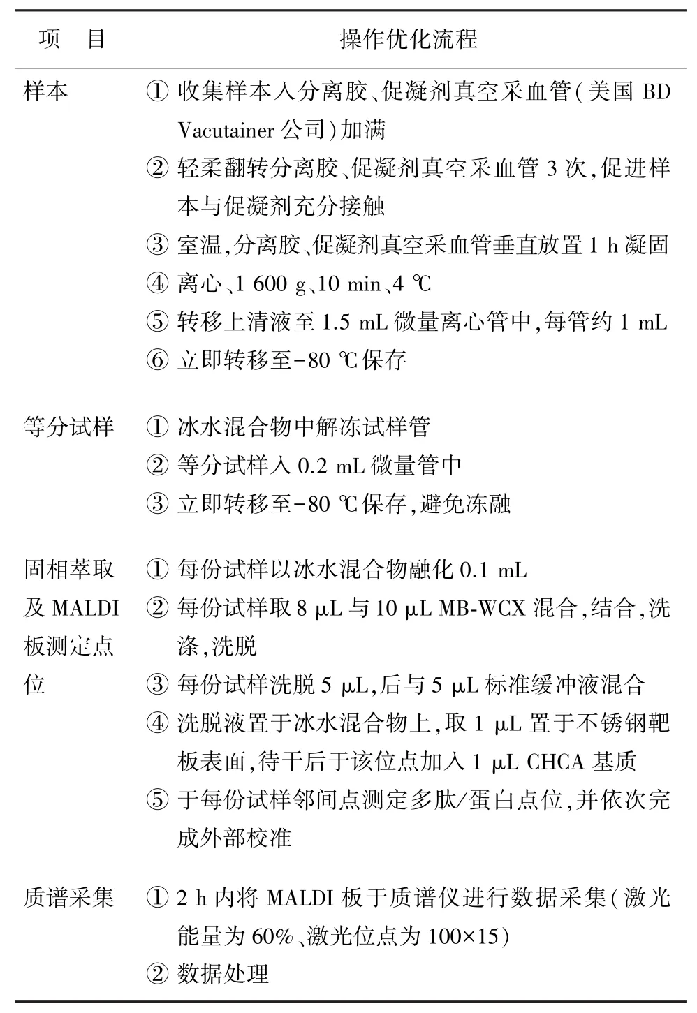
目前文献研究仅有对人血清样本的 MALDI-TOF MS 法操作优化后流程[7],尚无包括唾液在内的其他人体体液样本的操作优化流程(表 1)。

表1 人体体液样本的操作优化流程
当然,优化流程的方法不是唯一的,实验步骤的具体流程也不是唯一的。 在正式实验的过程中,研究者还需根据实验室温度、湿度、质谱仪性能、样本特点等客观条件适当调整实验步骤中各项条件的具体细节,以使实验条件对于实验结果的偏差及影响尽量减小,实验结果更为准确。 如样本与磁珠比例,不同实验室进行实验可能会因磁珠批号等存在差异而使最适样本磁珠比例有所改变,研究者可进行预实验以摸清实验条件,确定个性化的最优操作细节。
4 结论
作者对人体体液多肽组成分应用 MALDI-TOF MS法进行质谱成分分析实验中可能影响实验结果的偏差因素及部分实验操作方法的改进优化进行总结。 发现影响质谱分析结果的实验外部、内部偏差因素,对可改进的实验操作进行优化,可提高人唾液中多肽组成分质谱分析的准确性。 应用 MALDITOF MS 法对人体体液多肽组成分进行质谱分析是人体健康状况临床检查的新思路,其中特定多肽组成分有潜力成为监测人体相关部位健康状况的生物标志物。 对 MALDI-TOF MS 法实验步骤及操作细节的偏差、优化考量及研究证实是今后研究需重点关注的方面。
[1]Vitorino R, Barros AS, Caseiro A, et al.Evaluation of different extraction procedures for salivary peptide analysis [J].Talanta,2012,94(3):209-215.
[2]Bruegel M,Planert M,Baumann S,et al.Standardized peptidome profiling of human cerebrospinal fluid by magnetic bead separation and matrix-assisted laser desorption/ionization time-of-flight mass spectrometry[J].J Proteomics,2009,72(4):608-615.
[3]Fiedler GM, Baumann S, Leichtle A, et al.Standardized peptidome profiling of human urine by magnetic bead separation and matrix-assisted laser desorption/ionization time-of-flight mass spectrometry[J].Clin Chem, 2007,53 (3):421-428.
[4]Penno MA, Ernst M, Hoffmann P.Optimal preparation methods for automated matrix-assisted laser desorption/ ionization time-of-flight mass spectrometry profiling of low molecular weight proteins and peptides[J].Rapid Commun Mass Spectrom,2009,23(17):2656-2662.
[5]Karas M,Hillenkamp F.Laser desorption ionization of proteins with molecular masses exceeding 10, 000 daltons [J].Anal Chem,1988,60(20):2299-2301.
[6]Zhang X,Shi L,Shu S,et al.An improved method of sample preparation on AnchorChipTMtargets for MALDI-MS and MS/MS and its application in the liver proteome project[J].Proteomics,2007,7(14):2340-2349.
[7]Zeng X,Zhao L,Li Z,et al.Impact of experimental and demographic variables in serum peptide profiling based on magnetic bead and MALDI-TOF mass spectrometry[J]. Clin Chim Acta,2011,412(1):112-119.
[8]Krief G,Deutsch O,Gariba S,et al.Improved visualization of low abundance oral fluid proteins after triple depletion of alpha amylase,albumin and IgG[J].Oral Dis,2011,17 (1):45-52.
[9]Fiedler GM, Ceglarek U, Leichtle A, et al.Standardized preprocessing of urine for proteome analysis[J].Methods Mol Biol,2010,641:47-63.
[10]Hansen HG, Overgaard J, Lajer M, et al.Finding diabetic nephropathy biomarkers in the plasma peptidome by highthroughput magnetic bead processing and MALDI-TOF-MS analysis[J].Proteomics,2010,4(8/9):697-705.
[11]Guo N,Wen Q,Li ZJ,et al.Optimization and evaluation of magnetic bead separation combined with matrix-assisted laser desorption/ionization time-of-flight mass spectroscopy(MALDI-TOF MS)for proteins profiling of peritoneal dialysis effluent[J].Int J Mol Sci, 2014, 15(1):1162-1175.
[12]Fan NJ, Gao CF, Wang XL, et al.Serum peptidome patterns of colorectal cancer based on magnetic bead separation and MALDI-TOF mass spectrometry analysis[J].J Biomed Biotechnol,2012,2012:134-141.
[13]Dai Y,Hu C, Wang L, et al.Serum peptidome patterns of human systemic lupus erythematosus based on magnetic bead separation and MALDI-TOF mass spectrometry analysis[J].Scand J Rheumatol,2010,39(3):240-246.
[14]Voortman J, Pham TV, Knol JC, et al.Prediction of outcome of non-small cell lung cancer patients treated with chemotherapy and bortezomib by time-course MALDITOF-MS serum peptide profiling[J].Proteome Sci,2009,7(1):34-47.
[15]Ying X,Han S,Wang J,et al.Serum peptidome patterns of hepatocellular carcinoma based on magnetic bead separation and mass spectrometry analysis[J].Diagn Pathol,2013,8(1):130-135.
[16]Jia K,Li W,Wang F,et al.Novel circulating peptide biomarkers for esophageal squamous cell carcinoma revealed by a magnetic bead-based MALDI-TOFMS assay[J].Oncotarget,2016,7(17):23569-23580.
[17]Jimenez CR, Koel-Simmelink M,Pham TV, et al.Endogeneous peptide profiling of cerebrospinal fluid by MALDITOF mass spectrometry:optimization of magnetic beadbased peptide capture and analysis of preanalytical variables[J].Proteomics Clin Appl,2007,1(11):1385-1392.
[18]Fu G,Du Y,Chu L,et al.Discovery and verification of urinary peptides in type 2 diabetes mellitus with kidney injury[J].Exp Bio Med,2016,241(11):1186-1194.
[19]Li S, Wang L, Zhao S, et al.Preparation of phenyl-functionalized magnetic mesoporous silica microspheres for the fast separation and selective enrichment of phenyl-containing peptides[J].J Sep Sci,2015,38(22):3954-3960.
[20]Mahboob S, Mohamedali A, Ahn SB, et al.Is isolation of comprehensive human plasma peptidomes an achievable quest?[J].J Proteomics,2015,127(42):300-309.
[21]Yang X , Hu L , Ye M , et al.Analysis of the human urine endogenous peptides by nanoparticle extraction and mass spectrometry identification[J].Anal Chim Acta,2014,829: 40-47.
[22]Deng BG,Yao JH,Liu QY, et al.Comparative serum proteomic analysis of serum diagnosis proteins of colorectal cancer based on magnetic bead separation and maldi-tof mass spectrometry[J].Asian Pac J Cancer Prev,2013,14 (10):6069-6075.
[23]Schiffer E,Mischak H,Novak J.High resolution proteome/ peptidome analysis of body fluids by capillary electrophoresis coupled with MS[J].Proteomics,2006,6(20):5615-5627.
[24]Zhu W,Gallo RL,Huang CM.Sampling human indigenous saliva peptidome using a lollipop-like ultrafiltration probe: simplify and enhancepeptide detection for clinical mass spectrometry[J].J Vis Exp,2012,7(66):e4108.
[25]Fan NJ,Gao CF, Zhao G, et al.Serum peptidome patterns of breast cancer based on magnetic bead separation and mass spectrometry analysis[J].Diagn Pathol,2012,7:45. [26]Yang J,Song YC,Dang CX, et al.Serum peptidome profiling in patients with gastric cancer[J].Clinical Exp Med,2012,12(2):79-87.
[27]Gil GC, Brennan J, Throckmorton DJ, et al.Automated analysis of mouse serum peptidome using restricted access media and nanoliquid chromatography-tandem mass spectrometry[J].J Chromatogr B Analyt Technol Biomed Life Sci,2011,879(15/16):1112-1120.
[28]Xiao D, Meng FL, He LH, et al.Analysis of the urinary peptidome associated with Helicobacter pylori infection [J].World J Gastroentero,2011,17(5):618-624.
[29]Hu L,Ye M,Zou H.Recent advances in mass spectrometry-based peptidomeanalysis[J].Expert Rev Proteomics,2009,6(4):433-447.
[30]Hardt M,Thomas LR,Dixon SE,et al.Toward defining the human parotid gland salivary proteome and peptidome:identification and characterization using 2D SDS-PAGE, ultrafiltration,HPLC,and mass spectrometry[J].Biochemistry,2005,44(8):2885-2899.
[31]Iloro I, Gonzalez E, Gutierrez-de Juan V, et al.Non-invasive detection of drug toxicity in rats by solid-phase extraction and MALDI-TOF analysis of urine samples[J]. Anal Bioanal Chem,2013,405(7):2311-2320.
[32]Deutsch O, Fleissig Y, Zaks B, et al.An approach to remove alpha amylase for proteomic analysis of low abundance biomarkers in human saliva[J].Electrophoresis,2008,29(20):4150-4157.
Recent research on optimized mass spectrum analysis of human body fluids peptidome
GAO Lu1,2, GU Yan1,2, CHEN Feng1,3
(1.Beijing Oral Digital Medical Key Laboratory, National Oral Digital Medical Technology and Materials Engineering Laboratory, Beijing 100081, China; 2.Department of Orthodontic,Peking University Hospital of Stomatology, Beijing 100081, China; 3.Central Laboratory,Peking University Hospital of Stomatology, Beijing 100081, China)
Peptidome composition of human body fluid has gained more and more attention. Different human body fluids are excreta or secreta of different organs or cells in physiological or pathological status.By monitoring specific changes of peptidome composition in human body fluids, we can potentially be able to predict human health status, development of diseasess, curative effects of treatments and pharmaceutical toxicity and so on noninvasively early, sensitively and specifically. However, compositions of human body fluids are complicated, the body fluid systems are vulnerable to many factors, so it’ s a big challenge to concentrate, extract and separate peptidome composition which are of low molecular weight and measure specific peptidome composition accurately.Matrix-assisted laser desorption/ionization time-of-flight mass spectrometry(MALDI-TOF MS)method is widely used in researches of peptidome composition of human body fluids at present.Analysis of deviation in experiments and proposing of improved operation can increase accuracy when analyzing peptidome composition in human fluids.Analysing peptidome of human fluids by MALDI-TOF MS method is a new thought of examining human physical conditions clinically.Specific peptidome compositions in human fluids have the potential to be new biomarkers to monitor human physical condition of related body parts.
Body fluid; Peptidome; Matrix-assisted laser desorption/ionization time-of-flight mass spectrometry(MALDI-TOF MS)method; Deviation; Optimize
Q592;Q516
A
2095-3097(2017)01-0060-05
10.3969/j.issn.2095-3097.2017.01.016
2016-10-17 本文编辑:徐海琴)
100081 北京,口腔数字化医疗技术和材料国家工程实验室,口腔数字医学北京市重点实验室(高 璐,谷 岩,陈 峰);100081 北京,北京大学口腔医院正畸科(高 璐,谷 岩),中心实验室(陈 峰)
谷 岩,E-mail:guyan96@126.com;陈 峰,E-mail:molec ulecf@gmail.com
