Easy removal of rust rings formed after metal foreign bodies in cornea
2016-12-05ShuZhangGuoFanCaoJinLiu
Shu Zhang, Guo-Fan Cao, Jin Liu
1In-patient Department, Affiliated Eye Hospital of Nanjing Medical University, Nanjing 210029, Jiangsu Province, China2Department of Ophthalmology, The Second Affiliated Hospital of Nanjing Medical University, Nanjing 210011, Jiangsu Province, China
·Original article·
Easy removal of rust rings formed after metal foreign bodies in cornea
Shu Zhang1, Guo-Fan Cao1, Jin Liu2
1In-patient Department, Affiliated Eye Hospital of Nanjing Medical University, Nanjing 210029, Jiangsu Province, China2Department of Ophthalmology, The Second Affiliated Hospital of Nanjing Medical University, Nanjing 210011, Jiangsu Province, China
Received: 2016-03-29 Accepted: 2016-09-09
•AIM: To compare the effectiveness and outcome of two different methods for removing metal foreign bodies and rust rings from cornea.
•METHODS: Forty outpatients with cornea metal foreign body injuries were recruited. They were divided into two groups according to the methods used to remove the foreign body and the rust ring (group 1 using needle + forceps; group 2 using needle only). The effectiveness and outcome were compared across groups in term of the duration of the surgery, pain scores, and the recovery of epithelial damage, as well as the integrity of the removed rust rings, which was associated with the difficulty of the surgery.
•RESULTS: In group 1, rust rings were removed readily in one piece in one or two attempts, while in group 2, the rings were broken into several pieces before they were totally removed. The averaged surgery duration for patients in group 1 was 1.9min(37.3%)shorter than that in group 2 (P<0.001). The pain score of group 1 was also significantly lower than that of group 2 during and 1d after the surgery. In group 1, a full recovery of the corneal epithelium, indicated by negative fluorescent staining, was seen in 80% of cases 1d after the surgery, while in group 2, this was only 55%.
•CONCLUSION: The method in group 1 is easier to perform and results in better outcomes than the conventional method in group 2 for removing the rust rings formed by corneal metal foreign bodies.
metal corneal foreign body; rust ring; removal; epithelial healing
INTRODUCTION
Metal foreign body injuries to human corneas (usually caused by metal debris) are commonly seen in ophthalmology clinics[1-3]. Such injuries often lead to corneal erosions and the formation of rust rings around the foreign body within hours. The foreign body and rust ring must be clearly removed to ensure a full recovery of visual function. Usually, this is done with a small needle[3-6]. Although needle works well in removing foreign bodies, we have found that it is challenging to remove the rust ring smoothly and without causing additional damage with needle. This is largely due to the facts that 1) needle has a sharp tip and two cutting edges; 2) rust ring cannot be held by a needle, so the removing with needle requires repeated digging and picking, during which the ring is often broken into pieces before it can be removed completely. Repeated actions required also elongated the surgery duration.
With the development of micro-surgery equipment, a microscopic knot forceps is now available to handle fine surgical areas, such as the cornea.Inspired by it, we occasionally tried to use forceps to remove the rust ring in several difficult patients suffering from cornea metal foreign body injury, and we found that the outcome was surprisingly good. To the best of our knowledge, using forceps as the tool for this surgery has not been reported. Therefore we designed a controlled study as reported here to evaluate if forceps is a better method for cornea foreign body and rust ring removing. The criteria for this evaluation include surgery duration, pain score, and recovery of the cornea epithelium.
SUBJECTS AND METHODS
This was a prospective, comparative clinical study involving 40 outpatients (39 males, 1 female). The subjects were 18 to 56y old (average 32.2y) with no other ocular pathology or combined injuries. To check if the sample size was appropriate for the analysis, power analysis was done after all the samples were collected by using power analysis (GPower 3.1) software against the major index of the comparison between the two surgical approaches: the surgery time. Based upon the group means and SDs, the effective size of the sample is 0.739, and the minimal sample size to reach significant level of 0.05 and statistic power of 0.90 is 6 in each group. Each subject suffered from superficial corneal injury in one eye by metal debris that was not thermal. They were randomly (listed in advance by table of random numbers) assigned into the two groups, each containing 20 subjects. The time delay between injury and surgery in this sample ranged from 4 to 24h. The two groups were matched in age, the size of the corneal lesions, as well as the time delay (Table 1). Informed consent was obtained from every patient before the surgery and the experimental component of the surgery was explained to the patient.
Surgical Procedure In each patient, the anterior segment of the injured eye was examined carefully using a slit lamp before surgery. The eyes were kept open manually by the surgeon during the surgery. After obtaining topical analgesia with oxybuprocaine hydrochloride eye drops (Santen Pharmaceutical, Japan), a needle was placed tangentially to the eyeball with the bevel side facing upwards at the spot of the injury. Then, a levering action was used to remove the foreign body from the cornea. If the removal was incomplete after this action (with a remaining of a rust ring), the subject was further treated differently according to the group. In group 1, the residual material and rust ring were removed using a needle together with a forceps. The rim of the ring was elevated slightly by the needle tip so that it could be grasped easily with the forceps. Then, the entire ring was pulled away gently using the forceps (Figure 1). In group 2, the residual material and rust ring were removed using a needle alone. In this group, several trials of digging were usually required to remove the whole ring because it was often broken into pieces. All subjects were treated by one surgeon. Postoperatively, topical ointment antibiotics was applied and continued for 2-3d until the fluorescent staining test was negative, eye cover was not mandatory. All patients of both groups received the same outpatient review.
Table 1 Patient information
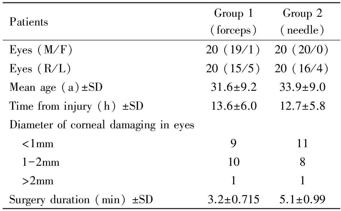
PatientsGroup1(forceps)Group2(needle)Eyes(M/F)20(19/1)20(20/0)Eyes(R/L)20(15/5)20(16/4)Meanage(a)±SD31.6±9.233.9±9.0Timefrominjury(h)±SD13.6±6.012.7±5.8Diameterofcornealdamagingineyes <1mm911 1-2mm108 >2mm11Surgeryduration(min)±SD3.2±0.7155.1±0.99

Figure 1 Image of an intact rust ring on a cotton swab after it was pulled out using forceps.
Outcome Evaluations A trained judge was employed to do the semi-quantitative evaluation. The information of grouping and days after the surgery were blinded from the judge to avoid bias. The integrity of the removed rust rings was compared between the two groups. If a removed rust ring was broken into two pieces or more, the integrity was considered as lost. The surgery duration in both groups was documented and analyzed. Patients were asked to rank the pain they experienced during, and 1 and 2 days after the surgery using a four-point scale (0=none; 1=little; 2=some; 3=much) (Table 2)[7-9]. The ranking of pain during the surgery was done immediately after the surgery.
The area of epithelial damage was examined with a micro-slit lamp (Haag-Streit 900, Gartenstadtstrasse, Switzerland) 24, 48, and 72h after the surgery, with blue light illumination after instilling fluorescent solution to show the lesion.
RESULTS
In 19 of 20 eyes in group 1, the rust ring was removed intact (Figure 1) in one or two actions with the forceps, while in group 2, all rings were broken into at least three pieces before they were removed completely.
The average surgery time for group 1 was 3.2±0.715min (mean±SD), as compared with 5.1±0.99min for group 2. The difference was 1.9min or 37.3%. The difference in surgery time was statistically significant [Student’st-test,t=6.958, degree of freedom (DF)=38,P<0.001].
Table 2 Pain scores as indicate by the number of eyes

EyesDuringoperative1dpostoperative2dpostoperativePainscores012301230123Group1711201550020000Group23494923018202P<0.001<0.001>0.05
Pare the results of post-hoc pairwise comparisons (Tukey) between groups.
Table 2 shows the pain scores given by the patients in both groups at the different time points. Two-way analysis of variance (ANOVA) in a general linear model showed significant group (F1=20.504,P<0.001) and time (F2=38.706,P<0.001) effects.Post-hoccomparisons (Tukey test) for the factor of group showed significantly (P<0.001) less pain in group 1 during and 1 day after the surgery. The difference was not significant 2 days after the surgery, which was likely due to the near-total recovery at this time (ceiling effect).
A full recovery of cornea was verified by the negative fluorescent staining in this study. As shown in Figure 2, such a full recovery was seen in 16 cases 1 day after the surgery in group 1, but was found only in eleven cases in group 2 at this time point. Again, between-group difference in the case of full recovery was not seen 3 days after the surgery due to the full recovery at this time.
DISCUSSION
Occupational eye injuries caused by metal debris are common in some industrial settings[1-4,6,10]. In addition to the direct mechanical damage to the cornea, metallic foreign bodies can also cause a deposit in the cornea epithelium and stroma due to the oxidative reaction of iron[11]. The reaction may result in corneal erosions and the formation of a rust ring surrounding the metal debris. This can happen within hours of the injury. If not treated promptly, the rust ring may cause corneal scarring and infection. Thus, to pursue complete removal of both the foreign body and the rust ring with minimal additional injury from the surgery is the goal of the clinical management of eye injury by metal debris[6,12].
Several tools are commonly used in this surgery, including needles, lancets, hand-held microdrills, and small chalazion curettes[5-6,13-14]. We have found that each of these tools is adequate for removing the foreign body itself, but they can cause damage to the surrounding tissue when they are used to remove the rust ring. Additional lesion is often created when repeated actions were taken to remove the residual ring when it was broken after first try.
Through preliminary use with surprisingly good result, we believed that forceps is more convenient for removing the rust ring than other tools. Therefore, a controlled comparison was conducted to confirm this advantage of forceps. In this study, we demonstrated that using forceps to remove the rust ring shortened the surgery and resulted in better outcomes. As soon as the rim of the ring being grasped by the forceps, it can be pulled out easily without further contact between the forceps and the cornea tissue and the ring can be pulled out in one piece. Unlike the surgery using needle, the cornea was barely contacted by the forceps. This is the reason of much less or no further lesion is created during the removal of the rust ring using forceps. In contrast, when using a needle, the rust ring was often broken into pieces before it was removed completely. This resulted in longer surgery time and additional damage.

Figure 2 Between group comparison of cornea recovery after the surgery. A full recovery was verified as negative fluorescent staining in the cornea. The number of eyes that were fully recovered was larger in group 1 at the first and the second day after the surgery.
After removing the rust ring, the lesion on the cornea in group 1 was limited to the area covered by the ring, while the corneal lesion in group 2 was much larger and resulted partially from the multiple attempts to remove the remaining pieces of the broken rust ring. Less corneal damage and shorter surgical duration resulted in less pain during and after the maneuver (Table 2), and quicker recovery of the corneal wounds[15-19]. The longer surgical time in group 2 reflected the greater difficulty in handling the rust ring and was associated with an additional wound, which also delayed the healing.
In summary, we demonstrated clear advantages of the needle forceps approach in removing metal foreign bodies with rust rings over the conventional needle-only approach.
1 Yokogawa H, Kobayashi A, Yamazaki N, Masaki T, Sugiyama K. Surgical therapies for corneal perforations: 10 years of cases in a tertiary referral hospital.ClinOphthalmol2014;8:2165-2170
2 Ozkurt ZG, Yuksel H, Saka G, Guclu H, Evsen S, Balsak S. Metallic corneal foreign bodies: an occupational health hazard.ArqBrasOftalmol2014;77(2):81-83
3 Gumus K, Karakucuk S, Mirza E. Corneal injury from a metallic foreign body: an occupational hazard.EyeContactLens2007;33(5):259-260
4 Welch LS, Hunting KL, Mawudeku A. Injury surveillance in construction: eye injuries.ApplOccupEnvironHyg2001;16(7):755-762
5 Health tips. Managing postnasal drip.MayoClinHealthLett2002;20(6):3
6 Beyer H, Cherkas D. Corneal foreign body removal using a bent needle tip.AmJEmergMed2012;30(3):489-490
7 Aslankurt M, Aslan L, Bakan AM, Aksoy A, Silay E, Yldz H. Pain and cooperation in patients having dominant-side or nondominant-side phacoemulsification.JCataractRefractSurg2014;40(2):199-202
8 Sauder G, Jonas JB. Topical anesthesia for penetrating trabeculectomy.GraefesArchClinExpOphthalmol2002;240(9):739-742
9 Kernt M, Cheuteu RE, Cserhati S, Seidensticker F, Liegl RG, Lang J, Haritoglou C, Kampik A, Ulbig MW, Neubauer AS. Pain and accuracy of focal laser treatment for diabetic macular edema using a retinal navigated laser (Navilas).ClinOphthalmol2012;6:289-296
10 Xu Z, Yu X, Li Z, Wang L. The role of in vivo confocal microscopy in the diagnosis of hidden corneal foreign bodies.JIntMedRes2014;42(1):145-152
11 Witherspoon SR, Hogan RN, Petroll WM, Mootha VV. Slit-lamp, confocal, and light microscopic findings of corneal siderosis.Cornea2007;26(10):1270-1272
12 Austin PE, Ljung M, Dunn KA. A new model for teaching corneal foreign body removal.AcadEmergMed1995;2(9):831-834
13 Egger SF, Huber-Spitzy V, Alzner E, Scholda C, Vecsei VP. Corneal wound healing after superficial foreign body injury: vitamin A and dexpanthenol versus a calf blood extract. A randomized double-blind study.Ophthalmologica1999;213(4):246-249
14 Clark WN. Embedded corneal foreign body in a child? Try a chalazion currette.CanJOphthalmol1999;34(7):394
15 Liu X, Zhao Y, Yang Y, Xu Y, Jiang Y, Dong F, Long Q. Mycobacterium massiliense keratitis.OptomVisSci2012;89(6):E944-E947
16 Yigit O, Yürüktümen A, Arslan S. Foreign body traumas of the eye managed in an emergency department of a single-institution.UlusTravmaAcilCerrahiDerg2012;18(1):75-79
17 Domniz Y, Lawless M, Sutton GL, Rogers CM, Meagher LJ. Successful treatment of Paecilomyces lilacinus endophthalmitis after foreign body trauma to the cornea.Cornea2001;20(1):109-111
18 Gupta S, Sehra S, Gogia V, Khokhar S, Agarwal T. Corneal nerve regeneration after foreign body removal on in vivo confocal microscopy.CanJOphthalmol2013;48(5):e125-e128
19 Wipperman JL, Dorsch JN. Evaluation and management of corneal abrasions.AmFamPhysician2013;87(2):114-120
取出角膜金属异物和锈环的改良方法
张 舒1,曹国凡1,刘 锦2
(作者单位:1210029 中国江苏省南京市,南京医科大学附属眼科医院住院部;2210011 中国江苏省南京市,南京医科大学第二附属医院眼科)
张舒,毕业于东南大学,本科,主任医师,副教授,研究方向:眼表疾病。
张舒.zhangs60@aliyun.com
目的:比较用两种方法取出角膜金属异物和锈环的效果。方法:将40例门诊角膜金属异物患者分为两组。第1组(20例)采用注射针头配合镊子取出异物和锈环;第2组(20例)采用注射针头取出异物和锈环。将两组间操作时间长短、患者疼痛程度、角膜上皮损伤修复时间和取出锈环的完整性进行比较。结果:第1组患者的角膜锈环经过一至两次尝试后完整取出;而在第2组中,锈环往往破碎成数块之后才能全部取出。第1组患者的平均手术时间较第2组短1.9min(37.3%)(P<0.001)。第1组患者术中和术后1d的疼痛评分也明显低于第2组。第1组患者80%的眼术后1d角膜上皮细胞已修复,荧光染色呈阴性;而在第2组,同期只有55%的眼角膜荧光染色呈阴性。结论:第1组取出角膜金属异物和锈环所采用的方法较第2组的传统方法更容易操作且疗效更好。
角膜金属异物;锈环;取出;上皮愈合
Shu Zhang. In-patient Department, Affiliated Eye Hospital of Nanjing Medical University, Nanjing 210029, China. zhangs60@aliyun.com
10.3980/j.issn.1672-5123.2016.12.03
:Zhang S, Cao GF, Liu J. Easy removal of rust rings formed after metal foreign bodies in cornea.GuojiYankeZazhi(IntEyeSci) 2016;16(12):2180-2183
引用:张舒,曹国凡,刘锦. 取出角膜金属异物和锈环的改良方法.国际眼科杂志2016;16(12):2180-2183
