Uterine Artery Embolization for Management of Primary Postpartum Hemorrhage Associated with Placenta Accreta
2016-10-13ZhiweiWangXiaoguangLiJiePanXiaoboZhangHaifengShiNingYangandZhengyuJin
Zhi-wei Wang, Xiao-guang Li, Jie Pan, Xiao-bo Zhang, Hai-feng Shi, Ning Yang, and Zheng-yu Jin*
Uterine Artery Embolization for Management of Primary Postpartum Hemorrhage Associated with Placenta Accreta
Zhi-wei Wang, Xiao-guang Li, Jie Pan, Xiao-bo Zhang, Hai-feng Shi, Ning Yang, and Zheng-yu Jin*
Department of Radiology, Peking Union Medical College Hospital, Chinese Academy of Medical Sciences & Peking Union Medical College, Beijing 100730, China
uterine artery embolization; postpartum hemorrhage; placenta accrete
Objective To evaluate the efficacy and safety of uterine artery embolization (UAE) in the management of primary postpartum hemorrhage associated with placenta accreta.
Methods We retrospectively reviewed the medical records of patients with placenta accreta between January 2010 and August 2014. Totally 18 women (mean age 30.8±4.2 years) of primary massive postpartum hemorrhage with diagnosis of placenta accrete received treatment of UAE after delivery. Images of DSA and medical records were reviewed. Technical success was defined as control of bleeding after embolization. The complications, control of hemorrhage and recurrent bleeding of the placenta left inside the uterus were retrospectively collected for assessment.
Results All patients underwent transcatheter embolization of bilateral uterine arteries. The technical success rate of embolization was 100%. Bleeding was controlled in 17 of 18 patients (94%) during follow-up period (median 18 months, 3-31months) without further bleeding recurred. One patient with placenta percreta undertook an emergent hysterectomy along with surgical bladder repair after UAE because of persistent uterine bleeding. Eight patients had postembolization syndrome and no other complications occurred.
Conclusion Uterine artery embolization is an effective and safe treatment for the management of primary postpartum massive hemorrhage associated with placenta accreta.
Chin Med Sci J 2016; 31(4):228-232
LACENTA accreta is a serious complication of pregnancy that is associated with maternal mortality and morbidity resulting from massive obstetric hemorrhage.1, 2The pathogenesis of placenta accrete was thought to be that deficiency of the decidua basalis at the endometrial scar results in invasion of chorionic villi into the myometrium.1Traditionally, abnormal placental adherence has been classified into placenta accreta, placenta increta, and placenta percreta based on depth of myometrial invasion: superficial, deep, and through serosa to the adjacent structures, respectively.3-8
Various treatments have been reported for manage- ment of placenta accrete in order to minimize blood loss after delivery and to preserve the uterus in women with placenta accreta after manual removal of the placenta, including uterine packing, prostaglandin administration, argon beam coagulation, methotrexate injection or uterine artery embolization (UAE).9-13UAE has been shown to be effective in the treatment of postpartum hemorrhage for various sources including placenta accrete.14-20However, there was limited literature regarding the role of UAE in treating placenta accrete. This retrospective studywas taken to evaluate the efficacy and safety of UAE based on our experience in the treatment of placenta accrete.
MATERIALS AND METHODS
Patients
This retrospective study was conducted in patients who underwent UAE for massive primary postpartum hemorr- hages resulting from placenta accrete between January 2010 and August 2014. Institutional review board approval was waived. Massive postpartum hemorrhage was defined as blood loss exceeding 500 ml. Primary postpartum hemorrhage was defined as hemorrhage occurring within the first 24 hours after delivery. The diagnosis of placenta accreta was made on the basis of the clinical and imaging as follows: (a) The placenta was not delivered spontane- ously after birth with difficult manual removal and heavy bleeding from the implantation site following forced placental removal; (b) Sonographic and MRI findings were suggestive of placenta accrete, including absence or thinning of the myometrial interface, lacunar spaces in the placental tissue or invasion into the myometrium or beyond the serosa.
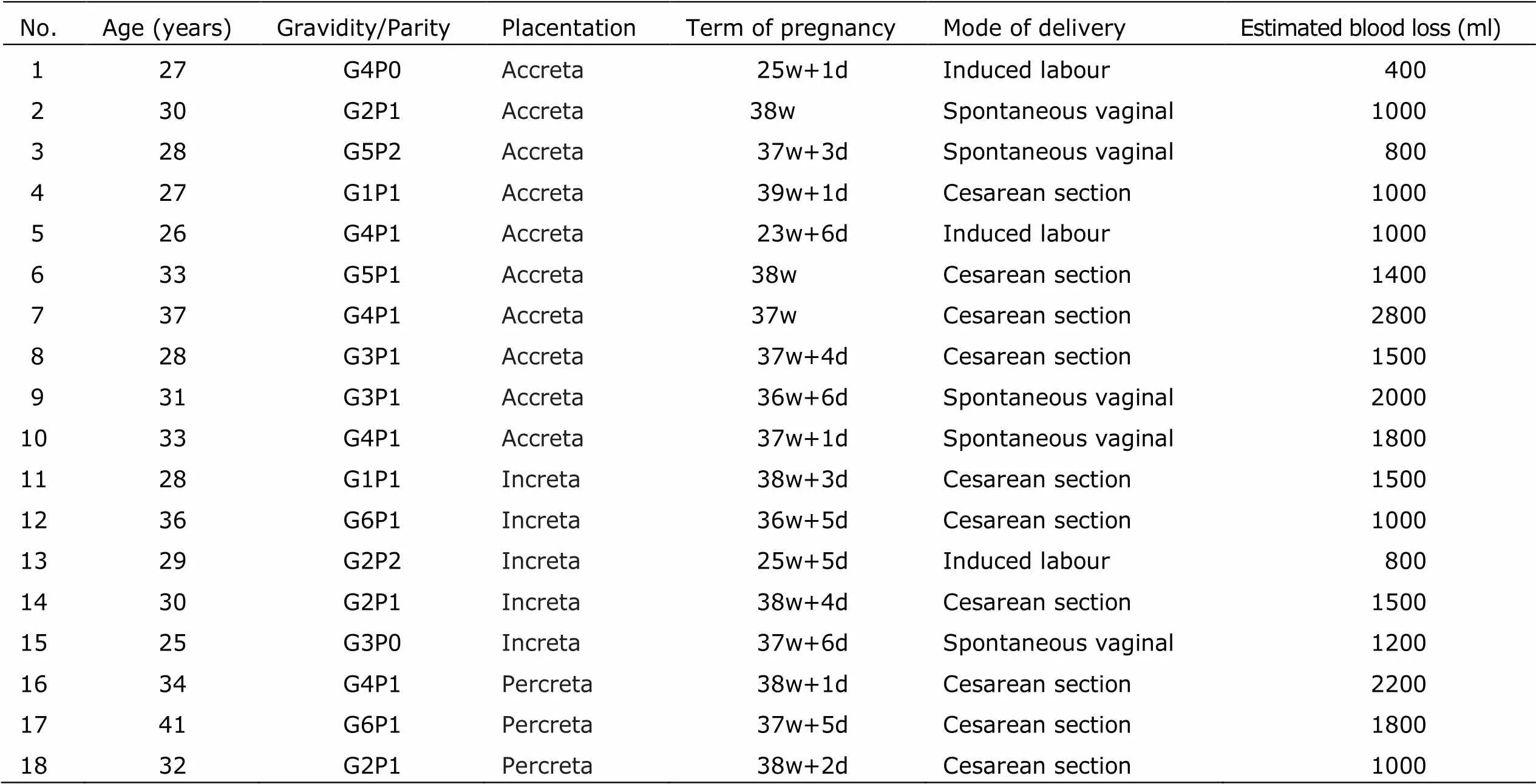
By reviewing medical records 18 women with placenta accreta who were treated byemergent UAE because of massive primary postpartum hemorrhage after delivery were included in the study. All patients were single-fetus pregnancies. The mean age was 30.8±4.2 years. According to clinical and sonographic findings, abnormal placentation were diagnosed: placenta accreta,=10; placenta increta,=5; placenta percreta,=3. The estimated blood loss exceeded 500ml in 17 of 18 enrolled patients. The mean estimated blood loss was 1372±587ml. The clinical features in detail were listed in Table 1.
Embolization procedure
Decisions to perform UAE for patients with persistent uterine bleeding were made by consensus of radiologist and obstetrician after discussion. Embolization procedures were performed by members from a group of six interven- tional radiologists who had 7 to 22 years of experience in pelvic embolization. Informed consent for the embolization procedure was obtained before UAE. With patient under local anesthesia, pelvic angiography using Omnipaque (350 mgI/ml, GE Healthcare, USA) (pressure: 500 atm; flow rate: 10 ml/s) was performed from the common femoral artery approach with a 5-Fr pigtail cathetera 5-Frvascularsheath (Cordis Corporation, USA). The pelvic Digital Subtraction Angiography (DSA, Siemens, German) provided images of vascular road map for the uterine arteries and excluded a potential site of bleeding outside uterus. A long reverse catheter (Beacon Tip Torcon NB Advantage Catheter COOK, USA) or a Cobra catheter (Terumo, Japan) was used to catheterize the uterine artery. Micro-catheters (Progreat Terumo, Japan) were used coaxially when Roberts uterine catheters or Cobra catheters could not advance into the distal uterine artery. In each patients embolization of the uterine arteries was performed using gelatin sponge particles in 700-900 μm or 1400-2000 μm (Hangzhou Alicon Pharm SCI&TEC Co. Ltd, China) or pledgets which were small pieces of gelfoam cutted into 1 to 2 mm in size.

Table 1. Clinical featured information of the 18 Cases in this study
The Gelatin sponge was soaked in the iodinated contrast medium and then was injected through microca- theter until complete stasis in the main uterine artery was achieved. After embolization, a pigtail catheter was placed at the level of renal arteries and an abdominal-pelvic aortography was performed. The technical success of embolization was achieved if uterine arteries wasnot filled by the contrast media on the completion of pelvic arteriography.
Assessment
Data on the control of hemorrhage after embolization were obtained retrospectively from the clinical medical records. Pain, infection, groin hematoma or uterine necrosis after UAE were documented as complications. Clinical success was defined as control of bleeding without repeat emboli- zation or surgical intervention such as laparotomy or hysterectomy.
Results
Angiography findings
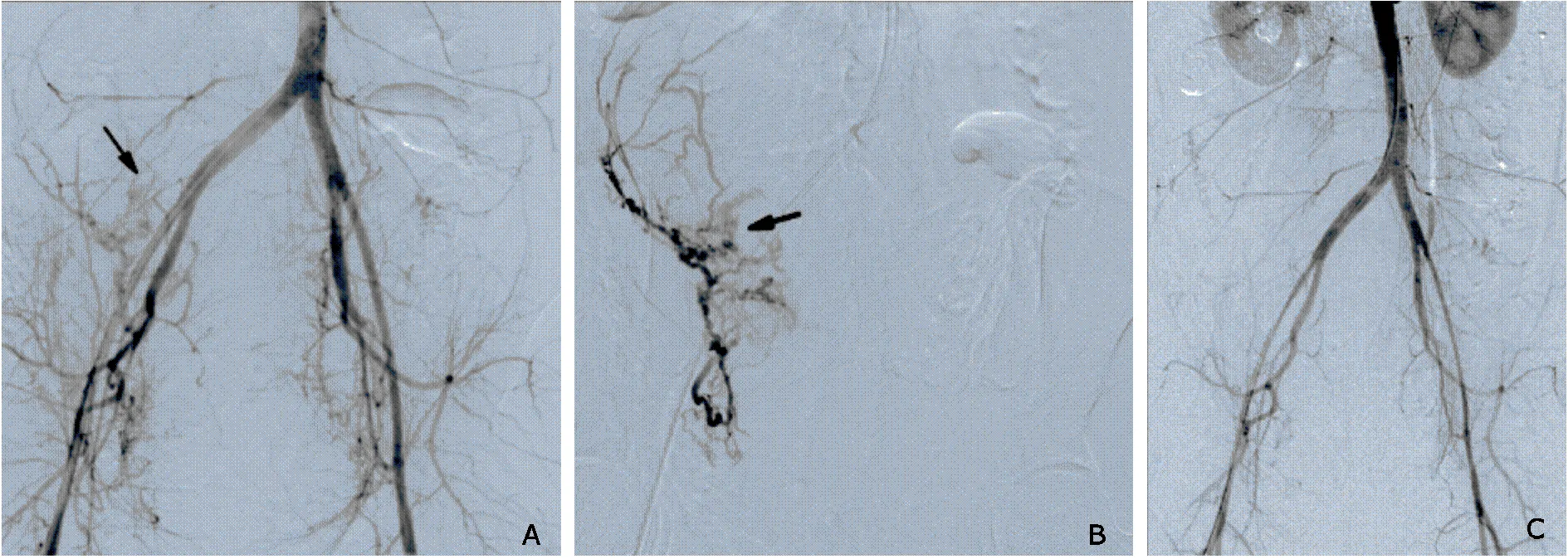
Angiography revealed contrast medium extravasation in the uterine cavities in 8 patients (Fig. 1A). In all cases, embolizations were technically successful in achieving stasis of the uterine artery(Fig. 1B,Fig. 1C). No collateral vessels were found.
Efficacy
In 17 of 18 patients, UAE controlled the bleeding successfully. There was no recurrent bleeding during the follow-up period (median18 months,3 -31 months). The total clinical success rate was 94% (17/18). One woman with placenta percreta required an emergent hysterectomy along with surgical bladder repair because of persistent uterine bleeding after UAE. The serumb-hCG decreased to normal in all patients within two months after the intervention.
Complications
Eight patients encountered fever for 1–2 days and lower abdominal pain after UAE due to post-embolization syndrome. No other complication was noted.

Figure 1. Digital subtraction angiography images of a 33-year-old woman with massive primary postpartum hemorrhage. A. Aortogram showed contrast medium extravasating (arrow) from the right uterine artery into the uterine cavity, indicating active bleeding. B. Right uterine arteriogram demonstrated contrast medium extravasating (arrow) from the right uterine artery. C. Abdominal and pelvic arteriogram after embolization of both uterine arteries with gelatin sponge, showing no uterine artery depicted and no collateral vessels to the uterus revealed.
Discussion
Incidence of placenta accreta has increased in the past a few years due to increasing rates of caesarean section, which is considered as one of the risk factor of placenta accreta for themyometrial damage associated to the pathogenesis of placenta accreta.3-8Women with placenta accreta, increta or percreta are at high risk of life-threatening hemorrhage. The maternal mortality rate was high in women with placenta percreta.21Traditionally the treatment option has been hysterectomy to control the bleeding in cases of percreta; however, conservative treatments, if possible, should be the favored options when future fertility is desired.9,10Several invasive conservative approaches, such as arterial ligation, UAE or internal iliac artery balloon occlusion have been developed to control severe primary postpartum hemorrhage.11-13
UAE has been shown to be associated with high technical success rates and good clinical outcomes for the treatment of primary postpartum hemorrhage.14–16However,the success rate of arterial embolization in patients with placenta accrete was relatively low in early studies, and sometimes hysterectomy had to be performed because of persistent bleeding.22In recent years, with advances of new technologies, catheterization of specific target vessels and interventionist expertise, some authors reported successful cases using UAE in the management of placenta accrete.16-20
This study showed that UAE was effective for controlling bleeding in patients who suffered from massive bleeding after dilivery. UAE offers several advantages: distal and specific catheterization may prevent bleeding through collateral circulation, and success of the procedure can immediately be verified by angiogram. Jung et al18reported the success rate was 82.4% for UAE in the emergent management of intractable postpartum hemorr- hage associated with placenta accreta. In the study of Soyer19UAE was shown to be effective to stop bleeding in 83.3% patients with placenta accreta, who conserved uterine successfully. Bros et al16performed a retrospective study to identify the predictive factor of recurrent bleeding within 24 h after UAE for postpartum hemorrhage. They found that the placenta accreta was likely to be a significant risk factor for postpartum hemorrhage, but was not associated with bleeding recurrence within 24 h after UAE,which indicating that UAE was effective for postpartum hemorrhage of placenta accrete. The success rate in our study was higher than that in the study of Jung18and Soyer.19The reason may lie in that there were less patients with placenta percreta (3/18, 16.7% ) in our study than that in the study of soyer which was 50%. The failure cases in both studies were all women with placenta percreta. So the depth of invasion may be a determinant of success.
The embolic agents used for UAE were gelatin sponge, microsphere and polyvinyl alcohol (PVA) particles.16-20Some authors preferred using absorbable gelatin sponges because gelatin sponges facilitate recanalization of the uterine artery, and thereby preserve the uterine function. The symptoms associated with post-embolization syn- drome, such as lower abdominal pain, fever and nausea, occur frequently and vary depend on the management after TAE. In our series, we used gelatin sponge as the embolic agent for temporary occlusion. Gelatin sponge seems to be sufficient to prevent further hemorrhage while making fewer complications.
Although this study has limitations such as relatively small sample and retrospectiveness,the results strongly support the clinical application of UAE in the management of primary postpartum hemorrhage which is associated with placenta accreta.
1. Publications Committee, Society for Maternal-Fetal Medicine, Belfort MA. Placenta accreta. Am J Obstet Gynecol 2010; 203: 430-9.
2. Angstmann T, Gard G, Harrington T, et al. Surgical man- agement of placenta accreta: a cohort series and sugge- sted approach. Am J Obstet Gynecol. 2010; 202:38.e1-9.
3. Choi SJ, Song SE, Jung KL, et al. Antepartum risk factors associated with peripartum cesarean hysterectomy in women with placenta previa. Am J Perinatol 2008; 25: 37–41.
4. Wu S, Kocherginsky M, Hibbard JU. Abnormal placentation: twenty-year analysis. Am J Obstet Gynecol 2005; 192: 1458–61.
5. Bauer ST, Bonanno C. Abnormal placentation. Semin Perinatol 2009; 33: 88–96.
6. Mehrabadi A, Hutcheon JA, Liu S, et al. Contribution of placenta accreta to the incidence of postpartum hemorr- hage and severe postpartum hemorrhage. Obstet Gynecol 2015; 125: 814-21.
7. Jin R, Guo Y, Chen Y. Risk factors associated with emergency peripartum hysterectomy. Chin Med J 2014; 127: 900-4.
8. Imudia AN, Awonuga AO, Dbouk T, et al. Incidence, trends, risk factors, indications for, and complications associated with cesarean hysterectomy: a 17-year experience from a single institution. Arch Gynecol Obstet 2009; 280:619-23.
9. Khan M, Sachdeva P, Arora R, et al. Conservative management of morbidly adherent placenta—a case report and review of literature. Placenta 2013; 34: 963-6.
10. D'Souza DL, Kingdom JC, Amsalem H, et al. Conservative Management of Invasive Placenta Using Combined Proph- ylactic Internal Iliac Artery Balloon Occlusion and Imme- diate Postoperative Uterine Artery Embolization. Can Assoc Radiol J 2015; 66: 179-84.
11. Teixidor Viñas M, Chandraharan E, Moneta MV, et al. The role of interventional radiology in reducing haemorrhage and hysterectomy following caesarean section for morbidly adherent placenta. Clin Radiol 2014; 69: e345-51.
12. Lin K, Qin J, Xu K, et al. Methotrexate management for placenta accreta: a prospective study. Arch Gynecol Obstet 2015; 291: 1259-64.
13. Kong MC, To WW. Balloon tamponade for postpartum haemorrhage: case series and literature review. Hong Kong Med J 2013; 19: 484-90.
14. Salazar GM, Petrozza JC, Walker TG. Transcatheter endovascular techniques for management of obstetrical and gynecologic emergencies. Tech Vasc Interv Radiol 2009; 12: 139-47.
15. Ganguli S, Stecker MS, Pyne D, et al. Uterine artery embolization in the treatment of postpartum uterine hemorrhage. J Vasc Interv Radiol 2011; 22: 169-76.
16. Bros S, Chabrot P, Kastler A, et al. Recurrent bleeding within 24 hours after uterine artery embolization for severe postpartum hemorrhage: are there predictive factors? Cardiovasc Intervent Radiol 2012; 35: 508-14.
17. Katz MD, Sugay SB, Walker DK, et al. Beyond hemostasis: spectrum of gynecologic and obstetric indications for transcatheter embolization. Radiographics 2012; 32: 1713-31.
18. Jung HN, Shin SW, Choi SJ, et al. Uterine artery embolization for emergent management of postpartum hemorrhage associated with placenta accreta. Acta Radiol 2011; 52: 638-42.
19. Soyer P, Morel O, Fargeaudou Y, et al. Value of pelvic embolization in the management of severe postpartum hemorrhage due to placenta accreta, increta or percreta. Eur J Radiol 2010; 11: 729-35.
20. Diop AN, Chabrot P, Bertrand A, et al. Placenta accreta: management with uterine artery embolization in 17 cases. J Vasc Interv Radiol 2010; 21: 644-8.
21. Silver RM. Abnormal Placentation: Placenta Previa, Vasa Previa, and Placenta Accreta. Obstet Gynecol 2015; 126: 654-68.
22. Inoue S, Masuyama H, Hiramatsu Y. Efficacy of transar- terial embolisation in the management of post-partum haemorrhage and its impact on subsequent pregnancies. Aust N Z J Obstet Gynaecol 2014; 54: 541-5.
for publication December 22, 2015.
Tel: 86-10-69155442, E-mail: pumchjinzhengyu@sina.com
杂志排行
Chinese Medical Sciences Journal的其它文章
- Expression of miRNA-140 in Chondrocytes and Synovial Fluid of Knee Joints in Patients with Osteoarthritis△
- Effects of Lianhua Qingwen on Pulmonary Oxidative Lesions Induced by Fine Particulates (PM2.5) in Rats
- The Effect of Sleep Deprivation on Coronary Heart Disease△
- Meta-analysis of aspirin-heparin therapy for un-explained recurrent miscarriage
- Pseudohyperkalemia with Myelofibrosis after Splenectomy
- A Case Report of Acute Arterial Embolization of Right Lower Extremity As the Initial Presentation of Nephrotic Syndrome with Minimal Changes△
