The inhibitory effect of dihydroartemisinin on the growth of neuroblastoma cells
2016-09-07LingQiYangYangYuCuiLiuTianXinZhuSongJinLinZangYuYingZhangKuangRenDepartmentofPathologyJilinMedicalUniversityJilin303ChinaDepartmentofPharmacologyJilinMedicalUniversityJilin303China
Ling Qi,Yang Yang,Yu-Cui Liu,Tian-Xin Zhu,Song Jin,Lin Zang,Yu-Ying Zhang,Kuang Ren*Departmentof Pathology,Jilin Medical University,Jilin,303,ChinaDepartmentof Pharmacology,Jilin Medical University,Jilin,303,China
The inhibitory effect of dihydroartemisinin on the growth of neuroblastoma cells
Ling Qi1,Yang Yang1,Yu-Cui Liu1,Tian-Xin Zhu1,Song Jin1,Lin Zang1,Yu-Ying Zhang1,Kuang Ren2*1Departmentof Pathology,Jilin Medical University,Jilin,132013,China
2Departmentof Pharmacology,Jilin Medical University,Jilin,132013,China
Original article http://dx.doi.org/10.1016/j.apjtb.2016.01.013
EDITOR’SNOTE
Nobelprizew inner Tu Youyou,w ith heracadem ic team found artem isinin(qinghaosu)and dihydroartem isinin(dihydroqinghaosu).“Herwork laid themost important foundation for the treatmentofmalariaby using artem isinin,gotvigorouspromotion by China and World Health Organization,savedmillionsof livesof patients suffered from malariaworld-w ide,especially those from developing countries,andmadeanoutstanding contribution to thetreatmentand controlof thisimportantparasitic disease.”[1].In recentyears,some scientists reported that dihydroartem isinin also hasanotheradvantageof killingmultiple cancer cells.The results of the presentstudy showed that dihydroartem isinin could inhibit the proliferation of neuroblastoma cells SH-SY5Y.
ARTICLE INFO
Article history:
Received in revised form 1Nov2015
Accepted 13 Jan 2016
Availableonline8Mar2016
Dihydroartem isinin
Neuroblastoma cells
Cyclin D1
Caspase-3
ABSTRACT
Ob jective:To evaluate the inhibitory effectof dihydroartem isinin on neuroblastoma cell line SH-SY5Y,explore the possible mechanism of dihydroartem isinin against neuroblastoma cells.
M ethods:The cellviability of dihydroartem isinin treated SH-SY5Y cellswas exam ined by MTT assay andmorphology of cellswas observed by using invertedm icroscope.Cell cycle was exam ined w ith fl ow cytometry assay,then cyclin D1 and caspase-3 proteins expression was detected by ELISA and western blotting assay.
Resu lts:MTT analysis results showed that cell viability signi fi cantly decreased after exposure to 0.05,0.50,5.00 and 50.00μmol/L dihydroartem isinin in a dose-dependent manner,and the lower density of cells was observed in treated groups.The number of cells in sub-G1 phase was increased after treatment w ith different doses of dihydroartemisinin compared w ith the controlgroup.Theexpression of cyclin D1 protein was decreased,while the expression of caspase-3 protein was increased in treated group.
Conclusions:Dihydroartem isinin could inhibit the proliferation through stopping the cell cycle and inducing the apoptosis in neuroblastoma SH-SY5Y cells.
1.Introduction
Neuroblastoma is one of themost common malignant solid tumors in infants and young children[2].So far,there has been no effective treatment.Surgery,chemotherapy and radiotherapy are the three main methods clinically.Among them, chemotherapy drugs could kill the tumor cells,but at the same time,they can also bring huge toxic side effects,and lead to serious impact on physical and mental health of children. Therefore,it is necessary to fi nd a kind of chemotherapeutic drugs w ith low toxicity and high ef fi ciency for neuroblastoma treatments.
Dihydroartem isinin(DHA),a main active metabolite extracted from artem isinin,has been w idely used as an antimalaria drug clinically.It possesses many advantages such as good absorption,wide distribution,rapid excretion and metabolism,high ef fi ciency and low toxicity,etc.In the study by Hou etal.,the resultshowed thatDHA has less effects on the grow th of normal cells[3],but it could signi fi cantly killmultiple cancer cells in vivo[4].
The present study aimed to explore the inhibition on neuroblastoma SH-SY5Y cells proliferation by DHA using MTT assay and morphology detection,thereby investigating the possible mechanisms responsible for DHA-induced inhibitionon SH-SY5Y cells by fl ow cytometry,ELISA and western blotting assay.
2.M aterials and methods
2.1.Drug and reagents
In this study,DHA was purchased from Chengdu Okay Co., Ltd.Dulbecco’smodi fi ed Eaglemedium was from Hyclone Co. (Logan,Utah,USA).Trypsin(1:250)and fetal bovine serum were from Invitrogen Co.(Carlsbad,CA,USA).Dimethylsulfoxide(DMSO)and MTT used in the experiment were from Sigma Chem icalCo.(St.Louis,MO,USA).Cyclin D1,caspase-3 andβ-actin antibodies were purchased from Santa Cruz Co. DHA was dissolved in DMSO solution at 50 mmol/L and reserved in refrigerator.
2.2.Cell line and cell culture
The human neuroblastoma cell line SH-SY5Y was provided by Scienti fi c Research Center of Jinlin Medical University.The experiments using human cell lines were approved by Jinlin Medical University Ethics Comm ittee.The cryopreserved SHSY5Y cells were placed in a 37°C water bath and vibrated untildissolved.A fter centrifugalization at1000 r/m in for 5m in, the cellsuspensionwas then cultured in theDulbecco’smodi fi ed Eagle medium supplemented w ith 10%fetal bovine serum at 37°C,in ahum idi fi ed atmosphere of 95%O2and 5%CO2.The cellswere subcultured every 2 or 3 days.
2.3.Cell viability assay
Cellswere cultured in 96-well plate at a density of 8×10³cells/well and incubated for 24 h.Various doses of DHA(0.00, 0.05,0.50,5.00 and 50.00μmol/L)were used to treat the cells for 24,48 and 72 h.A fter incubation,20μL MTT(5mg/m L) reagentwas added to each well for 4 h at37°C.Then the MTT liquid was replaced,and 150μL DMSO was added and the m ixture was agitated for 10 m in.Absorbance was read at a wavelength of 490 nm by using an EL×800 Universal M icroplate Reader(BIO-TEK,Norcross,GA,USA).Experiments were performed in triplicate,and cell viability was calculated as a percentage of the control(the group treated w ith 0.00μmol/L DHA).
2.4.Themorphology of SH-SY5Y cells
Cells were cultured overnight in 25 cm2fl ask,and then treated w ith 0.05,0.50,5.00 and 50.00μmol/L DHA for 24 h, respectively.Cells treated w ith vehicle served as control.Photos were taken under inverted m icroscope(Olympus Corporation, Japan).
2.5.Cell cycle assay
Cellswere collected after 24 h treatmentw ith DHA at0.05, 0.50,5.00 and 50.00μmol/L,respectively,and washed w ith phosphate-buffered saline solution,then resuspended in 80% ice-coldmethanoland incubated at−20°Covernight.Cellswere stained w ith buffered saline solution containing 20μg/m L propidium iodide for 30m in,fi ltered w ith 300-mesh nylon screen, and then analyzed w ith fl ow cytometry(Epics-XL,Beckman Coulter Inc.,Bria,CA,USA).
2.6.ELISA assay
The SH-SY5Y cellswere divided into 5 groups,and treated w ith 0.00,0.05,0.50,5.00 and 50.00μmol/L DHA for 24 h, respectively.The supernatantwas collected forexperiments,and then incubated with coating buffer at ratio of 1:1 overnight at 4°C.A fter closure,the cyclin D1 and caspase-3 antibodies (1:1000 dilution)were separately added,followed by continued incubation overnight at 4°C.Then the cells were incubated at 37°C for 1.5 h after adding secondary antibody(1:1000 dilution).Diam inobenzidine was added for 5 m in;when the color was brown,the absorbance value was determined by automatic m icroplate reader(MDC,USA)at 492 nm.
2.7.Western blotting
SH-SY5Y cells were collected after 0.05,0.50,5.00 and 50.00μmol/L DHA treatments,and lysed w ith a lysis buffer (50 mmol/L Tris,150 mmol/L NaCl,2 mmol/L ethylene diam ine tetraacetic acid,10%glycerol,1%Triton X-100,1% protease inhibitorm ixtureand 1mmol/L phenylmethanesulfonyl fl uoride).Fifty m icrograms of total protein from each lysate were separated through SDS-PAGE and transferred to membranes.Themembranes were incubated w ith the primary antibodies againstβ-actin,cyclin D1 and caspase-3.β-actin,as a loading control(1:1000;SANTA CRUZ,USA),was used overnight at 4°C.Primary antibody binding was detected w ith anti-rabbit immunoglobulin G conjugated to horseradish peroxidase and visualized by an enhanced chem ilum inescence detection system(Pierce,Rockford,IL,USA).Results were analyzed using Image-Pro Plus image analysis andmanagement systems(USA).
2.8.Statistical analysis
Datawereanalyzed by the SPSS 17.0 softwareand expressed asmean±SD.Statistical comparisons between differentgroups were done by using One-way ANOVA.The statistical signi ficance was determ ined at P<0.05.
3.Results
3.1.The effects of DHA on the proliferation of SH-SY5Y cells
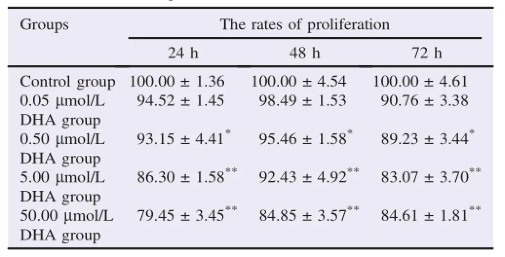
The results of MTT assay showed that different concentrations of DHA could inhibit the grow th of SH-SY5Y cells at24, 48 and 72 h,respectively;the proliferation was signi fi cantly decreased in 0.50,5.00 and 50.00μmol/L DHA treated groups (P<0.05,P<0.01).DHA could inhibit the grow th of SHSY5Y cells in a dose-dependent manner.The inhibition was themostobvious at24 h,therefore 24 h was taken as the proper time for the follow-up experiments(Table 1).
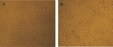
The morphology of cells was observed under the inverted m icroscope.The number of adherent cells in DHA treated groups was signi fi cantly decreased compared w ith the control group,and at the same time,the density of cellswas decreased w ith increasing concentrations of DHA(Figure 1).

Table1 Effects of DHA on the grow th of SH-SY 5Y cells.%.

Figure 1.DHA inhibited the grow th of SH-SY5Y cells(×200). A:Control group;B:Cells treated w ith 50μmol/L DHA.
3.2.The changes of cell cycle after DHA treatments
The number of SH-SY5Y cells in sub-G1 phase was increased w ith the increase in concentrations of DHA.In this phase,compared w ith the control group,the cells of 50μmol/L DHA groupwere signi fi cantly increased(P<0.05).Thenumber of SH-SY5Y cells in G0/G1 phasewas increased at fi rstand then decreased.Thenumberof SH-SY5Y cells in Sand G2/M phases was decreased(Table 2).

Table2 Effectof DHA treatmenton cell cycle of SH-SY 5Y cells.%.
3.3.The expression of cyclin D1 and caspase-3 proteins
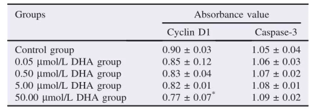
The results of ELISA showed that the secretion of cyclin D1 protein was decreased in cultured supernatant;when compared w ith control group,the secretion of cyclin D1 in 50μmol/L DHA group was signi fi cantly reduced(P<0.05).However,the secretion of caspase-3 protein was increased in all DHA treated groups compared w ith the control group w ithout signi fi cant difference(P>0.05)(Table 3).

Table 3 The secretion of cyclin D1 and caspase-3 proteins in supernatant.
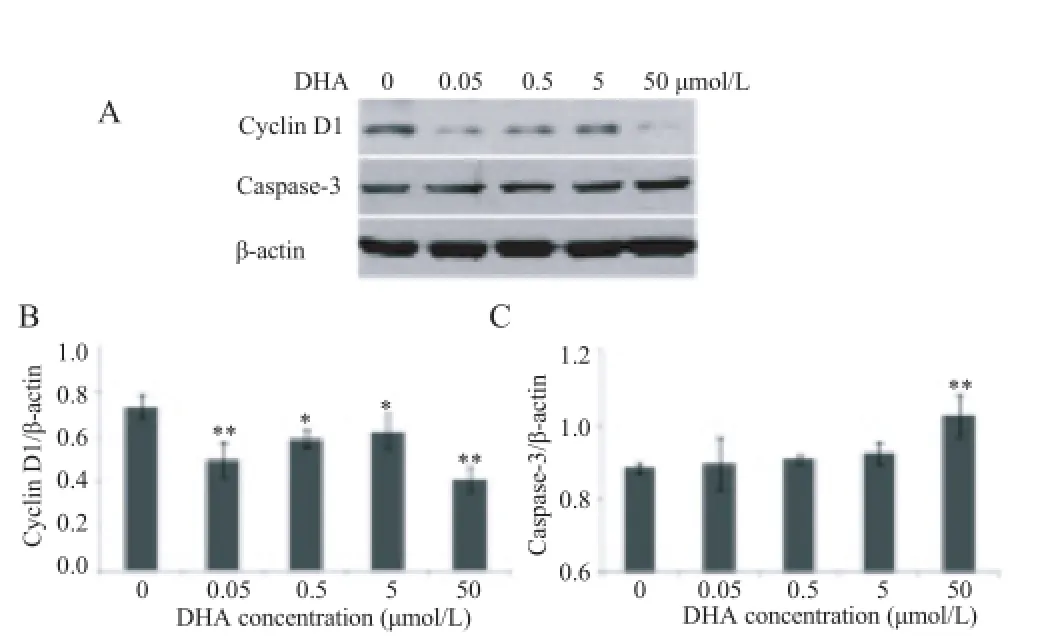
The results ofwestern blotting showed that the expression of cyclin D1 was decreased in DHA treated groups,and the difference was statistically signi fi cant(P<0.05,P<0.01),while caspase-3 protein was increased in different DHA doses treated groups when compared w ith control group,and the difference between 50μmol/L DHA group and control group was statistically signi fi cant(P<0.01)(Figure 2).

Figure 2.DHA affected the expression of cyclin D1 and caspase-3 proteins in SH-SY 5Y cells.A:The expression of cyclinD1 and caspase-3 proteins;B:The level of cyclin D1 protein expressed;C:The levelof caspase-3 protein expressed.*: P<0.05 compared w ith controlgroup;**:P<0.01 compared w ith control group.Data were expressed asmean±SD.
4.Discussion
Artem isinin is a kind of compound which contains the peroxide groups and is extracted from Artemisia annua L.It is an effective and famous natural product for clinical treatmentof malaria,otherw ise as a new anti-tumor agent[5].DHA is the main active metabolite of artem isinin in vivo,whose oral bioavailability is 10 times higher than that of artem isinin,and the anti-malaria effects are stronger than artem isinin by 4–8times;at the same time,DHA also has an obvious anti-tumor activity[5–8].However,the inhibitory effect of DHA on the grow th of SH-SY5Y cells is still unknown.
We studied how various doses of DHA affected the proliferation of SH-SY5Y cells,and thereby explored its anti-tumor function.The results revealed that different concentrations of DHA can signi fi cantly inhibit the grow th of neuroblastoma cells at indicated time points,and the inhibitory effectwas obviously dose-dependent.The density of cells was obviously decreased w ith increased doses of DHA.These results showed that DHA inhibited the grow th of SH-SY5Y cells in a concentration dependentmanner.
The results regarding cell cycle showed that the number of SH-SY5Y cells was increased in sub-G1 phase,increased and then decreased in G0/G1 phase,decreased in Sand G2/M phases w ith the increased concentrationsof DHA.As itis known to all, the grow th of the cells is regulated by cyclins and cyclindependent kinase inhibitor,among which cyclin D1 plays a positive regulatory role in the grow th of cells[8].Hosoya etal. and Mao etal.reported that DHA stopped the cell cycle atG0/ G1 phase,and made a problem in the conversion of G1/S junction[9,10].Cyclin D1 is an important protein which regulates the conversion of G1/S junction;blocking and knocking out cyclin D1 could stop the cell cycle at G0/G1 phase,and induce apoptosis[11].Therefore,our results suggested that DHA could down-regulate the expression and secretion of cyclin D1 protein to stop cell cycle and inhibit the proliferation of SH-SY5Y cells.
Apoptosis is a process of programmed cell death controlled by multiple factors,which is caused by a series of intracellular proteins.The caspases play an im portant role in the process of cell apoptosis[12].Although caspase is rarely expressed in most tumor tissues[13],it is the only way for the caspases cascade reaction,and it is also the key molecule in apoptosis.Casepase-3 is an important protein which exists in the form of proenzyme in cytoplasm;it is activated when apoptosis is induced in tumor cells[14].DHA induced the apoptosis through caspases pathw ay[15].Our results showed that there was an increase in the secretion and expression of caspase-3 protein by SH-SY5Y cells, which certi fi ed that DHA could induce apoptosis through caspase-3 activation.
In conclusion,DHA could induce apoptosis of neuroblastoma SH-SY5Y cells through down-regulating cylin D1 and activating caspase-3,thereby inhibiting the grow th of neuroblastoma cell.However,because there are many factors affecting the grow th of tumor cell,and among them,the cell cycle and apoptosis were prelim inarily studied in the present study,further investigation of the mechanism of DHA against tumor cells is still needed in the future study.
Con fl ict of interest statement
We declare thatwe have no con fl ictof interest.
Acknow ledgments
Theauthorssincerely appreciate the NationalNatural Science Foundation of China for the fi nancial support(No.81201671, No.81202953 and No.81202031).
References
[1]Com pilation team of Tu Youyou’s Biography.[Tu Youyou’s biography].Beijing:Beijing People’s Publishing House;2015, p.138-9.Chinese.
[2]Peifer M,Hertw ig F,Roels F,Dreidax D,Gartlgruber M, M enon R,etal.Telomerase activation by genomic rearrangements in high-riskneuroblastoma.Nature 2015;526(7575):700-4.
[3]Chen PQ,Yuan J,Du QY,Chen L,Li GQ,Huang ZY,et al.Effects of dihydroartemisinin on fi ne structure of srythrocytic stages of p lasm odium berghei ANKA strain.Acta Pharmacol Sin 2000; 21(3):234-8.
[4]Hou J,Wang D,Zhang R,Wang H.Experimental therapy of hepatomaw ith artemisinin and its derivatives:in vitro and in vivo activity,chem osensitization,and mechanisms of action.C lin Cancer Res 2008;14:5519-30.
[5]Dilshad E,Cusido RM,Ramirez Estrada K,Bon fi llM,M irza B.Genetic transformation of Artemisia carvifolia Buch w ith rol genesenhancesartem isinin accumulation.PLoSOne2015;10(10):e0140266.
[6]Li YJ,Zhou JH,Du XX,Jia de X,Wu CL,Huang P,etal.Dihydroartemisinin accentuates the anti-tumor effects of photodynamic therapy via inactivation of NF-κB in Eca109 and Ec9706 esophageal cancer cells.Cell Physiol Biochem 2014;33(5):1527-36.
[7]Zhang ZS,Wang J,Shen YB,Guo CC,Sai KE,Chen FR,et al. Dihydroartemisinin increases temozolomide ef fi cacy in glioma cells by inducing autophagy.Oncol Lett2015;10(1):379-83.
[8]Zhao S,Yi M,Yuan Y,Zhuang W,Zhang D,Yu X,et al. Expression of AKAP95,Cx43,CyclinE1 and CyclinD1 in esophageal cancerand theirassociationw ith the clinicaland pathological parameters.Int JClin Exp Med 2015;8(5):7324-32.
[9]Hosoya K,Murahari S,Laio A,London CA,Couto CG, KisseberthWC.Biologicalactivity of dihydroartem isinin in canine osteosarcoma cell lines.Am JVet Res 2008;69:519-26.
[10]Mao H,Gu H,Qu X,Sun J,Song B,GaoW,etal.Involvementof the mitochondrial pathway and Bim/Bcl-2 balance in dihydroartem isinin-induced apoptosis in hum an breast cancer in vitro.Int JMolMed 2013;31(1):213-8.
[11]Thoms HC,Dunlop MG,Stark LA.p38-M ediated inactivation of cyclinD1/cyclin-dependent kinase 4 stimulates nucleolar translocation of RelA and apoptosis in colorectal cancer cells.Cancer Res 2007;67(4):1660-9.
[12]Luparello C,Sirchia R,Lo Sasso B.M idregion PTHrP regulates Rip1and caspase expression in MDA-MB231 breast cancer cells. Breast Cancer Res Treat 2008;111(3):461-74.
[13]Nassar A,Lawson D,CotsonisG,Cohen C.Survivin and caspase-3 expression in breast cancer:correlation w ith prognostic parameters,proliferation,angiogenesis,and outcome.Appl Immunohistochem Mol Morphol 2008;16(2):113-20.
[14]Sui L,Dong Y,Ohno M,Watanabe Y,Sugimoto K,Tokuda M. Survivin expression and its correlation w ith cell proliferation and prognosisinepithelialovarian tumor.Int JOncol2002;21(2):315-20.
[15]Cao L,Duanmu W,Yin Y,Zhou Z,Ge H,Chen T,et al.Dihydroartemisinin exhibits anti-glioma stem cell activity through inhibiting p-AKT and activating caspase-3.Pharmazie 2014; 69(10):752-8.
20 Oct 2015
*Corresponding author:Kuang Ren,Departmentof Pharmacology,Jilin Medical University,Jilin,132013,China.
Tel:+86 432 64560301
E-mail:renkuang@sina.com
The experiments using hum an cell lines were approved by Jilin M edical University Ethics Comm ittee.
Foundation Project:Supported by the National Natural Science Foundation of China(NO.81201671,NO.81202953 and NO.81202031).
Peer review under responsibility of Hainan M edical University.The journal implements double-blind peer review practiced by specially invited international editorial boardmembers.
2221-1691/Copyright©2016 Hainan Medical University.Production and hosting by Elsevier B.V.This is an open accessarticle under the CC BY-NC-ND license(http:// creativecommons.org/licenses/by-nc-nd/4.0/).
杂志排行
Asian Pacific Journal of Tropical Biomedicine的其它文章
- Sudden death in a captive mee rkat(Suricata surica tta)w ith arterial m edial and m yocardial calcification
- Characteristics o f obese or overweight dogs visiting private Japanese vete rinary c linics
- Risk factors from HBV infection among blood donors:A system atic review
- Com putational in telligence in tropicalm edicine
- The African Moringa is to change the lives ofm illions in Ethiopia and far beyond
- Pediculosis capitis among p rimary and m idd le school children in Asadabad,Iran:An epidem iological study
