联合检测FHIT、Ki-67及PCNA在皮质醇增多症肾上腺皮质不同病变中的意义*
2016-07-24梁秀就黄高明蔡劲薇卢德成罗佐杰
冼 晶,梁秀就,黄高明,李 励,蔡劲薇,黄 松,卢德成,罗佐杰△
(广西医科大学:1.第一附属医院内分泌科;2.第一附属医院病理科;3.公共卫生学院流行病学与统计学教研室,广西南宁 530021)
·论 著·
联合检测FHIT、Ki-67及PCNA在皮质醇增多症肾上腺皮质不同病变中的意义*
冼 晶1,梁秀就2,黄高明3,李 励1,蔡劲薇1,黄 松1,卢德成1,罗佐杰1△
(广西医科大学:1.第一附属医院内分泌科;2.第一附属医院病理科;3.公共卫生学院流行病学与统计学教研室,广西南宁 530021)
目的 探讨皮质醇增多症肾上腺皮质不同病变中脆性组氨酸三联体(FHIT)、Ki-67及增殖细胞核抗原(PCNA)的表达水平及其在鉴别诊断中的作用。方法 应用免疫组织化学方法检测49例皮质醇增多症肾上腺皮质病变(腺癌14例、腺瘤26例、皮质增生9例)患者FHIT、Ki-67及PCNA的表达。结果 FHIT在肾上腺皮质增生表达最高,腺瘤次之,腺癌最低(分别为100.00%、96.15%、42.86%);Ki-67与PCNA在肾上腺皮质腺癌的表达最高,腺瘤次之,皮质增生最低(分别为85.71%、7.69%、0和100.00%、96.15%、77.78%)。FHIT、Ki-67及PCNA在皮质醇增多症肾上腺皮质病变中的表达两两相关,其中FHIT与Ki-67表达呈负相关(r=-0.718,P=0.00),FHIT与PCNA表达呈负相关(r=-0.449,P=0.001),Ki-67与PCNA表达呈正相关(r=0.387,P=0.006)。联合检测FHIT、Ki67及PCNA结果显示,腺癌表现为FHIT阴性或弱阳性、Ki-67强阳性、PCNA强阳性;腺瘤表现为FHIT阳性、Ki-67阳性、PCNA阳性;皮质增生表现为FHIT强阳性、Ki-67阴性、PCNA阴性。结论 联合检测FHIT、Ki-67及PCNA对肾上腺皮质腺癌、腺瘤及增生的鉴别诊断具有重要的价值。
皮质醇增多症;脆性组氨酸三联体;Ki-67;增殖细胞核抗原;联合检测
脆性组氨酸三联体(fragile histindine triad,FHIT)是一种肿瘤抑制基因,其蛋白表达的缺失与多种肿瘤的发生和发展密切相关[1]。肿瘤增殖抗原Ki-67,是一种存在于增殖细胞核的非组蛋白性核蛋白,与细胞增殖密切相关[2]。增殖细胞核抗原(proliferation cell nuclear antigen,PCNA),是一种仅在增殖细胞中合成或表达的核内多肽,其表达与合成与细胞周期有关,是判断各种恶性肿瘤细胞增殖及其恶性度的一种指标[3]。FHIT、Ki-67及PCNA为目前用于多种人类肿瘤发生、发展中的研究,但其在内分泌病变中的表达及和内分泌肿瘤的发生、发展的关系研究较少,亦少见三者联合检测用于肾上腺皮质腺瘤、腺癌及皮质增生鉴别诊断的报道。
1 资料与方法
1.1 一般资料 组织标本为广西医科大学第一附属医院2005~2012年手术切除、经病理证实的皮质醇增多症肾上腺皮质病变石蜡包埋标本49份,其中肾上腺皮质腺癌14份,腺瘤26份,增生9份[均为不依赖垂体促肾上腺皮质激素(ACTH)的双侧肾上腺小结节性增生]。其中,男19例,女30例;年龄5~68岁,平均(35.84±16.18)岁。患者均有皮质醇增多症典型的临床症状、体征和实验室改变,行小剂量地塞米松抑制试验均不被抑制,临床确诊为皮质醇增多症。研究获得本单位伦理委员会的审查批准及受试者签署的知情同意书。
1.2 方法
1.2.1 检测方法 兔抗人FHIT浓缩型多克隆抗体购自北京中山生物技术公司,鼠抗人Ki-67、PCNA单克隆抗体和EliVision 检测试剂盒为福州迈新生物技术公司产品。采用EliVision二步法,参照试剂盒说明书进行免疫组织化学染色,以PBS代替一抗作为阴性对照,用已知阳性切片作为阳性对照。主要染色步骤:切片脱蜡和水化后抗原修复,一抗FHIT(工作浓度1∶100)、Ki-67(即用型)、PCNA(即用型)37 ℃孵育60 min,然后滴加放大剂、多聚酶结合物,DAB显色,复染等。
1.2.2 结果判断 细胞核或细胞质呈棕褐色或棕黄色颗粒者为阳性细胞。FHIT蛋白定位于细胞质。Ki-67、PCNA阳性定位于细胞核。按染色强度评分:无色0分,浅黄色1分,棕黄色2分,棕褐色3分;再按阳性细胞所占的百分比评分:阳性细胞小于5%为0分,≥5%~25%为1分,>25%~50%为2分,>50%~75%为3分,>75%为4分。两者的乘积:0分为阴性(-),1~<5分为弱阳性(+),5~<9分为中等阳性(++),9~12分为强阳性(+++)。 Ki-67阳性定位于细胞核,阳性细胞数小于10%为阴性(-),阳性细胞数大于或等于10%为阳性:其中10%~25%为弱阳性(+),>25%~50%为中等阳性(++),>50%为强阳性(+++)[4]。
1.3 统计学处理 采用SPSS17.0统计学软件进行统计学分析,总阳性率比较采用χ2检验,如有实际数为零或不满足χ2检验条件,改用Fisher确切概率法;各指标间相关分析采用Spearman秩相关。以P<0.05为差异有统计学意义。
2 结 果
2.1 皮质醇增多症肾上腺皮质病变中FHIT、Ki-67及PCNA的表达
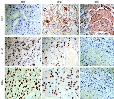
2.1.1 皮质醇增多症肾上腺皮质病变中FHIT的表达 腺癌的总阳性率均较腺瘤及增生低(χ2=16.272,P=0.000)。腺瘤与增生在总阳性率比较差异无统计学意义(P>0.05),在分级比较中,增生的表达全部为强阳性,腺瘤中16例为中阳性表达(61.54%),有9例为强阳性表达(34.62%)。腺癌仅6例表现为弱阳性(42.86%),其余全部为阴性。腺癌在阴性及弱阳性(-/+)的表达较腺瘤及增生高,见表1、图1。
2.1.2 皮质醇增多症肾上腺皮质病变中Ki-67的表达 腺癌Ki-67总阳性率的表达较腺瘤及增生高(χ2=29.187,P=0.000),腺瘤与增生Ki-67总阳性率比较差异无统计学意义(P>0.05)。在分级比较中,增生的表达全部为阴性,腺瘤有2例为弱阳性表达(7.69%)。腺癌在较高阳性(++/+++)的表达比腺瘤及增生高,见表2、图1。
2.1.3 皮质醇增多症肾上腺皮质病变中PCNA的表达 腺瘤、腺癌、增生三者的总阳性率在两两比较差异无统计学意义(χ2=3.794,P=0.147)。但在分级比较中,腺癌在较高阳性(++/+++)的表达较腺瘤、增生高,腺瘤、增生的表达多为阴性及弱阳性,差异有统计学意义(P<0.05),见表3、图1。

表1 FHIT在皮质醇增多症肾上腺病变中的表达[n(%)]
a:P<0.05,与腺癌比较。

表2 Ki-67在皮质醇增多症肾上腺病变中的表达[n(%)]
a:P<0.05,与腺癌比较。

表3 PCNA在皮质醇增多症肾上腺病变中的表达[n(%)]
a:P<0.05;与增生比较;b:P<0.01,与腺癌比较。

FHIT:腺癌阴性表达,腺瘤中等阳性,增生强阳性;Ki-67:腺癌强阳性,腺瘤弱阳性,增生阴性;PCNA:腺癌强阳性,腺瘤中等阳性,增生阴性 (箭头示阳性反应部位,免疫组化EliVisionTMsuper二步法,×400)。
图1 皮质醇增多症肾上腺皮质病变中FHIT、Ki-67及PCNA的表达
2.2 皮质醇增多症肾上腺皮质病变中FHIT、Ki-67及PCNA表达的关系 FHIT、Ki-67及PCNA在皮质醇增多症肾上腺皮质病变中的表达两两相关,其中FHIT与Ki-67表达呈负相关(r=-0.718,P=0.000),FHIT与PCNA表达呈负相关(r=-0.449,P=0.001),Ki-67与PCNA表达呈正相关(r=0.387,P=0.006),见表4~6。

表4 肾上腺皮质病变中FHIT与Ki-67表达的关系
2.3 联合检测FHIT、Ki67及PCNA结果 腺癌表现为FHIT阴性或弱阳性、Ki-67强阳性、PCNA强阳性;腺瘤表现为FHIT阳性、Ki-67阳性、PCNA阳性;增生表现为FHIT强阳性、Ki-67阴性、PCNA阴性。

表5 肾上腺皮质病变中FHIT与PCNA表达的关系

表6 肾上腺皮质病变中Ki-67与PCNA表达的关系
3 讨 论
皮质醇增多症肾上腺皮质病变的诊断包括定性诊断与病因诊断,在目前条件下,定性诊断一般不困难,但在病因诊断时不少病例在临床和病理上均不易明确区分,以致误诊及治疗方案选择困难。
FHIT基因异常与人类多种恶性肿瘤的发生、发展有关[1]。FHIT基因编码的蛋白质为FHIT蛋白,具有抑癌活性[5],在多种环境致癌物相关恶性肿瘤的发生中起重要作用[6]。在各种致癌因素影响下, 其表达受阻可使FHIT蛋白表达降低,恶性病变组织中检测到FHIT蛋白低表达或缺失,相反FHIT蛋白的高表达提示为良性。在多种人类肿瘤中可发现FHIT蛋白表达的缺失,其缺失的程度与其良、恶性及预后有关[7-9]。本研究发现在皮质醇增多症肾上腺皮质病变中,FHIT在腺癌的表达率最低,且全部为阴性或弱阳性,增生的表达率最高,且全部为强阳性,腺瘤的表达介于二者之间,提示FHIT基因转录产物异常及其蛋白表达的改变与肾上腺皮质病变的良、恶性有密切关系,腺癌较腺瘤和增生更为明显,其表达下调可能促进肿瘤的进展。另外发现FHIT在肾上腺皮质腺癌中除有57.14%(8/14)为阴性表达外,另有42.86%(6/14)为弱阳性表达,提示FHIT基因编码的蛋白质在恶性肿瘤中亦非全部缺失,部分仅为蛋白表达的减少,FHIT在肾上腺皮质腺癌中的部分低表达与此有关,其原因推测可能与肿瘤的恶性程度有关,部分腺癌的恶性程度较高,则FHIT蛋白的表达显示为缺失,而另一部分腺癌恶性程度较低,FHIT蛋白可部分表达。
Ki-67表达能可靠而迅速地反映恶性肿瘤增殖率,与多种肿瘤的发展、转移、预后有关,Ki-67高表达提示肿瘤的恶性程度较高,预示较差的预后[10-12]。Ki-67在肾上腺肿瘤的表达对皮质醇增多症肾上腺皮质病变的鉴别诊断有重要意义,但文献甚少这方面的报道。本研究发现皮质醇增多症肾上腺皮质病变中,Ki-67在腺癌的表达率最高,腺瘤较低,增生则不表达,提示皮质腺癌的细胞增殖较腺瘤和增生更为活跃,Ki-67的表达随增生、腺瘤、腺癌的细胞增殖程度的提高,表达的阳性强度增加,阳性率亦有增加。由于肿瘤的恶性程度与肿瘤细胞增殖分裂的程度密切相关,因此Ki-67表达的高低可反映肿瘤的恶性程度的高低[13]。
细胞过度增殖可能导致肿瘤的发生,而增殖是肿瘤浸润和转移的基础,因此肿瘤细胞增殖水平可反映肿瘤的恶性程度。PCNA是DNA合成必需的细胞周期蛋白,又仅在增殖细胞中表达,其在肿瘤细胞中的表达情况可以敏感地反映肿瘤细胞的增殖活性[3,14-15]。 较高的PCNA表达率反映了高水平的细胞增殖状态[16]。本研究发现在皮质醇增多症肾上腺皮质病变中,PCNA的表达在腺癌最高,腺瘤次之,增生最低。由于从增生、腺瘤到腺癌,细胞增殖水平的提高,PCNA表达的阳性强度亦随之增加。且由于增生和腺瘤的细胞增殖水平不同,PCNA表达高低也可以作为二者的判断指标。
FHIT基因蛋白具有抑癌活性, Ki-67和PCNA能反映细胞的增殖状况。本研究结果显示,FHIT、Ki-67及PCNA在皮质醇增多症肾上腺皮质病变中的表达两两相关。其中,FHIT与Ki-67表达呈负相关,即随着细胞增殖程度的不同,肿瘤抑制基因FHIT表达越高,Ki-67表达则越低,反之亦然。FHIT与PCNA表达呈负相关,即FHIT表达越高,PCNA表达则越低,反之亦然。而Ki-67与PCNA表达呈正相关,即随着细胞增殖程度的增加,肿瘤增殖抗原Ki-67表达升高,同时增殖细胞核抗原PCNA表达也升高,反之,Ki-67降低, PCNA也降低。
本研究发现 FHIT、Ki-67及PCNA在皮质醇增多症肾上腺皮质腺癌、腺瘤及增生的表达有一定的规律,FHIT阴性或弱阳性、Ki-67强阳性、PCNA强阳性的肾上腺皮质病变为腺癌;FHIT强阳性、 Ki-67阴性、PCNA阴性或弱阳性的肾上腺皮质病变为增生;而肾上腺皮质腺瘤介于腺癌和增生之间,FHIT阳性、Ki-67阳性、PCNA阳性的肾上腺皮质病变为腺瘤。因此,联合检测FHIT、Ki-67及PCNA对肾上腺皮质腺癌、腺瘤及增生的鉴别诊断具有重要的价值。根据FHIT、Ki-67及PCNA的不同表达结果,能对皮质腺癌、腺瘤及增生作出鉴别,FHIT、Ki-67及PCNA可作为皮质醇增多症肾上腺皮质病变鉴别诊断的较可靠的标记物。
[1]Karras JR,Paisie CA,Huebner K.Replicative stress and the FHIT gene:Roles in tumor suppression,genome stabiolity and prevention of carcinogenesis[J].Cancers (Basel),2014,6(2):1208-1219.
[2]Dowsett M,Nielsen TO,A Hern R,et al.Assessment of Ki67 in breast cancer:recommendations from the International Ki67 in Breast Cancer Working Group[J].J Natl Cancer Inst,2011,103(22):1656-1664.
[3]Kuang RG,Wu HX,Hao GX,et al.Expression and significance of IGF-2,PCNA,MMP-7 andα-actin in gastric carcinoma with Lauren classification[J].Turk J Gastroenterol,2013,24(2):99-108..
[4]Isidori AM,Kaltsas GA,Mohammed S,et al.Discriminatory value of the low-dose dexamethasone suppression test in establishing the diagnosis and differential diagnosis of cushing′s syndrome[J].J Clin Endocrinol Metab,2003,88(1):5299-5306.
[5]Huang Q,Liu Z,Xie F,et al.Fragile histidine triad(FHIT) suppresses proliferation and promotes apoptosis in cholangiocarcinama cells by blocking PI3K-Aktpathway[J].Sci World J,2014(1):179698.
[6]L loyd SM,Lopez M,El-Zein R.Cytokinesis-blocked micronucleus cytomeassay and spectral karyotyping as methods for identifying chromosome damage in a lung cancer case-control population[J].Genes Chromosomes Cancer,2013,57(7):694-707.
[7]Jeong YJ,Jeong HY,Lee SM,et al.Promotermethylation status of the FHIT gene and FHIT expression:association with HER2/neu status in breast cancer patients[J].Oncol Rep,2013,30(5):2270-2278.
[8]Bria E,De Manzoni G,Beghelli S,et al.A clinical-biological risk stratification model for resected gastric cancer:prognostic impact of Her2,FHIT and APC expression status[J].Ann Oncol,2013,24(3):693-701.
[9]Kapitanovic S,Cacev T,Loncar B,et al.Reduced FHIT expression is associated with tumor progression in sporaclic colon adenocarcinoma[J].Exp Mol Pathol,2014,96(1):92-97.
[10]HeWL,LiYH,YangDJ,etal.Combinedevaluation of centromere protein H and Ki67 as prognostic biomarker for patients with gastric carcinoma[J].Eur J Surg Oncol,2013,39(2):141-149.
[11]Gong P,Wang Y,Liu G,et al.New insight into Ki67 expression at the invasive front in breast cancer[J].PLoS One,2013,8(1):e54192.
[12]Wan Y,Xin Y,Zhang C,et al.Fermentation supernatants of Lactobacillus delbrueckii in hibit growth of human colon cancer cells and induce apoptosis through a caspase3-dependent pathway[J].Oncol Lett,2014,7(1):1738-1742.
[13]Inwald EC,Klinkhammer-Schalke M,Hofstadter F.Ki67 is a prognostic parameter in breast cancer patients:results of a large population-based cohort of a cancer registry[J].Breast Cancer Res Treat,2013,139(2):539-552.
[14]Ghalyl NR,Kotb NA,Nagy HM,et al.Toll-like receptor 9 in systemic lupus erythematosus,impact on glucocorticoid treatment[J].J Dermatol Treatm,2013,24(6):411-417.
[15]Lenert P.Nucleic acid sensing receptors in systemic lupus erythematosus:development of novel DNA and /or RNA like analogues for treating lupus[J].Clin Exp Immunol,2010,161(1):208-222.
[16]Zhu S,Sun P,Zhang Y,et al.Expression of c-myc and PCNA in Epstein-Barr virus-associated gastric carcinoma[J].Exp Ther Med,2013,5(4):1030-1034.
Significance of combined detection of FHIT,Ki-67 and PCNA among different adrenocortical diseases in hypercortisolism*
XianJing1,LiangXiujiu2,HuangGaoming3,LiLi1,CaiJingwei1,HuangSong1,LuDecheng1,LuoZuojie1△
(1.DepartmentofEndocrinology,FirstAffiliatedHospitalofGuangxiMedicalUniversity;2.DepartmentofPathology,FirstAffiliatedHospitalofGuangxiMedicalUniversity;3.DepartmentofEpidmiologyandStatistics,PublicHealthCollegeofGuangxiMedicalUniversity,Nanning,Guangxi530021,China)
Objective To study the expression of fragile histindine triad(FHIT),Ki-67 and proliferation cell nuclear antigen(PCNA) among different adrenocortical diseases in hypercortisolism and their role in the differential diagnosis.Methods The expressions of FHIT,Ki-67 and PCNA were detected by immunohistochemical staining in 49 cases of adrenocortical diseases,including 14 cases of adrenocortical adenocarcinoma,26 cases of adrenocortical adenoma and 9 cases of adrenocortical hyperplasia.Results The expression rate of FHIT in adrenocortical hyperplasia was highest,followed by adrenocortical adenoma and which in adrenocortical adenocarcinoma was lowest(100.00%,96.15% and 42.86%).The expression rate of Ki-67 in adrenocortical adenocarcinoma and adrenocortical adenoma was 85.71% and 7.69% respectively,while there was no expression at all in adrenocortical hyperplasia(0).The expression rate of PCNA in adrenocortical adenocarcinoma,adrenocortical adenoma and adrenocortical hyperplasia was 100.00%,96.15% and 77.78%,respectively.The expression of FHIT,Ki-67 and PCNA in adrenocortical diseases of hypercortisolism was pairwise correlated,in which the FHIT expression was negatively correlated with the Ki-67 expression (r=-0.718,P=0.00) and the FHIT expression was negatively correlated with the PCNA expression (r=-0.449,P=0.001),while the Ki-67 expression was positively correlated with the PCNA expression(r=0.387,P=0.006).The combined detect of FHIT,Ki-67 and PCNA expressions indicated that adrenocortical adenocarcinoma showed negative or weak positive FHIT,strongly positive Ki-67 and PCNA;adrenocortical adenoma was manifested by positive FHIT,Ki-67 and PCNA;adrenocortical hyperplasia was manifested by strong positive FHIT,negative Ki-67 and PCNA.Conclusion The combined detection of FHIT,Ki-67 and PCNA has an important value for the differential diagnosis of adrenocortical adenocarcinoma,adrenocortical adenoma and adrenocortical hyperplasia.
hypercortisolism;fragile histindine triad;Ki-67;proliferation cell nuclear antigen;combined detect
10.3969/j.issn.1671-8348.2016.05.002
国家自然科学基金资助项目(81060220);广西自然科学基金资助项目(2011GXNSFA018172);广西留学回国人员科学基金资助项目(桂科回0731019);广西青年科学基金资助项目(桂科青0728073);广西卫生厅自筹经费科研课题资助项目(Z2013027)。 作者简介:冼晶(1979-),主治医师,硕士研究生,主要从事内分泌肾上腺疾病诊治研究。△
,Tel:13607862298;E-mail:zluo888@163.com。
R586.2
A
1671-8348(2016)05-0580-04
2015-08-20
2015-10-14)
