纵向钢丝捆绑结合克氏针张力带治疗髌骨下极粉碎骨折
2016-06-25蒋协远黄晓文
张 健,蒋协远,黄晓文
(北京积水潭医院创伤骨科,北京 100035)
·论著·
纵向钢丝捆绑结合克氏针张力带治疗髌骨下极粉碎骨折
张健△,蒋协远,黄晓文
(北京积水潭医院创伤骨科,北京100035)
[摘要]目的:为达到稳定的骨折固定,牢固的骨性愈合,满足患者术后即刻开始功能康复的要求,使用纵向钢丝捆绑结合克氏针张力带的方法治疗未累及关节面的髌骨下极粉碎骨折,观察其并发症并评价疗效。方法: 回顾性研究了2013年1月至2015年1月使用纵向钢丝捆绑结合克氏针张力带治疗的15例髌骨下极粉碎骨折,其中男11例,女4例,平均年龄(54.5±6.9)岁,平均伤后(4.5±1.4) d行手术治疗。对患者的一般情况、受伤机制、局部软组织情况、骨折的类型等进行记录和分析,患者行术前CT检查以判断和测量下极骨折块的形态和大小,术后对患者进行连续随访,定期拍摄X线片,观察并记录骨折愈合情况及相关并发症并在最终随访时进行功能评分。结果: 随访时间为(13.1±2.1)个月,所有患者均恢复顺利,未出现感染、骨折不愈合、内固定物失效等并发症。患者膝关节平均活动范围为126.7°±6.9°,与对侧肢体相比,屈曲活动范围平均减少10.3°±8.8°。根据Böstman评分系统评判,优良率达100%,平均(28.9±1.1)分。结论: 对于未累及关节面的髌骨下极粉碎骨折,可首先通过纵向钢缆捆绑复位并且维持复位,同时结合克氏针张力带固定可提供非常可靠的骨性固定,操作简单、安全,能够满足术后即刻开始功能康复的要求,膝关节功能可得到很好的恢复,疗效满意。
[关键词]髌骨;骨折;骨折固定术,内
髌骨骨折是临床比较常见的骨折类型,髌骨下极的撕脱骨折并不多见,却可导致伸膝装置断裂,由于股四头肌收缩使骨折端移位非常明显,下极骨折块比较小且常伴有粉碎,即使手术治疗也很难稳定地固定骨折端、有效地重建伸膝装置,术后难以早期开始功能康复,可造成股四头肌肌力减退从而影响膝关节功能。目前多种固定方法都有使用,包括荷包缝合、局部经骨缝合结合减张钢丝、改良的荷包缝合、缝合锚、纵向钢丝捆绑以及特殊设计的钢板等。
本研究使用了纵向钢丝捆绑结合克氏针张力带的方法对粉碎移位的髌骨下极骨折进行复位和固定,观察患者早期功能锻炼情况,并随访患者术后的功能恢复情况。
1资料与方法
1.1患者一般资料
2013年1月至2015年1月,北京积水潭医院创伤骨科共手术治疗了185例髌骨骨折患者,其中23例(12.4%)为下极骨折,这23例中,3例因合并同侧股骨或胫骨骨折有可能影响膝关节功能评分而排除,4例因采用克氏针张力带结合缝合锚治疗而排除,1例因骨折极度粉碎使用减张钢丝结合局部缝合,术后使用支具固定而排除,因此,本研究共纳入15例髌骨下极骨折采用纵向钢丝捆绑结合克氏针张力带的方式进行手术治疗的患者。
对患者的一般情况、受伤机制、局部软组织情况、骨折的类型等进行记录,并对患者进行连续随访及功能评分(表1)。
本组病例中,男11例,女4例,年龄(54.5±6.9)岁,均为平地摔倒致伤,其中膝前皮肤擦伤4例,无开放骨折,伤后(4.5±1.4)d行手术治疗。合并症包括糖尿病4例,冠心病2例,5例患者有长期吸烟史。
所有患者均行术前CT检查,观察骨折位置、下极骨折块形态,并对下极骨折块的大小及移位的距离进行测量(图1)。通过CT检查发现,下极骨折线多为横行,骨折造成上、下两部分的移位距离为(19.0±2.7)mm,伸膝装置断裂明显,下极骨折块宽为(23.1±5.2)mm,高度为(15.4±3.0)mm。X线片显示9例下极骨折块完整,CT检查发现其中仅2例下极骨折块完整,其余7例均存在不同程度的骨折,包括冠状面骨折及矢状面骨折线,周围髌腱或其他软组织附着移位很小或无明显移位。

A, initial AP view of knee; B, initial lateral view of knee; C, CT measurement.图1 术前X线片及CT检查测量Figure 1 Initial radiographs and measurement
1.2手术技术
与髌骨张力带固定手术技术类似,患者仰卧位于透光床,使用气压止血带以减少术中出血。使用膝前纵向正中入路,向内、外侧全层皮瓣分离显露破裂的伸膝支持带,显露骨折端,清理关节内血肿及游离细小骨折块,由于下极骨折块多为粉碎,尤其应注意保护其软组织附着,避免骨折移位或形成游离骨折块。
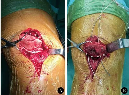
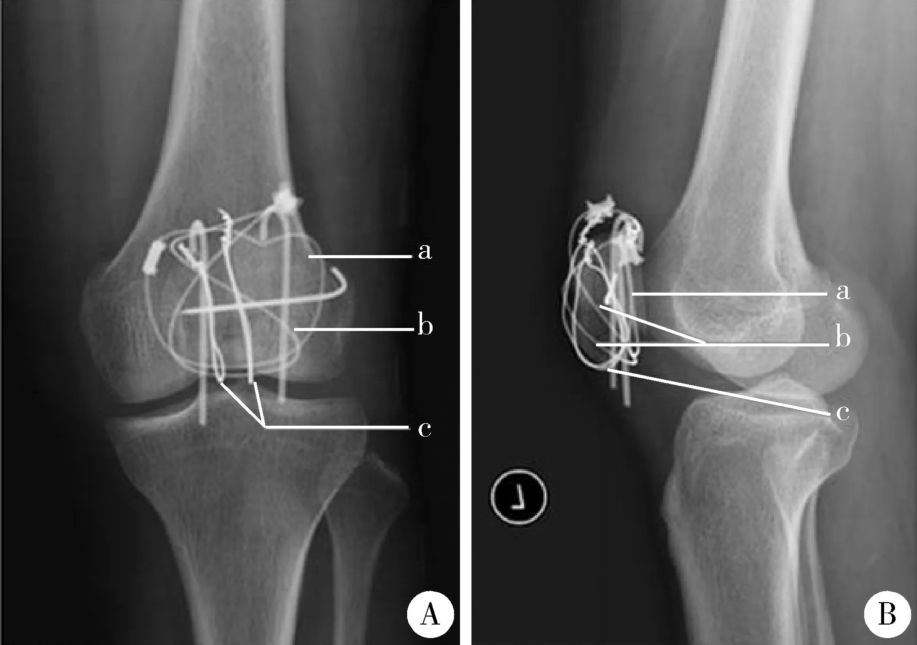
由于下极骨折块的宽度较小,本组病例均采用两枚纵向钢丝。如图2所示[1],首先使用2.0mm直径克氏针自近骨折端后方向髌骨的前、上方斜形钻孔,使用空心的套管针将直径1.2mm的钢丝带过针孔(图3),以相同方法使用套管针在紧贴下极骨折块下方穿过髌腱,并将钢丝带过。伸膝使远、近骨折端靠近,使用巾钳复位骨折端,采用标准技术将钢丝互相缠绕勒紧、加压以复位骨折端,并可使骨折端的复位得以维持。为减少内固定物对前方软组织的激惹反应,常规将钢丝的结置于髌骨上缘并埋于股四头肌腱内。X线透视下证实复位后,再使用标准的克氏针张力带技术,分别固定两枚平行克氏针及“8”、“O”型钢缆,固定后被动屈曲膝关节至130°以证实骨折端稳定性(图4)。

A, a number one Steinmann pin with a small hole at its end was inserted vertically from the antero-superior aspect of the patella to the most posterior aspect of the transverse osteotomy; B, a 0.75 mm diameter wire suture was passed through the hole in the Steinmann pin until the tip emerged from the osteotomy site and the Steinmann pin was then withdrawn with the wire; C, the distal end of the wire was passed through the patellar tendon as close as possible to the bone from the posterior aspect of the two bone fragments; D, the distal end of the wire was then pulled anteriorly, twisted, and tightened with the proximal end at the anterosuperior aspect of the patella. This procedure successfully reduced the number of fragments.图2 垂直钢缆操作示意图Figure 2 The separate vertical wiring technique

A, articular surface of patella was shown by turning around proximal fragment; B, vertical wires were passed through the hole drilled by Kirschner-wire; a, proximal fragment of patella fracture; b, inferior pole fragment of patella; white arrows showed the inferior margin of patella articula surface.图3 纵向钢丝捆绑结合克氏针张力带治疗髌骨下极粉碎骨折(上为头侧、下为尾侧)Figure 3 Separate vertical wiring combined with tension band and Kirschner-wire plus cerclage wire in the treatment of displaced inferior pole fractures of the patella (up is cranial, down is caudal)

图4 术后正位(A)、侧位(B)X线片,可见“O”型钢缆(a)、“8”字钢缆(b)及两枚纵向钢缆(c)Figure 4 Normal position (A) and lateral position (B) of postoperative radiographs showed: patella fracture was fixed by “O” shaped (a), “8” shaped (b) wires, and two vertical wires (c)
术后无需石膏制动,第3天开始功能康复,包括使用下肢关节功能恢复器(contionuouspassivemotion,CPM)进行被动屈膝训练及主动股四头肌肌力训练。术后第1周屈膝不超过45°,第2周逐渐增加至90°并允许部分负重,术后1个月复查X线片后开始逐渐增加负重,至术后6~8周达到完全屈曲。
2结果
术后1、2、3、6、12个月复查X线片判断骨折愈合情况,平均随访时间为13.1个月(12~19个月)。骨折线消失、连续骨小梁通过骨折部位判定为骨折愈合,平均愈合时间为9.8周(8~12周)。本组病例未出现感染、骨折不愈合、内固定物失效等并发症。9例患者于术后13.3个月(12~15个月)取出内固定,其中2例出现克氏针部分回退但无明显局部软组织刺激症状,另外7例无明显不适。
最终随访时,患者膝关节平均屈曲126.7°±6.9°,与对侧肢体相比,屈曲活动范围减少10.3°±8.8°。使用Böstman评分系统[2]对患者膝关节功能进行评定,该评分系统根据患者膝关节活动范围、疼痛、是否恢复伤前工作、大腿肌肉萎缩程度、行走是否需要辅助、跌倒病史、上楼梯等功能情况进行评分,满分30分,28~30分为优,20~27分为良,本组病例优良率达100%,平均(28.9±1.1)分。
3讨论
髌骨下极是髌骨前缘皮质的延续,并不带有关节面,此部位的骨折多是由于间接暴力造成的撕脱骨折,常同时合并直接暴力造成远端骨折块存在隐性骨折,这在本组病例中也得到了证实。髌骨下极骨折占所有髌骨骨折的9.3%~22.4%[3]。多数研究认为,在屈膝90°位置时髌骨位于股骨髁滑车内,位置相对比较固定,容易发生髌骨下极撕脱骨折[2]。由于局部骨折块较小、常伴有粉碎且骨质并不坚硬,因此解剖复位并维持复位和使用常规方式固定均比较困难。此种骨折治疗的主要目标应是重建伸膝装置连续性,而不是解剖复位骨性损伤结构,且重建后的伸膝装置要有足够的强度以耐受术后早期的功能训练。

表1 患者病历资料
ROM,rangeofmotion.
本组病例中下极骨折块宽为(23.1±5.2)mm,高为(15.4±3.0)mm,CT检查发现13例患者下极骨折块存在骨折线,因此克氏针张力带或空心螺钉结合张力带的方法很难有效固定,或因固定后不稳定而无法早期功能训练。目前仍没有针对髌骨下极粉碎骨折的理想治疗方式,常用的方法包括以下两种:(1)下极骨折块固定;(2)下极骨折块切除,髌腱起点重建。
采用下极骨折块切除,经骨隧道缝合[4]或使用缝合锚缝合[5]重建髌腱起点的方法在以往文献中均有报道,但对于髌腱起点重建的部位是贴近关节面还是贴近前侧皮质争议较多[4-6],除此之外,髌腱起点重建成功的关键是术后使用石膏或支具制动以保护重建部位,避免因股四头肌收缩造成重建失败。Kastelec等[7]使用骨块切除、髌腱起点重建的方法治疗了14例髌骨下极骨折,尽管最终随访疗效满意,但术后平均使用石膏固定患侧膝关节6.5周。为了能够早期功能锻练,一些研究建议贯穿髌骨、胫骨结节使用“O”或“8”型减张钢丝以保护重建部位[4,6-7],但精确恢复髌腱的生理长度比较困难,并可能引起髌骨倾斜,增加髌骨股骨关节应力,常导致钢丝断裂,远期可引发疼痛、加速关节退变等问题[6-7]。
为了使愈合更快、更坚固的“骨愈合”替代“腱-骨愈合”,尽量保留髌骨下极骨质,骨科医生发明了新的固定方法,使用篮状钢板固定粉碎髌骨下极骨折,Matejcic等[3]对其进行了改进,钢板远端加入了骨钩以更好地把持髌骨下极骨质,近端通过螺钉稳定固定于近端髌骨骨折块,固定稳定并可早期负重。但是,目前国内并无此类产品,而且由于钢板体积较大,髌骨前方软组织较薄,术后容易出现内固定物刺激症状,这也限制了此种方法的使用。
多个纵向钢缆捆绑固定最早由Yang等[1]提出,他们在尸体骨上制作了髌骨下极粉碎骨折的模型,分别使用两枚纵向钢缆捆绑与经骨减张缝合的方法固定,生物力学测试显示,纵向钢缆的强度明显高于经骨减张缝合。Yang等[1]使用此方法治疗了25例患者,平均随访22个月,骨折愈合率达到100%,无内固定物断裂。Song等[8]使用纵向钢缆捆绑结合环形减张钢丝的方法,固定强度优于单独使用纵向钢缆捆绑,但这种方法治疗的21例患者中出现4例减张钢丝断裂。Oh等[9]使用纵向钢缆捆绑结合Krackow方法编织缝合髌腱加强固定治疗了11例髌骨下极骨折患者,能够满足术后功能锻炼的要求,也取得了比较好的疗效,平均愈合时间10周,膝关节功能恢复满意。
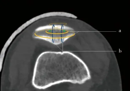
我们认为,本组病例采用的两枚纵向钢缆捆绑结合传统克氏针张力带的方法固定稳定性好,而且手术操作简便,首先,复位后可通过两枚纵向钢缆捆绑维持髌骨下极骨折的复位,重建伸膝装置的连续性,且不影响髌腱长度;其次,通过克氏针、张力带的方式使复位得到进一步加强,纵向钢缆与横行的张力带在髌骨下极组成类似网状结构(图5),可以非常有效地固定下极骨折块,对抗股四头肌腱的牵拉,因此,尤其适用于髌骨下极骨折块较小或骨折粉碎,其他方法难于有效固定而影响术后早期功能锻炼的病例。

图5 髌骨下极内固定示意图(仰视图)可见两横、两纵形成网状结构,a为“8”及“O”型钢缆,b为垂直钢缆Figure 5 A diagram (upward view) of the internal fixation in inferior pole of patella showed that a mesh structure was composed of the “O” shaped and “8”shaped wires (a), and two vertical wires (b)
本组病例在术中固定结束后,均做被动完全屈膝以确认固定的稳定性,术后即可主动、被动屈膝无需制动。随访未发现骨折复位丢失或内固定物断裂,膝关节屈伸功能恢复满意,患者满意度高。
本研究不足之处在于病例数不够充分,如能设计为前瞻性对照研究,说服力会更强。
综上所述,对于未累及关节面的髌骨下极粉碎骨折,可首先通过纵向钢缆捆绑复位并且维持复位,同时结合克氏针张力带固定,可提供非常可靠的骨性固定,操作简单、安全,术后膝关节功能恢复优良,疗效满意。
参考文献
[1]YangKH,ByunYS.Separateverticalwiringforthefixationofcomminutedfracturesoftheinferiorpoleofthepatella[J].JBoneJointSurgBr, 2003, 85(8): 1155-1160.
[2]BöstmanO,KiviluotoO,NirhamoJ.Comminuteddisplacedfracturesofthepatella[J].Injury, 1981, 13(3): 196-202.
[3]MatejcicA,PuljizZ,ElabjerE,etal.Multifragmentfractureofthepatellarapex:basketplateosteosynthesiscomparedwithpartialpatellectomy[J].ArchOrthopTraumaSurg, 2008, 128(4): 403-408.
[4]SaltzmanCL,GouletJA,McClellanRT,etal.Resultsoftreatmentofdisplacedpatellarfracturesbypartialpatellectomy[J].JBoneJointSurgAm, 1990, 72(9): 1279-1285.
[5]AnandA,KumarM,KodikalG.Roleofsutureanchorsinmanagementoffracturesofinferiorpoleofpatella[J].IndianJOrthop, 2010, 44(3): 333-335.
[6]MarderRA,SwansonTV,SharkeyNA,etal.Effectsofpartialpatellectomyandreattachmentofthepatellartendononpatello-femoralcontactareasandpressures[J].JBoneJointSurgAm, 1993, 75(1): 35-45.
[7]KastelecM,VeselkoM.Inferiorpatellarpoleavulsionfractures:osteosynthesiscomparedwithpoleresection[J].JBoneJointSurgAm, 2004, 86-A(4): 696-701.
[8]SongHK,YooJH,ByunYS,etal.Separateverticalwiringforthefixationofcomminutedfracturesoftheinferiorpoleofthepatella[J].YonseiMedJ, 2014, 55(3): 785-791.
[9]OhHK,ChooSK,KimJW,etal.InternalfixationofdisplacedinferiorpoleofthepatellafracturesusingverticalwiringaugmentedwithKrachowsuturing[J].Injury, 2015, 46(12): 2512-2515.
(2016-01-15收稿)
(本文编辑:任英慧)
Separate vertical wiring combined with tension band and Kirschner-wire plus cerclage wire in the treatment of displaced inferior pole fractures of the patella
ZHANG Jian△, JIANG Xie-yuan, HUANG Xiao-wen
(DepartmentofTraumaticOrthopedics,BeijingJishuitanHospital,Beijing100035,China)
ABSTRACTObjective:To investigate the clinical efficacy and outcomes of two separate vertical wiring combined with tension band and Kirschner-wire plus cerclage wire in the treatment of displaced inferior pole fractures of the patella. Methods: From January 2013 to January 2015, 15 consecutive patients (mean age 54.5 years) with inferior pole fractures of the patella were retrospectively included in this study. All the patients underwent open reduction and internal fixation by separate vertical wiring combined with tension band and Kirschner-wire plus cerclage wire through longitudinal incision, 4.5 d (range: 3.1-5.9 d) after initial injury. A safety check for early knee range of motion was performed before wound closure. The complications including infection, nonunion, loss of fixation and any wire breakage or irritation from implant were recorded. Anteroposterior and lateral views of the knee joint obtained during the follow-up were used to assess bony union based on the time when the fracture line disappeared. At the time of the final outpatient follow up, functional evaluation of the knee joint was conducted by Böstman system. Results: The follow-up time was 13.1 months (range: 12-19 months) after surgery on average, immediate motion without immobilization in all the cases was allowed and there was no case of reduction loss of the fracture and wire breakage. There was no case of irritation from the implant. At the final follow-up, the average range of motion (ROM) arc was 126.7° (range: 115°-140°), the average ROM lag versus contralateral healthy leg was 10.3° (range: 0°-35°). The mean Böstman score at the last follow-up was 28.9 (range: 27-30), and graded excellent in most cases. Conclusion: Two separate vertical wiring is an easy and effective method to reduce the displaced inferior pole fracture of patella. Augmentation of separate vertical wiring with tension band and Kirschner-wire plus cerclage wire in these patients provides enough strength to protected the early exercise of the knee joint and uneventful healing. By this surgical treatment, excellent results in knee function can be expected for cases of displaced inferior pole fractures of the patella.
KEY WORDSPatella; Fractures, bone; Fracture fixation, internal
[中图分类号]R683.42
[文献标志码]A
[文章编号]1671-167X(2016)03-0534-05
doi:10.3969/j.issn.1671-167X.2016.03.027
△Correspondingauthor’se-mail,zjzjbmu@139.com
网络出版时间:2016-5-1213:38:21网络出版地址:http://www.cnki.net/kcms/detail/11.4691.R.20160512.1338.034.html
