Relationship between arterial atheromatous plaque morphology and platelet-associated miR-126 and miR-223 expressions
2015-12-08HengSongTianQingGuoZhouFangShaoDepartmentofCardiologyAffiliatedHospitalofXinxiangMedicalCollegePingMeiShenmaMedicalGroupGeneralHospitalPingdingshanHenanProvinceChina
Heng-Song Tian, Qing-Guo Zhou, Fang ShaoDepartment of Cardiology, Affiliated Hospital of Xinxiang Medical College, Ping Mei Shenma Medical Group General Hospital, Pingdingshan, Henan Province, China
Relationship between arterial atheromatous plaque morphology and platelet-associated miR-126 and miR-223 expressions
Heng-Song Tian*, Qing-Guo Zhou, Fang Shao
Department of Cardiology, Affiliated Hospital of Xinxiang Medical College, Ping Mei Shenma Medical Group General Hospital, Pingdingshan, Henan Province, China
ARTICLE INFO
Article history:
Received 15 January 2015
Received in revised form 20 February 2015
Accepted 15 March 2015
Available online 20 April 2015
Platelets
miR-126
miR-223
Coronary heart disease
Atherosclerotic plaque
Objective: To study the expression of miR-126 and miR-223 in platelet of rabbit arterial plaque models, and explore its correlation with plaque morphology. Methods: Rabbit arterial plaque models were established, peripheral blood of models and control animals was collected. Plaque morphologies were divided into type Ⅰ, type Ⅱ and type Ⅲ based on angiography plaque morphology and Ambrose method. Platelet isolation kit was applied to isolate and purify peripheral blood platelets, CD45 immunomagnetic beads were used to remove the residual white blood cells. The miRNAs of platelets was extracted by miRNA Isolation Kit, and expressions of miR-126 and miR-223 of the platelets samples were detected by Real-time PCR. The correlation between plaque morphology and platelet-associated miR-126 and miR-223 expressions were analyzed. Expressions of target gene VCAM-1 and P2Y12 receptors of miR-126 and miR-223 in the atherosclerosis plaque of rabbit model were detected by Western blot. Results: Relative expression levels of miR-126 and miR-223 in the model group were 0.27 ±0.10 and 0.71±0.14, respectively. Plaque morphology was divided into types Ⅰ, Ⅱ and Ⅲ; and miR-126 and miR-223 expression levels were detected in each type. Expression levels of miR-126 in each type were 0.42±0.07, 0.17±0.11 and 0.22±0.15, respectively; and expression levels of miR-223 in each type are 0.68±0.02, 0.57±0.06 and 0.88±0.10, respectively. Relative to the control group, miR-126 and miR-223 known target genes in VCAM-1 and P2Y12 receptors increased platelets in rabbit atherosclerotic plaque models (P<0.05). Conclusions: Relative to normal control animals, miR-126 and miR-223 platelets were reduced in the rabbit atherosclerotic plaque model group (P<0.05). In the type Ⅱ plaque morphology group, miR-126 was greatly reduced; and there is no significant correlation between miR-223 and plaque morphology.
1. Introduction
Coronary heart disease is caused by coronary atherosclerosis, which leads to the occurrence of coronary artery atherosclerotic disease; and further causes myocardial ischemia, angina and myocardial infarction. Coronary heart disease has become a serious hazard to human health diseases. In recent years, studies for prevention and significant treatment of coronary heart disease have been undertaken from an anatomic and biochemical level of depth to the cell molecular level[1-3]. During hematopoiesis, miRNAs is an important class of negative regulator, which may be obtained by complementary matching the 3'-UTR region of target gene mRNA; leading to the inhibition received target mRNA translation; and furthermore participate in regulation of blood cell development, differentiation and apoptosis, hormone secretion, as well as biological processes such as signal transduction. Studies have
shown that thousands of human protein-coding genes are performed by miRNA regulation, demonstrating that miRNAs plays various important roles in cell growth and regulation during the development process[4,5].
Latest studies have shown that a number of miRNA expressions participated in regulating related gene expressions in platelets[6,7], and that platelets is an important factor in coronary heart disease and its development process. Therefore, studies conducted on platelet-associated miRNA expressions and their target gene regulatory process have important significance for understanding the involvement of platelets in relevant cardiovascular disease processes at the molecular level.
With vascular function, miR-126 and miR-223 are closely related miRNAs[8,9], which could express in vascular endothelial cells and platelets. There is a direct relationship between the abnormal expressions of numbers of related vascular cell proteins with the occurrence of atherosclerosis. Further, miRNA expressions may be different between different types of atherosclerotic plaque morphologies.
In this study, a rabbit atherosclerotic plaque model was established to investigate the relevance of platelet-associated miR-126 and miR-223 expressions with atherosclerotic plaque morphology; further understanding and providing some ideas on the role of miRNAs in the development of coronary artery diseases.
2. Materials and methods
2.1. Establishment of animal model
One hundred, healthy, adult, male New Zealand white rabbits purchased from Shanghai SLRC Laboratory animals Co., weighing 2.0-2.5 kg. Rabbits were randomly divided into two groups: 80 are for the atherosclerotic plaque group and 20 for the control group. Animals were fed and were allowed to freely consume water (high fat diet group); they were kept in standard animal cages with ventilation, natural day and night lighting, and a maintained temperature at 18-25 ℃.
2.2. Drug reagents and equipment
Maverick 2 balloon (Boston-38928-0920); Platelet pure separation kit (ShangHai HaLin g Biological Technology Co.Ltd. -HL10366.2); Dynabeads CD45 (lifetechnologies-11153D); miRNA Isolation Kit(mirVana-AM1560); miRNA Reverse Transcription Kit (TaqMan-4366596); Bulge-LoopmiRNA qRT-PCR Primer Set (Guangzhou RiboBio Co., Ltd.); Real-time PCR Fluorescence Quantitative Diagnosis Kits (SsoAdvanced SYBR Green Supermix, Bio-Rad-172-5264); ReadyPrep Protein Extraction Kits (Bio-Rad); P2Y12 antibody(Santa Cruz Biotechnology-sc27152); VCAM-1antibody (Santa Cruz Biotechnology-sc1504); Horseradish peroxidase (HRP) labeled secondary antibody (Wuhan Boster Biological Technology Co., Ltd.); ECL Color Development Kit (Millpore); PVDF Membrane (Polyvinylidene fluoride-Millpore); Color Doppler Ultrasonic Diagnosis Apparatus (Philips IU Elite); MultiSkan FC enzyme mark instrument (Thermo Scientific); Fluorescence RTPCR detect system CFX96 Touch (Bio-Rad).
2.3. Establishing the animal model
The rabbit model commenced after one week in adaptive feeding. Control animals were fed with a normal diet, while model animals were fed with a high fat diet (containing 2% cholesterol). Animals were fed according to a 150 g/day amount of feeding, continuously fed for eight weeks, and lipids were measured to determine high cholesterol formation. The rabbits were anaesthetized with 3% sodium pentobarbital by intravenous infusion. The balloons were inserted through the external carotid artery to induce intimal injury. Antibiotic was administered intramucularly to prevent postoperative infection, then continued to fed for 4 weeks; Philips IU Elite color Doppler ultrasound was used to examine the left and right carotid arteries. Transverse scans were conducted along the vessel, so as to observe whether there is intimal thickening and the size of plaque and characteristics of echo. Puncture needles were introduced percutaneously into the femoral artery or radial artery, a special catheter was sent along the descending aorta retrograde to the aortic root, and the catheter was placed in the left and right coronary ostia, respectively. Then, Iopromide was injected and X-ray video was turned on at the same time. There are 74 cases of coronary angiography for atherosclerosis or vascular blockage for the study. According to the Ambrose aliquot type method[10,11], plaques were divided into three types according to morphological expressions: type Ⅰ, concentric or eccentric plaques with smooth surface and plaque with a wide base; type Ⅱ, plaque has a narrow base or cuspshaped (both surface smooth or matte), plaque surface have shadow niches, plaque surface uneven, or were wavy or crater-like (radial or bias); type Ⅲ, long segments of irregularly narrow plaques.
2.4. Platelet isolation and purification
Venous blood containing anticoagulants were collected in blood collection tubes; and stored at room temperature after obtaining platelets by centrifugation, according to high purity platelet isolation kit instructions. Dynabeads CD45 MACS kit was used to purify peripheral blood platelets according to manufacturer's instructions; precipitation was obtain after removal of platelet leukocytes.
2.5. Expressions of miR-126 and miR-223 by Real-time PCR
Platelet total miRNA was extracted by miRNA Isolation Kit and stored at -80 ℃. RNA solution A260 was detected by UV spectrophotometer and RNA concentration was calculated, RNA purity was evaluated by OD260/OD280ratio. According to the TaqMan miRNA Reverse Transcription Kit, miRNA-specific primers were used to conduct reverse transcription. Reverse transcription was only conducted to mature miRNA instead of pre-mirna, and its transcript CDNA was used as a template. Expressions of miR-126 and miR-223 were detected by Real-time PCR. 2-△Ctvalue method was used to calculate relative expression levels of target genes, average of three replicate experiments was the Ct value for each sample, △Ct =Ct(Target gene)-Ct(internal reference ), △△Ct= △Ct(sample) -△Ct (contro1), so relative expression levels of target gene = 2-△△Ct, relative expression of the control group is 20 = 1, reference gene is U6.miR-126, miR-223 and U6-specific primers were all purchased from Guangzhou Ribobio company. CFX Manager software was used to analyze Real-time PCR results. PCR reaction system is showed in Table 1.
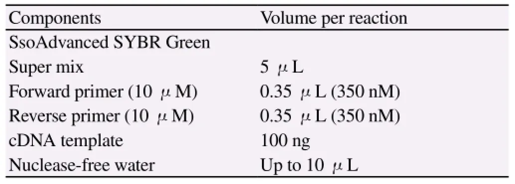
Table 1 PCR reaction system.
2.6. Western blot
Platelet samples were collected and washed two times with PBS. Cells were filtered with 1 mL pipette, and the supernatant was removed by centrifugation. Platelet precipitation was lysed by ReadyPrep protein extraction kit lysate, then protease inhibitor cocktail was added, mixed by pipetting. After the samples were placed on ice for 30 minutes, cells were lysed by ultrasonic waves. The lysed mixture was placed at 4 ℃ and centrifuged at 13 000 r/ min for 20 minutes; protein concentrations in the new supernatant were determined using a Protein Assay Kit. Protein samples extraction was performed by SDS-PAGE electrophoresis. The gel was immersed into transfer buffer equilibrium for 10 minutes, and plugged the electrode at 100 V for 45-60 minutes. After the film was transferred, PVDF membrane was rinsed with TBS for 10-15 minutes. The film was placed in a TBS/T blocking buffer containing 5% (w/v) skimmed milk powder, shaking for one hour at room temperature, appropriate dilution of primary antibody [containing 1% (w/v) skimmed milk powder in TBS/T dilution] was added, incubated at room temperature for two hours, and TBST rinsed the membrane three times every 5-10 minutes. The film was incubated in a TBST diluted secondary antibody (1:10 000, HRP labeled) containing 0.05% (w/v) nonfat dry milk at room temperature for one hour. TBS/T rinsed the film three times every 5-10 minutes, exposed, took a photo, and experiment results were saved. Repeated experiment for three times. Quantity one v4.62 software molecular bands were used for gray value (Trace Tracking), drawing the optical density curve according to different subunit bands. Areas under the optical density curves were calculated as a quantity reference for electrophoretic bands and results were statistically analyzed.
2.7. Statistical analysis
SPSS 17.0 statistical software was applied to the experimental data, and results were expressed as mean±SD. t-test was used to compare test the two groups. P<0.05 was considered statistically significant.
3. Result
3.1. Expressions of miR-126 and miR-223 in platelet samples were detected by real-time PCR
A total of 74 rabbits atherosclerotic plaque model were successfully established and 20 rabbits were used as healthy controls. Platelets were obtained after isolation and purification of peripherals. Total miRNAs was extracted by kit, and then reverse transcribed into cDNA. Expressions of miR-126 and miR-223 were detected by Real-time PCR. Figure 1 is PCR reaction amplification curve (Amplification curve of control group and model group, miR-126 group and miR-223 group respectively). The result was shown in Figure 2.
Figure 3 showed the data distribution of miR-126 and miR-223 in three kinds of plaque morphology, and correlation between plaque morphology and miR-126 and miR-223 expressions. That is,miR-126 expression is the lowest in Type Ⅱ plaque morphology group; however, miR-126 expression is evenly distributed in type Ⅰ plaque morphology group, there is no significant correlation, and there is no significant correlation between miR-223 and three plaque morphology. A, Relative expression of miR-126 and miR-223 (2-△△Ctmethod),wherein relative expression levels of miR-126 and miR-223 in the model group were 0.27±0.1 and 0.71±0.14, respectively. B-C. Plaque morphology was divided into types Ⅰ, Ⅱ and III; and miR-126 and miR-223 expression levels were detected in each type.
Expression levels of miR-126 in each type are 0.42±0.07, 0.17± 0.11 and 0.22±0.15, respectively; and expression levels of miR-223 in each type are 0.68±0.02, 0.57±0.06 and 0.88±0.10, respectively. D, Detected the internal reference U6 expression in samples of each group.
3.2. Detection of VCAM-1 and P2Y12 receptors in the plaque by Western blotting
Total protein of platelet samples in each group was extracted by kit,and expressions of target gene VCAM-1 and P2Y12 receptors of miR-126 and miR-223 were detected by Western blot. As shown in figure 3, the VCAM-1 and P2Y12 receptors in the healthy control group were in a low level, however,the results showed that VCAM-1 and P2Y12 receptors in platelets were significantly increase in the model group (P<0.05). Table 2 is analysis of relative signal value of the target protein through protein bands signal value of internal reference by Quantity one software.
4. Discussion
Coronary heart disease is caused by coronary atherosclerosis, which leads to the occurrence of coronary artery atherosclerosis; and further cause myocardial ischemia, angina and myocardial infarction. The pathogenesis of coronary heart disease remains unclear; thus, there is a lack of effective treatment. Thrombosis is an important incentive for cardiovascular disease, which is currently widely accepted in cardiovascular research[12,13]. With the in-depth study of thrombosis, the role played by thrombosis in platelets would be clearer. A reliable animal model of atherosclerosis has important significance to ascertain the pathogenesis of atherosclerosis and for the research and development of prevention and treatment drugs. Rabbits are herbivores; and under normal conditions, high cholesterol rarely occurs. In this study, high-fat diet caused high cholesterol. To establish a rabbit atherosclerotic plaque model, a balloon was used and inserted into the artery. This caused intimal tear during the experiment. Postoperative infection is the main reason for the failure of this model. Some animals also failed to induce plaque formation. miRNAs are short and small non-coding RNA molecules that are similar to highly conservative siRNA molecules. miRNAs are equivalent 20-25 nucleotides in length and regulate posttranscriptional mRNA expressions, which are usually complementary to miRNAs binding sequence in the 3 ‘untranslated region (3'-UTR); and leads to silence suppression and gene conversion. Studies have
shown that thousands of human protein-coding genes are carried out by miRNA regulation, indicating that miRNAs plays an important role in a variety of processes in cell growth and development. As miRNAs research techniques continues to develop, platelet miRNA, as the central gene expression regulatory mechanism, has presently become a focus in cardiovascular disease[14-16]. Platelets are released into the peripheral circulation of blood, after bone marrow megakaryocyte shedding. Their seedless characteristics determine the synthesis and mechanism of action of platelet miRNAs, which may differ from the regulatory mechanisms of miRNAs in nucleated cells. The study shows that miRNAs is indeed involved in regulating expressions in a number of genes in platelets; therefore, we believe platelets miRNAs can be used as a cardiovascular disease platelet activation level for monitoring, diagnosis, prognosis and efficacy evaluation index, while clinical gene therapy for thrombotic diseases is also possible.
According to the classification method of Ambrose et al[10,11], these were divided into three types according to plaque morphology performance. Platelet samples were isolated by platelets isolation kit and residual leukocytes were removed using CD45 magnetic beads to purify the platelets. Total miRNAs were extracted from platelet samples and reverse transcribed into cDNA. Expression levels of miR-126 and miR-223 in platelets were detected by real-time PCR. From the results, we found that miR-126 in animal platelets of the model group significantly reduced (P<0.01) and miR-223 has also shown a downward trend (P<0.05); suggesting that the abnormal expression of miR-126 and miR-223 can be related to the occurrence of atherosclerotic plaques. Thus, we also further detected VCAM-1 and P2Y12 receptors in miR-126 and miR-223 known target genes by Western blotting. The results showed that VCAM-1 and P2Y12 receptors in platelets had different degrees of increase in the model group. The negative correlation in Real-time PCR results prompted the low expressions of miR-126 and miR-223; and raised the target gene expressions of VCAM-1 and P2Y12 in platelets; thus, exerting its biological effects. Although the regulatory processes of miRNAs are complex, same miRNAs may regulate the expression of multiple genes; simultaneously, a gene is often regulated by multiple miRNAs[17]. However, VCAM-1 and P2Y12 genes in platelets in the animal model group significantly increase, which may be caused by the abnormal expressions of miR-126 and miR-223. Using simulated endogenous miRNAs: miRNA mimic, and miRNAs inhibitors: to have a clearer understanding of miRNA inhibitors in regulating miR-126 and miR-223 in platelets; this would be the focus of our future research.
In order to investigate the correlation of miR-126 and miR-223 expression levels with plaque morphology, we grouped platelet samples according to different plaque morphology and detected the expressions of miR-126 and miR-223. From the results, we can conclude that miR-126 was underexpressed in platelets in the model group; and their expressions were not the same in different plaque morphologies. Type Ⅱ had the lowest (P<0.01), type Ⅰ had the highest (P<0.05) and type Ⅲ had intermediate expression levels of miR-126 in the different plaque morphology groups. This may be related to the miR-126 regulation of vascular endothelial growth factor (VEGF) expression[18,19]. VEGF can nourish atherosclerotic plaques proliferation during blood vessel growth; thus, miR-126 has a certain correlation with coronary stenosis. In this study, miR-126 target gene VCAM-1 was detected by triggering local immunity and release of various cytokines; thus, promoting the formation of early atherosclerotic plaques[20]. In the type Ⅲ plaque morphology group, miR-223 expressed in higher; and the remaining difference between the two groups were not significant. As one of their target genes in P2Y12 platelets, ADP receptor is a central regulating terminal of platelet activation control[21]. Thus, the abnormal expression of miR-223 was the same in platelets in the model group; suggesting that this plays an important role in the occurrence and development of atherosclerotic plaque formations.
With platelet miRNAs findings and their regulatory mechanisms of gene expression revealed, further studies on platelet effects on cardiovascular disease provides new ideas; and also for building a new platform for platelet internal molecular mechanisms. Identifying the important functions platelet miRNAs and function of its target genes would become an important research direction in platelet research.
Conflict of interest statement
We declare that we have no conflict of interest.
[1] Lesiak M, Araszkiewicz A. “Leaving nothing behind”: is the bioresorbable vascular scaffold a new hope for patients with coronary artery disease? Postepy Kardiol Interwencyjnej 2014; 10(4): 283-288.
[2] Rossi MA, Tanowitz HB, Malvestio LM, Celes MR, Campos EC, Blefari V, et al. C oronary microvascular disease in chronic Chagas cardiomyopathy including an overview on history, pathology, and other proposed pathogenic mechanisms. PLoS Negl Trop Dis 2010; 4(8): pii: e674.
[3] Münzel T, Schulz E. Treatment of coronary heart disease with nitric oxide donors. Pharm Unserer Zeit 2010; 39(5): 359-368.
[4] Chen B, Li H, Zeng X, Yang P, Liu X, Zhao X, et al. Roles of miRNA on cancer cell metabolism. J Transl Med 2012; 10: 228.
[5] Zhu L, Liu J, Cheng G. Role of miRNAs in schistosomes and
schistosomiasis. Front Cell Infect Microbiol 2014; 4: 165.
[6] Schmidt Y, Simunovic F, Strassburg S, Pfeifer D, Stark GB, Finkenzeller G. miR-126 regulates platelet-derived growth factor receptor-α expression and migration of primary human osteoblasts. Biol Chem 2015; 396(1): 61-70.
[7] Duan X, Zhan Q, Song B, Zeng S, Zhou J, Long Y, et al. Detection of platelet microRNA expression in patients with diabetes mellitus with or without ischemic stroke. J Diabetes Complications 2014; 28(5): 705-710.
[8] Qiang L, Hong L, Ningfu W, Huaihong C, Jing W. Expression of miR-126 and miR-508-5p in endothelial progenitor cells is associated with the prognosis of chronic heart failure patients. Int J Cardiol 2013; 168(3): 2082-2088.
[9] Zhao Y, Samal E, Srivastava D. Serum response factor regulates a muscle-specific microRNA that targets Hand2 during cardiogenesis. Nature 2005; 436(7048): 214-220.
[10] Ambrose JA, Winters SL, Stern A, Eng A, Teichholz LE, Gorlin R, et al. Angiographic morphology and the pathogenesis of unstable angina pectoris. J Am Coll Cardiol 1985; 5(3): 609-616.
[11] Lo YS, Cutler JE, Blake K, Wright AM, Kron J, Swerdlow CD. Angiographic coronary morphology in survivors of cardiac arrest. Am Heart J 1988; 115(4): 781-785.
[12] Zander T, Medina S, Montes G, Nuñez-Atahualpa L, Valdes M, Maynar M. Endoluminal occlusion devices: technology update. Med Devices (Auckl) 2014; 7: 425-436.
[13] Burke EL, Walvekar RR, Lin J, Hagan J, Kluka EA. Common agents used to unblock blood clots within tympanostomy tubes: an ex vivo study and review of literature. Int J Pediatr Otorhinolaryngol 2009; 73(12): 1725-1728.
[14] Luo M, Ren MP, Wu JB. The platelet micro RNA research pogress. Chin J Cardiol 2012; 40(8).
[15] Leidinger P, Backes C, Dahmke IN, Galata V, Huwer H, Stehle I, et al. What makes a blood cell based miRNA expression pattern disease specific? - A miRNome analysis of blood cell subsets in lung cancer patients and healthy controls. Oncotarget 2014; 5(19): 9484-9497.
[16] Landry P, Plante I, Ouellet DL, Perron MP, Rousseau G, Provost P. Existence of a microRNA pathway in anucleate platelets. Nat Struct Mol Biol 2009; 16(9): 961-966.
[17] Lu TP, Lee CY, Tsai MH, Chiu YC, Hsiao CK, Lai LC, et al. miRSystem: an integrated system for characterizing enriched functions and pathways of microRNA targets. PLoS One 2012; 7(8): e42390.
[18] Hong F, Li Y, Xu Y. Decreased placental miR-126 expression and vascular endothelial growth factor levels in patients with pre-eclampsia. J Int Med Res 2014; 42(6): 1243-1251.
[19] Zhu X, Li H, Long L, Hui L, Chen H, Wang X, et al. miR-126 enhances the sensitivity of non-small cell lung cancer cells to anticancer agents by targeting vascular endothelial growth factor A. Acta Biochim Biophys Sin (Shanghai) 2012; 44(6): 519-526.
[20] Kim E, Cook-Mills J, Morgan G, Sredni ST, Pachman LM. Increased expression of vascular cell adhesion molecule 1 in muscle biopsy samples from juvenile dermatomyositis patients with short duration of untreated disease is regulated by miR-126. Arthritis Rheum 2012; 64(11): 38093-3817.
[21] Buriakina TA, Zateĭshchikov DA. The role of direct-acting P2Y12 inhibitors in acute coronary syndrome. Kardiologiia 2012; 52(4): 74-79.
ment heading
10.1016/S1995-7645(14)60336-9
*Corresponding author: Heng-Song Tian, Associate Chief Physician, Department of Cardiology, Affiliated Hospital of Xinxiang Medical College, Ping Mei Shenma Medical Group General Hospital ,Pingdingshan, Henan Province, China.
Tel: 13903750327
E-mail: ths-2008@163.com
Foundation project: This project is supported by scientific and technological project of Henan province, No. : 132102310095.
杂志排行

Asian Pacific Journal of Tropical Medicine的其它文章
- A brief review on biomarkers and proteomic approach for malaria research
- Trigonelline protects the cardiocyte from hydrogen peroxide induced apoptosis in H9c2 cells
- In vitro cholinesterase inhibitory and antioxidant effect of selected coniferous tree species
- Monascus pilosus-fermented black soybean inhibits lipid accumulation in adipocytes and in high-fat diet-induced obese mice
- Antiprotozoal assessment and phenolic acid profiling of five Fumaria (fumitory) species
- Profile and geographical distribution of reported cutaneous leishmaniasis cases in Northwestern Saudi Arabia, from 2010 to 2013