V型根管的分布
2015-11-21余顺兰王斯思王笑喆
余顺兰, 王斯思, 王笑喆
(北京大学口腔医学院预防科, 北京 100081)
V型根管的分布
余顺兰, 王斯思, 王笑喆
(北京大学口腔医学院预防科, 北京 100081)
目的: 观察Ⅴ型根管的牙位分布。方法:选取2013-12—2015-02进行CBCT检查的患者198例共2 984个牙为研究对象,对具有Ⅴ型根管的牙位进行观察、记录和分析。结果:1 306个前牙中,下颌切牙和尖牙Ⅴ型根管出现率分别为3.36%和2.24%,而上颌前牙均未出现;897个前磨牙中,Ⅴ型根管出现比率最高的为上颌第二前磨牙5.83%,其次为下颌第一前磨牙4.46%;781个磨牙中,Ⅴ型根管出现率第二磨牙(5.88%)较第一磨(0.52%)牙高。结论:Ⅴ型根管在较多牙位都可能出现,上颌第二前磨牙、下颌第一前磨牙以及第二磨牙出现Ⅴ型根管的可能性最高。
CBCT; 根管治疗; Ⅴ型根管
[DOI] 10.15956/j.cnki.chin.j.conserv.dent.2015.12.008
[Chinese Journal of Conservative Dentistry,2015,25(12): 736]
Vertucci分类法[1]分析的根管构型中,Ⅴ型根管(1-2型)在釉牙骨质界水平只有1个根管,在根中1/3或根尖1/3分成2个根管。这种根管类型在临床根管治疗中易被误认为是I型根管,常因根管遗漏而导致根管治疗失败。因此,本研究通过CBCT分析Ⅴ型根管的临床牙位分布,以期为临床根管治疗提供参考。
1 临床资料和方法
1.1 临床资料
选取我院自2013-12—2015-02进行CBCT检查的患者198例共2 984个牙为研究对象,对具有Ⅴ型根管的牙位进行观察、记录和分析。其中男性92例,女性106例;年龄17~80岁,平均年龄41岁;前牙1 306个,前磨牙897个,磨牙781个。 纳入标准:①根尖发育完成;②图像清晰可辨;③牙列基本完整,牙位可准确辨析。排除第三磨牙。
1.2 方法
图像均采用 NewTom VG CBCT (QR,意大利) 进行拍摄,技术参数:电压110 kV,自动曝光模式,图像重建的层厚和层间距均为0.15 mm。将CBCT图像轴位唇、舌侧釉质影像消失的平面,以及矢状位唇、舌侧釉质颈部下缘连线判定为釉牙骨质界。从釉牙骨质界往根尖方向连续观察,分析并记录Ⅴ型根管出现的牙位及牙根分布。
2 结果
CBCT图像分析结果可见,Ⅴ型根管在上颌第二前磨牙、下颌第一前磨牙和第二磨牙以及下颌前牙出现的比率相对较高(表1~3)。附典型CBCT影像(图1~4)。
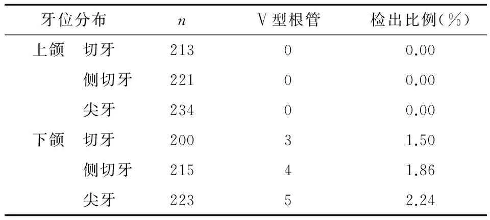
表1 前牙组Ⅴ型根管分布 (牙)
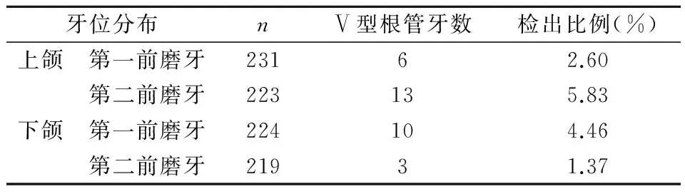
表2 前磨牙组Ⅴ型根管的牙齿分布 (牙)

表3 磨牙组Ⅴ型根管的牙齿分布 (牙)

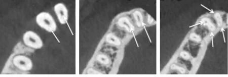
a.根颈1/3,1个根管 b.根中1/3,1个根管 c.根尖1/3,2个根管
图1 25 Ⅴ型根管


a.根颈1/3,1个根管 b.根中1/3,1个根管c.根尖1/3,2个根管
图2 44 Ⅴ型根管

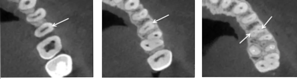
a.根颈1/3,颊根1个根管 b.根中1/3,颊根2个根管c.根尖1/3:颊2个根管
图3 27颊根Ⅴ型根管

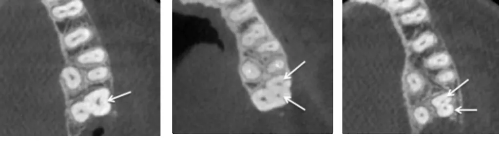
a.根颈1/3,1个根管 b.根中1/3,1个根管c.根尖1/3,2个根管
图4 42、43 Ⅴ型根管
3 讨论
本结果显示,前牙组中,638个下颌前牙中有13个出现Ⅴ型根管,而上颌前牙均未出现。Han和赵莹等[2-3]在对下颌前牙的CBCT图像进行观察时,也证实下颌前牙都有可能出现Ⅴ型根管,但检出比率和本结果略有不同。
前磨牙组中,每个前磨牙都可能出现Ⅴ型根管,出现比率最高的是上颌第二前磨牙(5.83%, 13/223),其比率在本研究的所有牙位里也是最高的。Yang等[4]在利用CBCT研究上颌第二前磨牙的根管构型时观察到的Ⅴ型根管比率与此相似,为6.4%。其次是下颌第一前磨牙(4.46%, 10/224), 而下颌第二前磨牙Ⅴ型根管出现最少(本研究中219个下颌第二前磨牙只有3个出现Ⅴ型根管)。与此相似,Yu等[5]利用CBCT研究下颌前磨牙时,178个下颌第二前磨牙中,出现Ⅴ型根管的有3个;而174个单根的下颌第一前磨牙中,则有17个牙出现Ⅴ型根管。
磨牙组中, Ⅴ型根管主要出现在上颌第二磨牙和下颌第二磨牙,而第一磨牙很少出现。6例Ⅴ型根管出现的上颌第二磨牙中,有4例出现在颊腭两根的颊根,2例出现在具有3根的近中颊根,未在腭根发现Ⅴ型根管。这个结果与Kim等[6]研究的775个上颌第二磨牙根管构型的结果近似。5例Ⅴ型根管出现的下颌第二磨牙,均出现在近中根。Blige等[7]在观察下颌第二磨牙的根管构型时发现,Ⅴ型根管主要出现在近中根,1 165个下颌第二磨牙中,近中根出现Ⅴ型根管的共有60个,而远中根只有3个出现Ⅴ型根管。第一磨牙较少出现Ⅴ型根管,本研究只检出1个下颌第一磨牙的近中根呈现Ⅴ型根管。
在本研究未检出Ⅴ型根管的牙位和牙根中,也有出现Ⅴ型根管的罕见个案病例报道和研究。Calvert[8]发现1例Ⅴ型根管的右上切牙;Holderrieth等[9]在临床根管治疗中发现两例上颌第二磨牙的腭根出现Ⅴ型根管;另有研究[10-12]发现,下颌第一、二磨牙的远中根也可能出现Ⅴ型根管等。
综上所述,Ⅴ型根管可能出现的牙位分布比较广泛,其中以上颌第二前磨牙、下颌第一前磨牙以及第二磨牙Ⅴ型根管出现率最高,对于这些牙位,临床根管治疗时应仔细观察初诊根尖片、初尖锉方向的偏斜角度,并使用根管显微镜观察根管中下部的细微结构,必要时拍摄CBCT,避免遗漏,以达到完善的根管治疗效果。
[1]Vertucci FJ. Root canal anatomy of the human permanent teeth [J].OralSurgOralMedOralPathol, 1984, 58(5): 589-599.
[2]Han T, Ma Y, Yang L,etal. A study of the root canal morphology of mandibular anterior teeth using cone-beam computed tomography in a Chinese subpopulation [J].JEndod, 2014, 40(9): 1309-1314.
[3]赵莹, 董颖韬, 王晓燕,等. 4674颗下颌前牙根管构型的锥形束CT分析 [J] . 北京大学学报(医学版), 2014, 46(1): 95-99.
[4]Yang L, Chen X, Tian C,etal. Use of cone-beam computed tomography to evaluate root canal morphology and locate root canal orifices of maxillary second premolars in a Chinese subpopulation [J].JEndod, 2014, 40(5): 630-634.
[5]Yu X, Guo B, Li KZ,etal. Cone-beam computed tomography study of root and canal morphology of mandibular premolars in a western Chinese population [J].BMCMedImaging, 2012, doi:10.1186/1471-2342-12-18.
[6]Kim Y, Lee SJ, Woo J. Morphology of maxillary first and second molars analyzed by cone-beam computed tomography in a Korean population: variations in the number of roots and canals and the incidence of fusion [J].JEndod, 2012, 38(8): 1063-1068.[7]Bilge GN, Evren O, Mustafa A,etal. Evaluation of the root and canal morphology of mandibular permanent molars in a southeastern Turkish population using cone-beam computed tomography [J].EurJDent, 2014, 8(2): 154-159.
[8]Calvert G. Maxillary central incisor with type V canal morphology: case report and literature review [J].JEndod, 2014, 40(10): 1684-1687.
[9]Holderrieth S, Gernhardt CR. Maxillary molars with morphologic variations of the palatal root canals: a report of four cases [J].JEndod, 2009, 35(7): 1060-1065.
[10]Peiris HR, Pitakotuwage TN, Takahashi M,etal. Root canal morphology of mandibular permanent molars at different ages [J].IntEndodJ, 2008, 41(10): 828-835.
[11]Harris SP, Bowles WR, Fok A,etal. An anatomic investigation of the mandibular first molar using micro-computed tomography [J].JEndod, 2013, 39(11): 1374-1378.
[12]Filpo-Perez C, Bramante CM, Villas-Boas MH,etal. Micro-computed tomographic analysis of the root canal morphology of the distal root of mandibular first molar [J].JEndod,2015, 41(2): 231-236.
Distribution of Vertucci type Ⅴ canal configuration
YU Shun- lan, WANG Si- si, WANG Xiao- zhe
(DepartmentofPreventiveDentistry,PekingUniversitySchoolandHospitalofStomatology,Beijing100081,China)
AIM: To investigate the distribution of Vertucci type Ⅴ canal configuration . METHOD: 2984 teeth in 198 patients were examined by cone- beam computed tomographic (CBCT) imaging and the distribution of Vertucci type Ⅴ canal configuration was recorded and analyzed. RESULTS: Type Ⅴ canal configuration was found in both mandibular incisors(3.36%) and canines(2.24%), but not in maxillary anterior teeth. Of the premolars, the prevalence in the second maxillary premolars (5.83%) was the highest, followed by the first mandibular premolars (4.46%). Of the molars,type Ⅴ canal configuration was frequently found in the second molars(5.88%), but rarely in the first molars(0.52%). CONCLUSION: Type Ⅴ canal configuration can be found in many teeth, especially in maxillary second premolars, mandibular first premolars and second molars.
cone- beam computed tomography(CBCT); root canal treatment; vertucci type Ⅴ canal configuration
2015-03-04
余顺兰(1978-),女,汉族,福建人。本科,主治医师
R780.4
A
1005-2593(2015)12-0736-04