COX-2与Survivin蛋白在人成釉细胞瘤中的表达及意义
2015-08-23李乐张兴乐王鹏刘雨
李乐,张兴乐,王鹏,刘雨
COX-2与Survivin蛋白在人成釉细胞瘤中的表达及意义
李乐1,张兴乐1,王鹏1,刘雨2
目的研究环氧化合酶-2(COX-2)和生存素(Survivin)在人成釉细胞瘤(AB)中的表达情况。方法收集60例AB患者的肿瘤组织和正常口腔黏膜组织。60例AB患者中原发40例,复发20例;组织学分型:棘皮瘤型12例,滤泡型18例,丛状型18例,基底细胞型6例,促结缔组织增生型6例。采用免疫组化法和蛋白质免疫印迹法检测AB和正常口腔黏膜中COX-2和Survivin的表达,比较不同组织及分型的AB表达情况。结果AB中COX-2蛋白的阳性表达率(82.7%)高于正常口腔黏膜(10.0%),Survivin表达阳性率(83.3%)高于正常口腔黏膜(16.7%)。AB 中COX-2蛋白表达水平为0.781±0.142,高于正常口腔黏膜组织的0.179±0.056;Survivin蛋白表达水平为0.447± 0.139,高于正常口腔黏膜组织的0.072±0.017,差异均有统计学意义。COX-2与Survivin的表达呈正相关(r=0.778,P<0.05)。不同组织学分型AB中的COX-2和Survivin表达阳性率差异无统计学意义。结论COX-2、Survivin在AB中高表达,可能与AB的发生发展有关。
成釉细胞瘤;环氧化合酶-2;生存素;免疫组织化学;印迹法,蛋白质
人成釉细胞瘤(ameloblastoma,AB)是常见的牙源性肿瘤,其发生发展的分子生物学机制尚不明了[1]。有研究发现,在胰头癌、胃癌、结肠癌、肺癌、乳腺癌中及口腔鳞癌中均有环氧化合酶-2(cyclooxygenase-2,COX-2)和生存素(Survivin)蛋白的表达,两者皆可以促进肿瘤细胞的增殖,抑制肿瘤细胞凋亡,参与肿瘤血管形成,与肿瘤的发生发展关系密切[2]。但有关这2种蛋白在AB中的研究较少,其与AB发生发展的关系尚不明确。本实验采用免疫组化法和蛋白免疫印迹法(Western Blot)法检测AB组织和正常口腔黏膜中COX-2、Survivin的蛋白表达,探讨两者在AB发生发展过程中起到的作用。
1 资料与方法
1.1一般资料选择2007年1月—2013年12月在我院口腔颌面外科经手术治疗的AB患者60例(AB组),男33例,女27例,年龄12~70岁,平均(45.1±5.2)岁。60例AB患者中,原发40例,复发20例(未选择相同患者)。部位:下颌骨42例,上颌骨18例。组织学分型:棘皮瘤型12例,滤泡型18例,丛状型18例,基底细胞型6例,促结缔组织增生型6例。以WHO在2002年发布的《头颈部肿瘤病理学与遗传学》中牙源性肿瘤分类作为本研究的病理诊断标准[3],所有患者均未患其他肿瘤,所有肿瘤HE切片均经我院病理科2位教授共同复读确认。选择同期行唇裂手术患者的正常唇红黏膜组织60例作为对照组。
1.2主要试剂SP-9000试剂盒、浓缩型DAB显色剂、0.01 mol/L的PBS缓冲液、BCA蛋白浓度测定试剂盒均购自福州迈新生物技术开发公司。鼠抗人COX-2、Survivin、β-actin单克隆抗体均购自美国Santa Cruz公司。二抗购自北京博奥森公司。PVDF膜、化学发光液购自美国Milipore公司。
1.3方法
1.3.1免疫组化法测COX-2、Survivin在AB和正常口腔黏膜中的表达标本经10%中性福尔马林固定,常规石蜡包埋,4 μm连续切片。采用免疫组化S-P法检测AB和正常口腔黏膜组织中COX-2和Survivin的蛋白表达,DAB法显色。阳性对照为口腔鳞癌,阴性对照采用PBS代替一抗。于显微镜下观察切片,胞浆中出现黄色颗粒为阳性着色,读取任意5个高倍镜视野进行判断。A(细胞显色深浅评分):0分为细胞无色;1分为浅黄色;2分为棕黄色;3分为棕褐色。B(显色瘤细胞比例评分):1分为≤25%;2分为>25%~75%;3分为>75%。最终每例积分=A×B[4],按积分高低分为:阴性(-),积分0分;弱阳性(+),积分1~4分;中度阳性(++),积分>4~7分;强阳性(+++),积分>7。
1.3.2Western Blot法测COX-2、Survivin在AB和正常口腔黏膜中的表达取-80℃保存的AB和正常口腔黏膜组织各100 mg放入含RIPA裂解液的研磨器中,用剪刀剪碎后研磨,静置40 min后4℃离心,取其上清。用BCA法测定蛋白浓度。SDS-PAGE电泳、转膜、封闭。加入一抗(鼠抗人COX-2、Survivin抗体稀释浓度为1∶1 000,鼠抗人β-actin抗体稀释浓度为1∶5 000),4℃冰箱过夜,第2天加入二抗(山羊抗鼠1∶5 000)。暗室中涂布化学发光液,以X线片曝光并常规显影、定影,以β-actin为内参。采用FlourChem V 2.0凝胶成像分析软件分析,记录每条蛋白电泳带的蛋白吸光度容积比率。
1.4统计学方法数据以SPSS 19.0统计软件进行分析。定性资料采用χ2检验分析。定量资料以±s表示,采用t检验分析。COX-2与Survivin表达量的关系行Pearson相关。以P<0.05为差异有统计学意义。
2 结果
2.1COX-2、Survivin蛋白水平的免疫组化结果COX-2、Survivin蛋白在丛状型AB的中间星网状层细胞与外周柱状或立方状细胞质中呈中、高度表达,见图1;在滤泡型AB的外周层细胞核中也中、高度表达,见图2;而在人正常口腔黏膜中却几乎未见表达,见图3。AB组中COX-2、Survivin蛋白的阳性表达率均高于对照组(P<0.05),原发AB和复发AB中COX-2、Survivin蛋白的阳性表达率差异无统计学意义,见表1。各组织学分型间COX-2和Survivin蛋白阳性表达率差异无统计学意义,见表2。
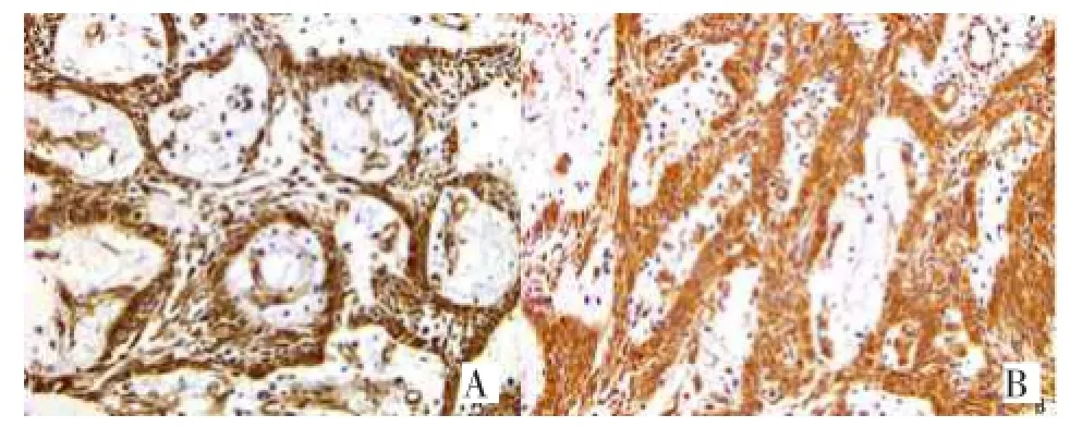
Fig.1 The expression of COX-2(A)and Survivin(B)protein in plexiform AB(SP,×200)图1 COX-2蛋白(A)和Survivin蛋白(B)在丛状型AB中的表达(SP,×200)
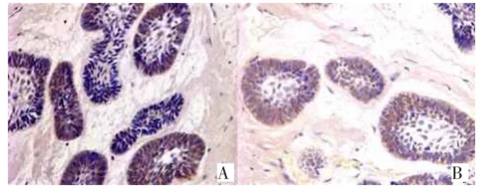
Fig.2 The expression of COX-2(A)and Survivin(B)protein in follicular AB(SP,×400)图2 COX-2蛋白(A)和Survivin蛋白(B)在滤泡型AB中的表达(SP,×400)

Fig.3 The expression of COX-2(A)and Survivin(B)protein in normal oral mucosa(SP,×400)图3 COX-2蛋白(A)和Survivin蛋白(B)在正常口腔黏膜中的表达(SP,×400)
2.2COX-2、Survivin蛋白水平的Western Blot结果肿瘤组织中COX-2蛋白、Survivin蛋白表达水平明显高于正常口腔黏膜组织(P<0.05),见表3、图4。Survivin蛋白与COX-2蛋白呈正相关(r= 0.778,P<0.05)。
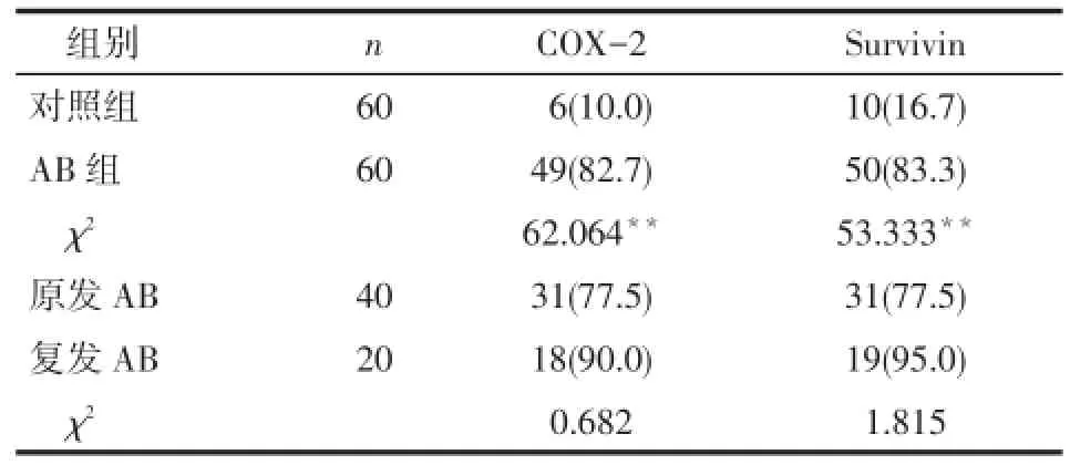
Tab.1 The positive expression of COX-2 and Survivin protein in AB and normal oral mucosa groups表1COX-2、Survivin蛋白在AB组和对照组的阳性表达情况 例(%)
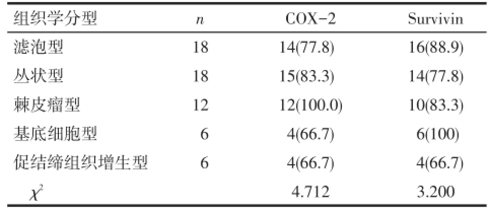
Tab.2 COX-2 and Survivin protein expression in various types of AB groups表2COX-2、Survivin蛋白在各型AB中的表达例(%)
Tab.3 Comparison of the expression of COX-2 and Survivin between AB and normal oral mucosa groups表3COX-2、Survivin蛋白在AB组和对照组中的表达水平比较 (±s)

Tab.3 Comparison of the expression of COX-2 and Survivin between AB and normal oral mucosa groups表3COX-2、Survivin蛋白在AB组和对照组中的表达水平比较 (±s)
**P<0.01
组别AB组对照组t n 60 60 COX-2 0.781±0.142 0.179±0.056 17.65**Survivin 0.447±0.139 0.072±0.017 11.976**
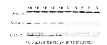
Fig.4 The expression of COX-2 and Survivin protein图4 COX-2、Survivin蛋白的表达
3 讨论
COX-2是一种诱导型酶,在多种肿瘤组织中高表达,但在正常组织中无表达,其与肿瘤的发生、发展、侵袭和转移密切相关[5]。COX-2的过度表达具有以下作用:(1)抵抗肿瘤细胞凋亡。(2)刺激肿瘤细胞增殖。(3)促进肿瘤的浸润和转移。(4)促进肿瘤组织及其周围组织的血管形成[6]。本研究结果显示COX-2蛋白在AB组织中的阳性表达率高于正常口腔黏膜组织,表明AB相对于正常口腔黏膜组织具有更强的增殖活性。COX-2蛋白主要表达于中间星网状层细胞与外周柱状或立方状细胞质中,这可能说明星网状层细胞与外周柱状或立方状细胞的增殖活性更强,是肿瘤的生发中心。Sun等[7]发现COX-2可明显抑制细胞凋亡,同时上调抗凋亡因子bcl-2的水平。既往有研究表明,bcl-2在成釉细胞瘤中阳性率高,且表达位置与本实验中COX-2的表达位置一致[8]。由此提示,AB中COX-2可能通过刺激bcl-2的表达从而抑制AB细胞凋亡,促进肿瘤细胞的增殖。COX-2还可通过提高肿瘤组织中血管内皮细胞生长因子(VEGF)的表达而促进肿瘤及周围组织中的血管生成[9]。以往的研究显示,AB为典型的血管依赖性病变,VEGF在AB为强阳性表达,并伴随AB生物学行为的不同(复发和恶变)其表达递增[10]。本实验Western Blot结果显示COX-2在AB组织中表达水平明显高于正常口腔黏膜组织,与VEGF的表达趋势相同,提示AB中COX-2可能通过调节VEGF的表达促进血管生成,进一步促进肿瘤的发展,与既往结果一致[11],COX-2在AB的发生发展过程中发挥了很重要的作用。
上皮-间质诱导存在于AB的发生、发展及其构成的临床生物学行为中[11],本实验证实survivin不但表达于中间星网状层细胞与外周柱状或立方状细胞质中,在纤维细胞、血管内皮细胞、炎性淋巴细胞等AB的间质成分中也都有survivin的阳性表达,这提示survivin在AB的起始阶段可能就开始发挥了调节作用。Survivin蛋白是迄今为止分子质量最小、抗凋亡作用最强的凋亡抑制蛋白,有研究发现survivin可通过抑制促凋亡基因Bax和Fas下游的反应而抑制细胞凋亡[12]。AB中Bax虽然在外周层和星网状层细胞中都有表达,但表达以弱阳性为主,在恶变组中表达明显降低[7]。结合本实验免疫组化结果提示,可能正是由于survivin在AB的外周层和星网状层细胞中的高表达,对Bax的表达起抑制作用,从而抑制了肿瘤细胞的凋亡。本实验60例AB中有50例survivin呈阳性表达,且表达程度较高,正常人口腔黏膜上皮细胞、颗粒性变细胞和角化退变样细胞中survivin蛋白的表达为阴性或弱阳性,而Endoh等[13]研究证实,由于survivin的过度表达,凋亡关卡失去了在细胞正常增殖周期中的限制作用,并通过有丝分裂促进转化细胞的异常增殖,这说明由于survivin的过度表达促进了AB的发展。本实验的Western Blot结果显示survivin在AB中的表达水平明显高于正常口腔黏膜组织中的表达水平,结合免疫组化结果提示,survivin在AB中可能通过抑制促凋亡基因Bax的表达来增强肿瘤细胞的抗凋亡作用,并通过抑制凋亡关卡的限制作用而促进AB中肿瘤细胞的增殖。
[1]Huang HZ,Tao Q.Clinical and basic research into ameloblastomas [J].Chinese Journal of Practical Stomatology,2009,2(2):82-86.[黄洪章,陶谦.成釉细胞瘤的临床及基础研究[J].中国实用口腔科杂志,2009,2(2):82-86].
[2]Pomianowska E,Schiolberg AR,Clausen OP,et al.Cyclooxygenase-2 overexpression in resected pancreatic head adenocarcinomas correlates with favourable prognosis[J].BMC Cancer,2014,20(14): 458-465.
[3]Poi MJ,Knobloch TJ,Sears MT,et al.Coordinated expression of cyclin-dependent kinase-4 and its regulators in human oral tumors. [J].Anticancer Res,2014,34(7):3285-3292.
[4]Zhong M,Zhang LZ,Zhang B,et al.Expression of proteinkinase C-αand telomerase in human ameloblastoma[J].J Pract Stomatol,2006,22(5):696-699.[钟鸣,张陆荘,张波,等.人成釉细胞瘤中蛋白激酶c-α表达与端粒酶活性的关系[J].实用口腔医学杂志,2006,22(5):696-699].
[5]Nozoe T,Ezaki T,Kabashima A,et al.Significance of immunohistochemical expression of cyclooxygenase-2 in squamous cell carcinoma of the esophagus[J].Am J Surg,2005,189(1):110-115.
[6]Liang K,Geng M.Expression and significance of PTEN、COX-2 and Survivin in gastric carcinoma[J].Chinese Journal of Gerontology,2009,29(8):987-989.[梁堃,耿明.PTEN、COX-2、Survivin在胃癌中的表达及意义[J].中国老年学杂志,2009,29(8):987-989].
[7]Sun Y,Tang XM,Half E,et al.Cyclooxygenase-2 overexpression reduces apoptotic susceptibility by inhibiting the cytochrome c-dependent apoptotic pathway in human colon cancer cells[J].Cancer Res,2002,62(21):6323-6328.
[8]Wang J,Zhong M,Liu JD,et al.Expression of bax and its significance in human ameloblastoma[J].J Chin Med Uni,2006,35(1):45-46.[王洁,钟鸣,刘敬东,等.bax蛋白在成釉细胞瘤中的表达和意义[J].中国医科大学学报,2006,35(1):45-46].
[9]Yang S,Han H.Effect of cyclooxygenase-2 silencing on the malignant biological behavior of MCF-7 breast cancer cells[J].Oncol Lett,2014,8(4):1628-1634.
[10]Chen WL,Ouyang KX,Li HG,et al.Expression of inducible nitric oxide synthase and vascular endothelial growth factor in ameloblastoma[J].J Craniofac Surg,2009,20(1):171-175;discussion 176-177.
[11]Li L.Expression and significance of cyclooxygenase-2 in ameloblastoma[D].Shen Yang:China Medical University,2007.[李乐.人成釉细胞瘤中环氧化合酶-2的表达和意义[D].沈阳:中国医科大学,2007].
[12]Cheng XL,Li MK.Effect of topiramate on apoptosis-related protein expression of hippocampus in model rats with Alzheimers disease [J].Eur Rev Med Pharmacol Sci,2014,18(6):761-768.
[13]Endoh T,Tsuji N,Asanuma K,et al.Survivin enhances telomerase activity via up-regulation of specificity protein 1-and c-Myc-mediated human telomerase reverse transcriptase gene transcription[J]. Exp Cell Res,2005,305(2):300-311.
(2014-08-29收稿2014-10-23修回)
(本文编辑闫娟)
The expression of COX-2 and Survivin in ameloblastoma and its clinical significance
LI Le1,ZHANG Xingle1,WANG Peng1,LIU Yu2
1 Department of Stomatology,2 Operation Room,the Affiliated Hospital of Chengde Medical College,Chengde 067000,China
ObjectiveTo investigate the expression of cyclooxygenase-2(COX-2)and Survivin in ameloblastoma(AB)tissues.MethodsA total of 60 AB samples(primary AB 40 cases,recurrent AB 20 cases)and 60 normal oral mucosas were collected from the Affiliated Hospital of Chengde Medical College.The AB samples included 12 acanthomatous types,18 follicular patterns,18 plexiform patterns,6 basal cell ameloblastomas and 6 desmoplastic ameloblastomas.The expression levels of COX-2 and Survivin were detected by immunohistochemistry and Western blot methods in AB and normal oral mucosa.The expressions of COX-2 and Survivin were compared between different types and tissues of AB.ResultsThe positive rate of COX-2 was significantly higher in AB(82.7%)than that in normal oral mucosa(10.0%).The positive rate of Survivin was significantly higher in AB(83.3%)than that in normal oral mucosa tissues(16.7%).The expression of COX-2 (0.781±0.142)was higher in AB than that of normal oral mucosa tissues(0.179±0.056).The expression of Survivin(0.447± 0.139)was significantly higher in AB than those in normal oral mucosa tissues(0.072±0.017).There was a positive correlation between the expression of COX-2 and Survivin(r=0.778,P<0.05).There were no significant differences in the positive expressions of COX-2 and Survivin between different types of AB(P>0.05).ConclusionCOX-2 and Survivin were overexpressed in AB,which may be involved in the occurrence and development of AB.
ameloblastoma;cyclooxygenase-2;survivin;immunohistochemistry;blotting,Western
R739.8
ADOI:10.11958/j.issn.0253-9896.2015.03.013
河北省科技支撑计划项目(201121054)
1承德医学院附属医院口腔科(邮编067000),2手术部
李乐(1980),男,主治医师,硕士,主要从事口腔颌面部肿瘤研究
