Mantle Cell Lymphoma in a Lacrimal Gland in a Female and a Review of the Literature
2014-09-17JianhaoCaiZeyiLiYuanshengZhou
Jianhao Cai, Zeyi Li, Yuansheng Zhou
Joint Shantou International Eye Center of Shantou University and the Chinese University of Hong Kong,Shantou 515041, China.
Introduction
Case report
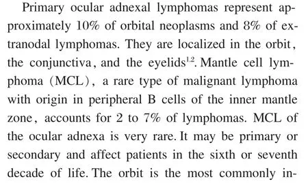
Figure 1 The appearance of the patient.Right supraorbital swelling with round lymphadenopathy in the lacrimal gland regions.
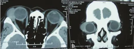
Figure 2 Computerized tomography (CT) scan.Mass in bilateral lacrimal gland region, more significant in right eye(a.horizontal position, b.coronal position)


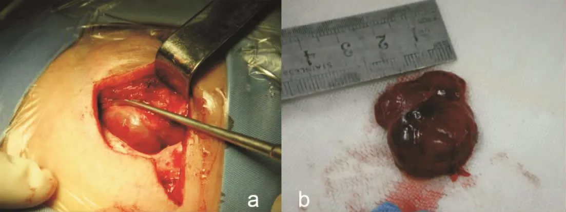
Figure 3 The general appearance of lacrimal gland mass.a.The mass located in the lacrimal gland fossa.b.The mass conformed to the shape of the globe with black-violet color,soft texture,and measured about 24 mm×26 mm×6 mm.

Discussion
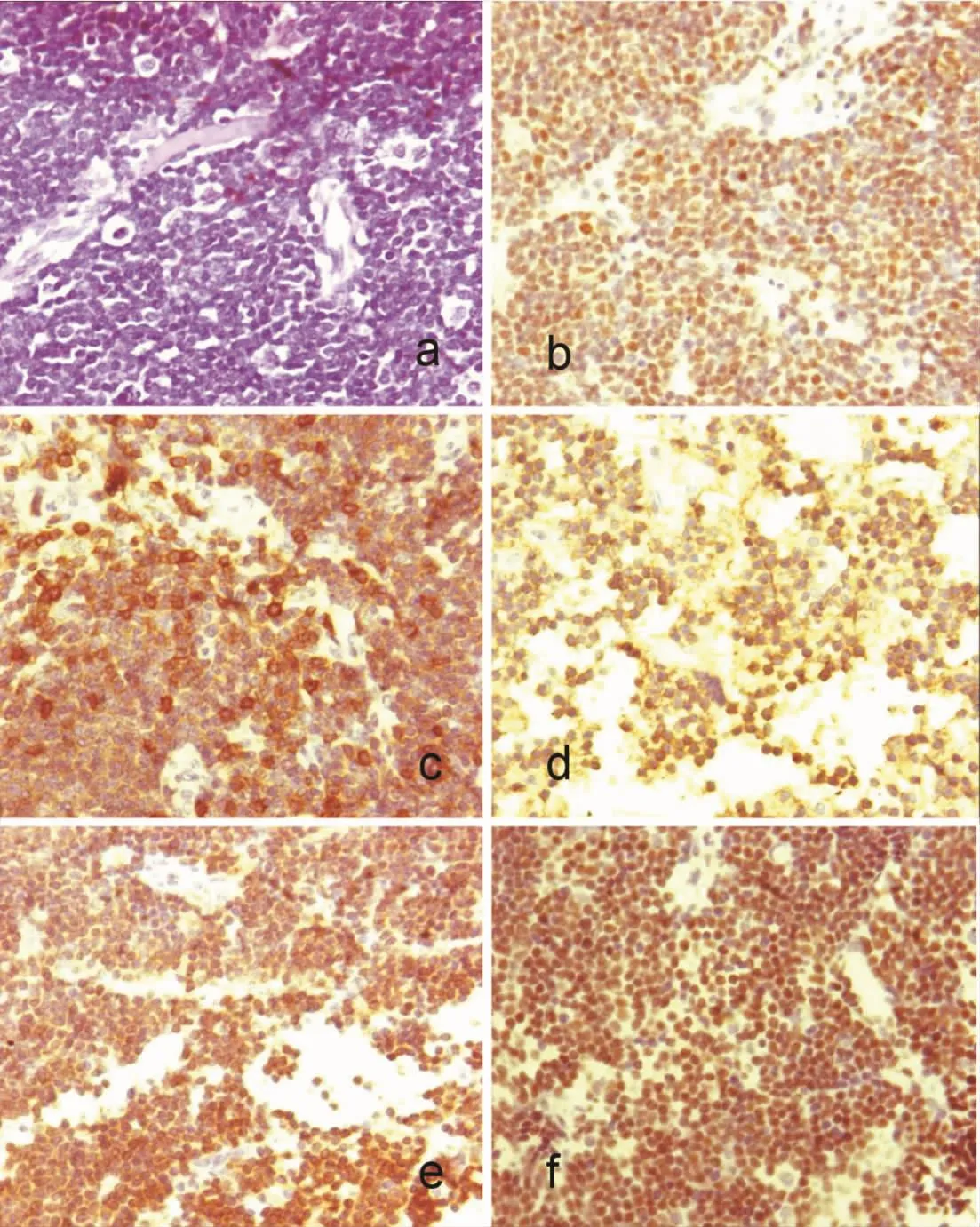
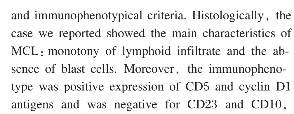
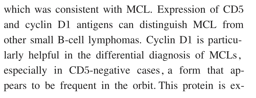
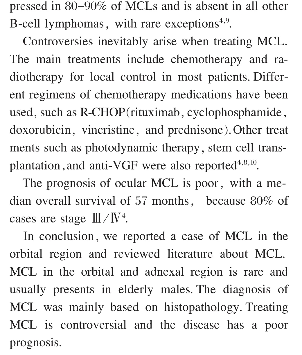
Figure 4 histological examination of the mass.HE stain shows that infiltration by monomorphic cell population composed of small-to medium-sized lymphocytes with irregular, indented, or cleaved nuclei, condensed chromatin, and scant cytoplasm.The growth pattern was diffuse and partially nodular (a,200×).Immunohistochemical study revealed that tumor cells were CyclinD1(b), CD5(c), CD20(d), CD79a(e), and Pax-5(f) positive(400×).
Acknowledgement

杂志排行

眼科学报的其它文章
- Progress of Application of Sedation Technique in Pediatric Ocular Examination
- Prevention and Control of Perioperative Incision Infection in Patients Undergoing Day Cataract Surgery
- Herniation of the Retina in the Central Macula in an Adult after Iridocyclitis
- Favorable Outcome in Open Globe Injuries with Low OTS Score
- Follow-up of a Case of Vitelliform Macular Dystrophy Over an 8-year Period
- Clinical Observation of Transepithelial Corneal Collagen Cross-linking by Iontophoresis of Riboflavin in Treatment of Keratoconus