Herniation of the Retina in the Central Macula in an Adult after Iridocyclitis
2014-09-17QingGuoYuliPiTingGao
Qing Guo, Yuli Pi, Ting Gao
Department of Ophthalmology, First Hospital Affiliated to Chinese PLA General Hospital, Beijing 100048,China
Introduction


Case report


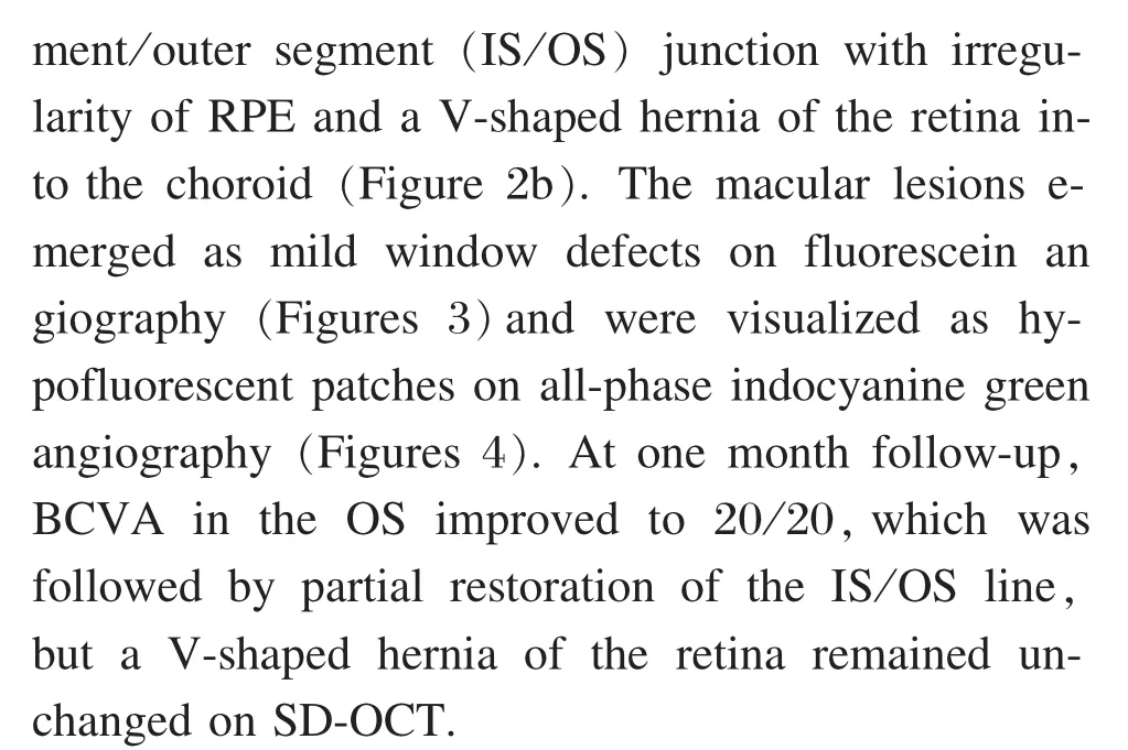

Figure 1 Spectral domain optical coherence tomography(SD-OCT) line scan through the macular lesion showing unremarkable features in this patient with iridocyclitis
Discussion
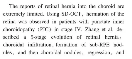
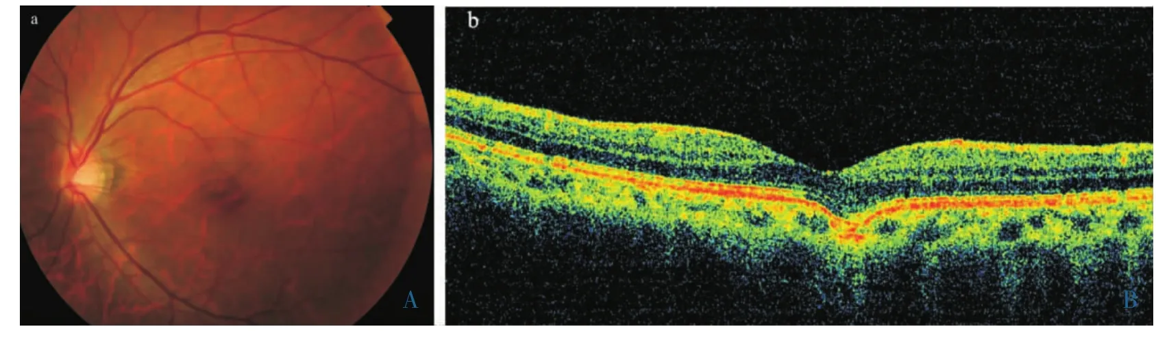
Figure 2 a.Fundus color photograph showing yellowish spots in the macular area 2 weeks following resolution of iridocyclitis in the left eye.b.Spectral domain optical coherence tomography (SD-OCT) line scan through the macular lesion showing a V-shaped hernia of the retina into the choroid.
Figure 3 Fluorescence angiography (FA) showing mild window defects in the macular area.(a) hyperfluorescent spots presentation on early-stage FA.(b) hyperfluorescent spots wane in late-stage FA.

Figure 4 Indocyanine green angiography(ICGA) showing hypofluorescent patches in the macular area.(a) early-stage ICGA.(b) late-stage ICGA.


杂志排行

眼科学报的其它文章
- Progress of Application of Sedation Technique in Pediatric Ocular Examination
- Prevention and Control of Perioperative Incision Infection in Patients Undergoing Day Cataract Surgery
- Mantle Cell Lymphoma in a Lacrimal Gland in a Female and a Review of the Literature
- Favorable Outcome in Open Globe Injuries with Low OTS Score
- Follow-up of a Case of Vitelliform Macular Dystrophy Over an 8-year Period
- Clinical Observation of Transepithelial Corneal Collagen Cross-linking by Iontophoresis of Riboflavin in Treatment of Keratoconus