Follow-up of a Case of Vitelliform Macular Dystrophy Over an 8-year Period
2014-09-17ShizhouHuangLezhengWuFengWenGuangweiLuoFutianJiang
Shizhou Huang,Lezheng Wu,Feng Wen,Guangwei Luo,Futian Jiang
State Key Laboratory of Ophthalmology, Zhongshan Ophthalmic Center, Sun Yat-sen University, Guangzhou 510060,China
Introduction


Case report

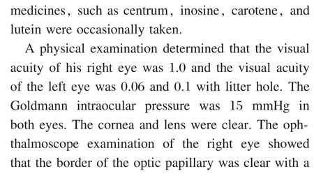


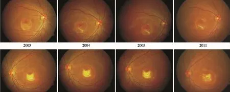
Figure 1 The fundus color photography at the initial visit, and at 1, 2, and 8 years of follow-up (the photographs of 2007,2009, 2010 were neglected).
The follow-up of visual acuity

The follow-up by color photography

The follow-up of fundus fluorescein angiography

The follow-up of OCT


Figure 2 The fundus fluorescein angiography at the initial visit and after 2 and 8 years of follow-up (the photographs from 2007, 2009 were not included)
The test of EOG

The follow-up of multi-focal electroretinography

Discussion


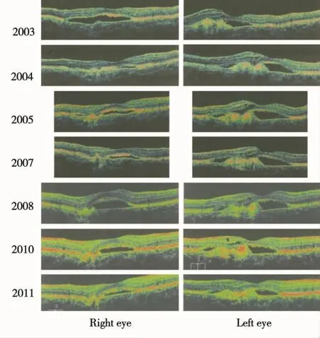
Figure 3 OCT in the initial visit and follow-up with downup scan (the photographs of 2005 and 2007 were 5 mm scanning, the other photographs were 6 mm scanning).All of the scans passed through the fovea.

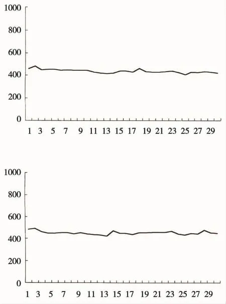
Figure 4 EOG at the initial visit.Abscissa: minutes;ordinate: μV (Upper: right eye; Lower: left eye)

Table 1 The results of EOG


Figure 5 The mfERGs in 2, 8 year follow-up.Upper: right eye; lower: left eye

杂志排行
眼科学报的其它文章
- Relationship between Refractive Error and Ocular Biometrics in Twin Children:the Guangzhou Twin Eye Study
- Ophthalmic Evaluation of Children from the Tibet Plateau with Congenital Heart Disease
- Ten-year Etiologic Review of Chinese Children Hospitalized for Pediatric Cataracts
- Clinical Features and Differential Diagnosis of Acute Idiopathic Blind Spot Enlargement Syndrome
- Relationship between Foxp3-3279 (rs376158) Polymorphism and Dust Mite Allergic Conjunctivitis
- Comparison of Postoperative Pain Following Laser-assisted Subepithelial Keratectomy and Transepithelial Photorefractive Keratectomy:a Prospective,Random Paired Bilateral Eye Study
