危及器官边界在估计心脏亚结构照射剂量中的作用*
2014-09-10黎艳萍王晓红李俊玉谭文勇胡德胜
黎艳萍 王晓红 李 莹 李俊玉 谭文勇 胡德胜
危及器官边界在估计心脏亚结构照射剂量中的作用*
黎艳萍 王晓红 李 莹 李俊玉 谭文勇 胡德胜
目的:探讨心脏亚结构(CS)的计划危及体积(PRV)在左乳癌调强放疗(IMRT)中估计CS照射剂量的作用。方法:勾画23例左乳腺癌保乳后IMRT患者的CS,以CS的平均运动幅度为外放边界建立PRV。设计2个不同的IMRT计划并计算CS及PRV的体积、平均剂量、最大剂量(D2%)和标准差,并计算CS和其PRV的平均剂量、D2%的差别。结果:与CS本身相比,心脏和左心室PRV体积增加50%~80%,冠状动脉主干及主要分支PRV体积增加18.7~42.6倍。在两个不同IMRT计划中,心脏、心脏前壁区域(AMT)、前降支及相应的PRV的平均剂量分别为9.4~11.4 Gy、11.0~17.5 Gy、22.6~27.8 Gy,其D2%分别为24.5~36.2 Gy、28.2~38.8 Gy、36~45 Gy。冠状动脉左右主干、右缘支和左旋支的平均剂量为8.6~14.9 Gy,D2%为12.5~23.7 Gy。与CS的剂量相比,相应的PRV的平均剂量差别为-2.5%~12.5%,D2%增加了8.0%~43.1%。多数CS的PRV剂量的标准差明显增大。结论:在左乳癌保乳后IMRT中CS和相应PRV的平均剂量差别<12%。
乳腺癌 放射疗法 调强放疗 器官运动 心脏病
可治愈的恶性肿瘤如乳腺癌和霍奇金淋巴瘤在接受胸部放疗后,放射相关性心脏病越来越多的得到关注[1-3],其发生可能与心脏照射的体积和/或剂量相关[2]。实际上,胸部放疗时心脏所接受的照射剂量并不均匀,如乳腺癌术后放疗时心脏的前部接受的剂量高于其他部位。为此在Tan等[4-6]以前的一系列研究中,提出了心脏前壁区域(anterior myocardial territory,AMT)的概念,并推荐在左侧乳腺癌保乳术后的调强放疗(intensity-modulated radiotherapy,IMRT)中,用AMT取代心脏作为一个独立的危及器官(or-gan at risk,OAR)可更好的保护心脏。在Tan等[4-5]研究中AMT包括多个心脏亚结构(cardiac substructure,CS)如左右冠状动脉主干和分布在心脏前表面的冠状动脉,及大的分支如前降支、右缘支、左旋支。此外Tan等[6]也报道了CS由于心脏搏动所致的器官运动范围为3~8 mm。本研究的目的是探讨CS危及计划体积(planning risk volume,PRV)及估计左侧乳腺癌IMRT时CS的照射剂量的作用,为今后准确估计心脏剂量、评估放疗相关的心脏并发症风险提供参考。
1 材料与方法
1.1 IMRT计划
23例左侧乳腺癌接受保乳手术后放疗入组研究,采用左半胸的5野逆向动态调强技术,其处方剂量为50 Gy,25次。详细的靶区定义,IMRT计划的照射野、剂量体积限制参数等在Tan等[4-5]的研究中已有详细的描述。
1.2 心脏亚结构的定义
心脏包括所有的心肌,排除大血管;左心室包括左心室的心肌及室间隔;AMT为心脏的前表面及其后1 cm后的心肌组织,同时也包括冠状动脉主干及心脏前表面的主要分支如前降支、右缘支、左旋支。这些冠状动脉的分支均起始于各自从冠状动脉主干分支处到左心室的心室内面的最下的层面为止[6]。CS的详细定义见以前的研究[4-6]。在勾画冠状动脉及分支时,主要根据影像解剖判断,对于部分层面难于确定,在这些层面上不直接勾画,采用通过上下层面自动插置的方法以减少勾画的不确定性。
1.3 心脏亚结构的剂量体积参数
所有的IMRT计划中OAR包括左右肺、右侧乳腺、心脏或AMT。每个患者的计划选择2个IMRT计划:将包括心脏 的IMRT计划,称为IMRT(H)计划(目前的标准方法);将包括AMT的IMRT计划称为IMRT(AMT)计划(本研究所推荐的方法)。在完成CS的勾画后,并根据以前的所测的CS运动幅度[6]作为外放边界建立相应的PRV。在IMRT(H)计划和IMRT(AMT)计划中分别计算CS及其PRV的体积、平均剂量和D2%(最大剂量,2%的CS或PRV体积接受的照射剂量),CS或PRV照射剂量的标准差(standard deviation,SD)为剂量均匀性的替代指标[7]。
1.4 统计学分析
采用配对t检验比较同一个IMRT计划中的CS和其PRV的照射剂量、SD的差别,同样采用t检验比较IMRT(H)计划和IMRT(AMT)计划中CS和PRV的剂量差别。所有的检验以P<0.05作为有统计学意义,所有的统计学分析通过SPSS 20.0软件包完成。
2 结果
2.1 CS的体积
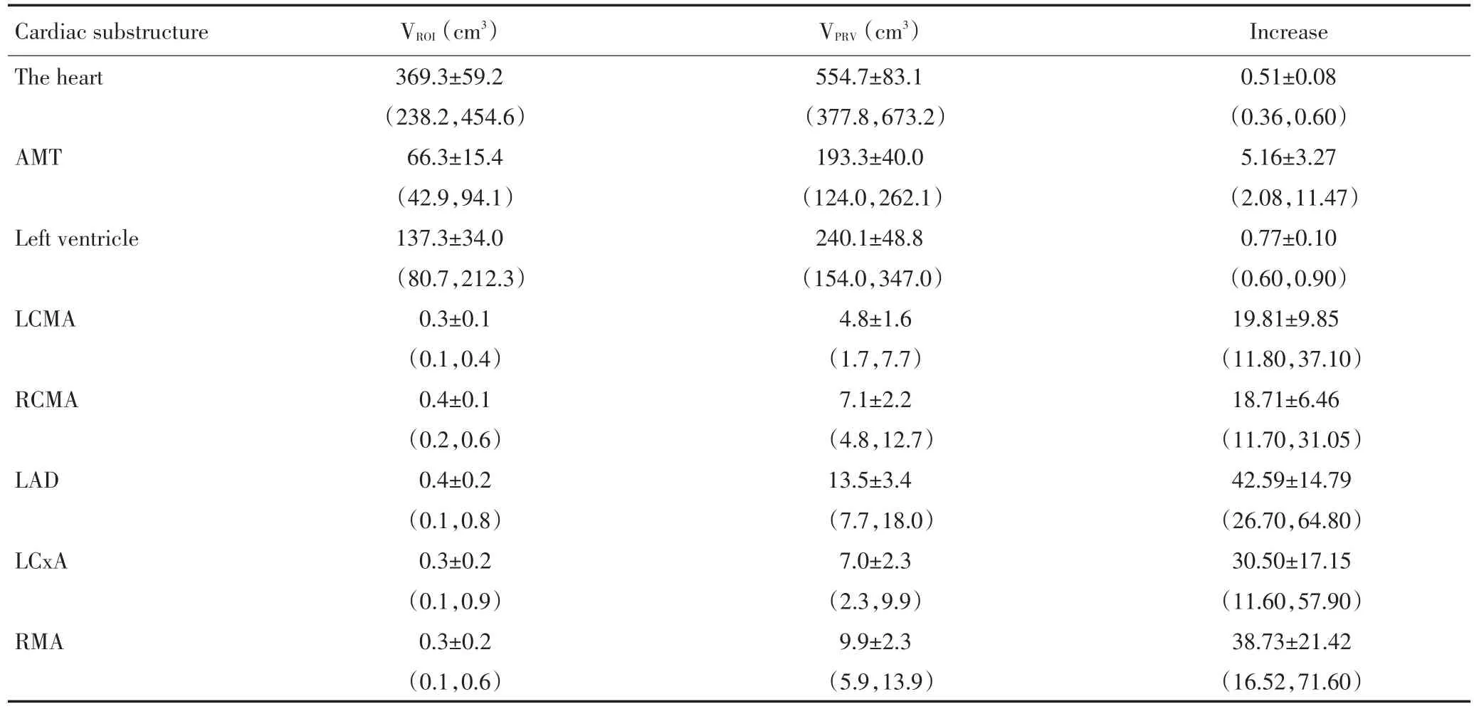
通过外放一定的边界得到PRV后,心脏和左心室的体积增加50%~80%,AMT的体积增加5.2倍,而冠状动脉主干及主要分支的体积均在0.5 cm3以下,而其相应的PRV体积达到4.8~9.9 cm3,增加了18.7~42.6倍(表1)。
2.2 IMRT(H)计划中CS的照射剂量
在IMRT(H)中心脏和PRV的平均剂量分别为11.0 Gy和11.4 Gy,D2%为33.5 Gy和36.2 Gy,与心脏本身的剂量相比,心脏PRV的平均剂量和D2%分别增加5.5%和12.1%(表2)。AMT和其PRV的平均剂量分别17.5 Gy和16.0 Gy,其D2%分别为35.9 Gy和38.8 Gy,与AMT本身的剂量相比,其PRV的平均剂量下降了8.4%,而D2%增加了9.4%均具有统计学意义。左心室PRV的平均剂量和D2%均较左心室本身的剂量有不同程度的增加。在冠状动脉及其分支中,前降支及PRV的平均剂量分别为27.8 Gy和26.2 Gy,D2%分别为41.5 Gy和45.8 Gy,高于其他动冠状动脉分支。其他的冠状动脉及分支包括左右主干、左旋支、右缘支的平均剂量为(9.4~14.2)Gy,D2%为(12.5~19.5)Gy;外放PRV边界后,PRV的平均剂量变化明显,所有冠状动脉的D2%均增加(12.1~33.2)%,有明显统计学意义(表2)。
2.3 IMRT(AMT)中CS的照射剂量
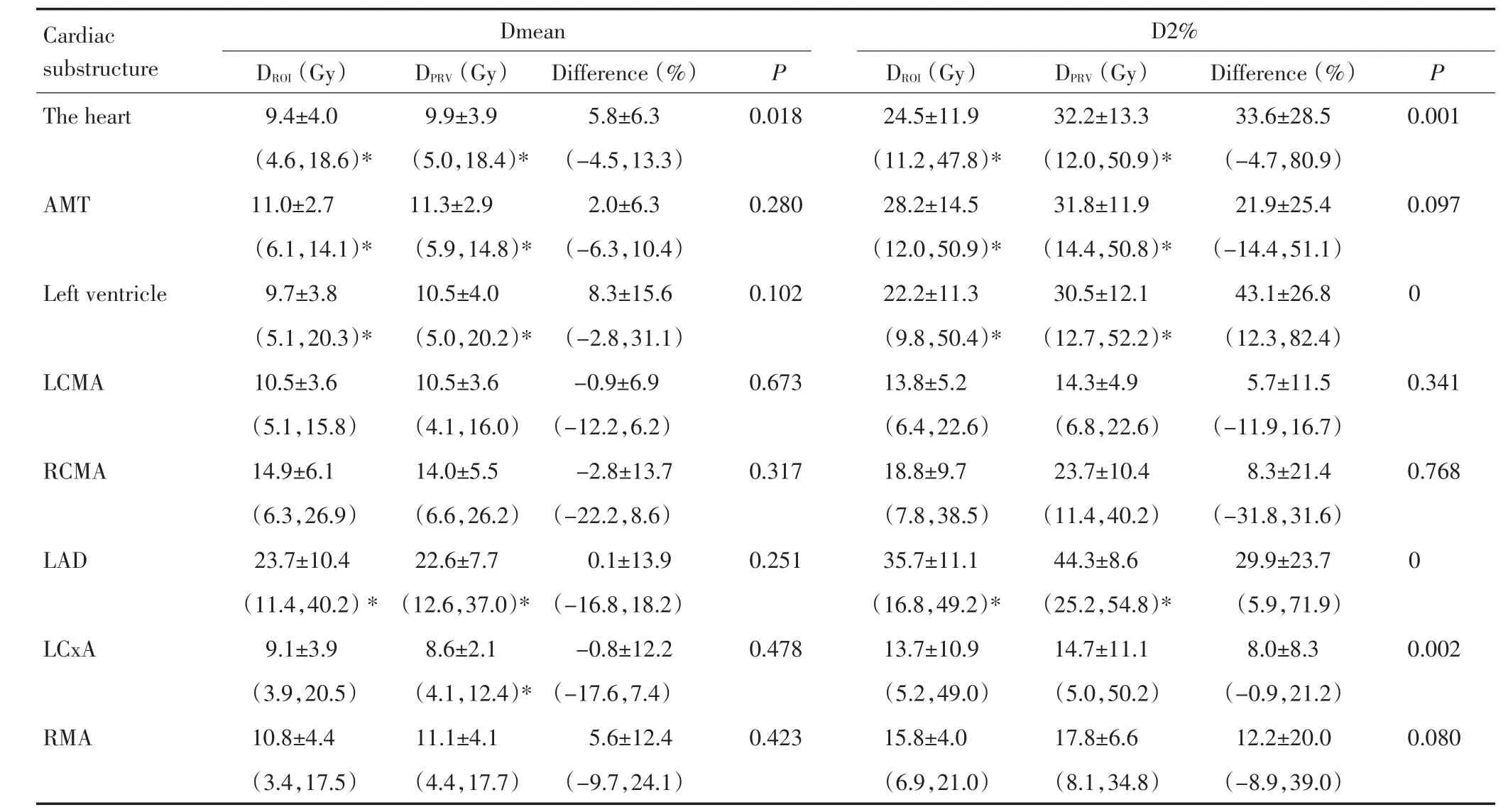
在IMRT(AMT)中心脏和PRV的平均剂量分别9.4 Gy和9.9Gy,D2%为24.5 Gy和32.2 Gy,与心脏本身的剂量相比,心脏PRV的平均剂量和D2%分别增加5.8%和33.6%,有明显统计学意义(表3)。AMT和其PRV的平均剂量分别为11.0 Gy和11.3 Gy,D2%分别为28.2 Gy和31.8 Gy;与AMT本身的剂量相比,其PRV的平均剂量和D2%分别增加了2.0%和21.9%。与左心室本身的剂量相比,其PRV的平均剂量和D2%均分别增加8.3%和43.1%。在冠状动脉及其分支中,前降支及PRV的平均剂量分别为22.6 Gy和23.7 Gy,D2%分别为35.7 Gy和44.3 Gy,明显高于其他动脉。其它的冠状动脉及分支包括左右主干、左旋支、右缘支的平均剂量为(8.6~14.9)Gy,D2%为(13.7~23.7)Gy;在CS外放建立PRV后,PRV的平均剂量变化明显,前降支和左旋支的D2%升高有显著统计学意义(表3)。
与IMRT(H)计划相比,IMRT(AMT)计划中的心脏、AMT和左心室的平均剂量和D2%均明显降低(表3);前降支的平均剂量和D2%分别降低(15.0±10.7)%(95%CI:0.5%,30.6%)和(15.0±13.8)%(95%CI:2.2%,30.6%),其PRV的平均剂量和D2%分别降低(14.0±10.4)%(95%CI:0.5%,29.3%)和(3.4±5.1)%(95%CI:1.6%,11.6%),均有明显的统计学意义(表3)。
2.4 其PRV剂量体积参数的标准差
心脏、左心室、右冠状动脉主干、前降支和右缘支的PRV剂量的SD在两个IMRT计划中都明显增加(表4)。AMT的SD在IMRT(AMT)计划中明显增加,而左冠状动脉主干在的SD在IMRT(H)计划中明显增加。
表1 各个心脏亚结构及相应的危及计划体积 (±s)Table 1 Volume of cardiac substructure and its planning risk volume (±s)

表1 各个心脏亚结构及相应的危及计划体积 (±s)Table 1 Volume of cardiac substructure and its planning risk volume (±s)
Abreaviations:SD:standard deviation;VROIand VPRVwere the volume of cardiac substructure and its planning risk volume;AMT:anterior myocardial territory;LCMA=left coronary artery;RCMA=right coronary artery;LAD=left anterior descending artery;LCxA=left circumflex artery;RMA=right marginal artery.95%confidence interval was shown in the brackets
Cardiac substructure The heart AMT Left ventricle LCMA RCMA LAD LCxA RMA VROI(cm3)369.3±59.2(238.2,454.6)66.3±15.4(42.9,94.1)137.3±34.0(80.7,212.3)0.3±0.1(0.1,0.4)0.4±0.1(0.2,0.6)0.4±0.2(0.1,0.8)0.3±0.2(0.1,0.9)0.3±0.2(0.1,0.6)VPRV(cm3)554.7±83.1(377.8,673.2)193.3±40.0(124.0,262.1)240.1±48.8(154.0,347.0)4.8±1.6(1.7,7.7)7.1±2.2(4.8,12.7)13.5±3.4(7.7,18.0)7.0±2.3(2.3,9.9)9.9±2.3(5.9,13.9)Increase 0.51±0.08(0.36,0.60)5.16±3.27(2.08,11.47)0.77±0.10(0.60,0.90)19.81±9.85(11.80,37.10)18.71±6.46(11.70,31.05)42.59±14.79(26.70,64.80)30.50±17.15(11.60,57.90)38.73±21.42(16.52,71.60)
表2 IMRT(H)计划的剂量体积参数 (±s)Table 2 Dose-volume parameters in IMRT(H)planning (±s)

表2 IMRT(H)计划的剂量体积参数 (±s)Table 2 Dose-volume parameters in IMRT(H)planning (±s)
Abreaviations:SD:standard deviation;Dmean:the mean dose;DROIand DPRVwere the planned radiation dose of cardiac substructure and its planning risk volume;AMT:anterior myocardial territory;LCMA=left coronary artery;RCMA=right coronary artery;LAD=left anterior descending artery;LCxA=left circumflex artery;RMA=right marginal artery
Cardiac substructure The heart Dmean AMT Left ventricle LCMA RCMA LAD LCxA RMA DROI(Gy)11.0±4.0(4.8,20.5)17.5±6.7(8.7,31.5)12.8±4.6(6.7,22.8)10.3±3.0(5.5,14.9)14.2±5.3(8.0,27.8)27.8±11.3(13.6,42.6)9.6±2.8(4.8,14.5)11.7±2.8(8.6,17.4)DPRV(Gy)11.4±3.9(6.1,20.2)16.0±6.0(7.8,29.0)13.0±4.4(6.6,22.5)10.5±3.2(5.0,15.4)14.2±4.9(8.1,26.8)26.2±7.9(7.9,16.1)9.4±2.5(5.0,13.7)12.9±4.2(5.9,20.8)Difference(%)5.5±8.0(-3.1,16.8)-8.4±5.1(-16.5,-2.5)1.9±3.8(-3.1,8.1)1.6±4.6(-6.1,7.6)0.6±4.9(-5.6,9.1)-1.1±14.3(-17.5,19.6)-1.1±6.1(-9.1,8.9)12.5±39.7(-18.6,69.6)D2%P DROI(Gy)33.5±12.1(12.2,49.6)35.9±11.8(12.0,51.4)32.7±12.1(9.8,50.5)12.4±3.7(6.4,18.6)16.8±7.4(8.9,38.5)41.5±9.9(26.8,53.9)12.5±4.9(5.2,25.2)15.1±4.4(6.9,21.0)DPRV(Gy)36.2±11.2(15.3,51.5)38.8±11.5(14.5,52.3)37.3±11.7(12.7,51.8)14.5±4.5(6.8,20.2)19.5±7.7(10.4,40.9)45.8±8.5(25.2,54.0)14.3±4.6(8.8,26.7)16.2±3.8(9.0,21.2)difference(%)12.1±19.7(-20.6,35.7)9.4±7.9(1.5,23.5)16.6±11.5(2.2,34.7)19.7±31.2(-6.3,68.1)17.7±9.7(6.3,33.4)12.1±13.3(-2.4,34.7)24.5±55.7(1.0,110.5)33.2±34.6(-17.1,73.7)P 0.012 0 0.185 0.188 0 0 0.0920.016 0.8690 0.1640.002 0.3200.039 0.2290.050
表3 IMRT(AMT)计划的剂量体积参数 (±s)Table 3 Dose-volume parameters in IMRT(AMT)planning (±s)

表3 IMRT(AMT)计划的剂量体积参数 (±s)Table 3 Dose-volume parameters in IMRT(AMT)planning (±s)
Abreaviations:SD:standard deviation;Dmean:the mean dose;DROIand DPRVwere the planned radiation dose of cardiac substructure and its planning risk volume;AMT:anterior myocardial territory;LCMA=left coronary artery;RCMA=right coronary artery;LAD=left anterior descending artery;LCxA=left circumflex artery;RMA=right marginal artery.*:The dose-volume parameter between IMRT(H)and IMRT(AMT)plan was statistically different with the P<0.05.95%confidence interval was shown in the brackets
Cardiac substructure The heart Dmean AMT Left ventricle LCMA RCMA LAD LCxA RMA DROI(Gy)9.4±4.0(4.6,18.6)*11.0±2.7(6.1,14.1)*9.7±3.8(5.1,20.3)*10.5±3.6(5.1,15.8)14.9±6.1(6.3,26.9)23.7±10.4(11.4,40.2)*9.1±3.9(3.9,20.5)10.8±4.4(3.4,17.5)DPRV(Gy)9.9±3.9(5.0,18.4)*11.3±2.9(5.9,14.8)*10.5±4.0(5.0,20.2)*10.5±3.6(4.1,16.0)14.0±5.5(6.6,26.2)22.6±7.7(12.6,37.0)*8.6±2.1(4.1,12.4)*11.1±4.1(4.4,17.7)Difference(%)5.8±6.3(-4.5,13.3)2.0±6.3(-6.3,10.4)8.3±15.6(-2.8,31.1)-0.9±6.9(-12.2,6.2)-2.8±13.7(-22.2,8.6)0.1±13.9(-16.8,18.2)-0.8±12.2(-17.6,7.4)5.6±12.4(-9.7,24.1)D2%P P 0.0180.001 0.2800.097 0.1020 0.6730.341 0.3170.768 0.2510 0.4780.002 0.423 DROI(Gy)24.5±11.9(11.2,47.8)*28.2±14.5(12.0,50.9)*22.2±11.3(9.8,50.4)*13.8±5.2(6.4,22.6)18.8±9.7(7.8,38.5)35.7±11.1(16.8,49.2)*13.7±10.9(5.2,49.0)15.8±4.0(6.9,21.0)DPRV(Gy)32.2±13.3(12.0,50.9)*31.8±11.9(14.4,50.8)*30.5±12.1(12.7,52.2)*14.3±4.9(6.8,22.6)23.7±10.4(11.4,40.2)44.3±8.6(25.2,54.8)*14.7±11.1(5.0,50.2)17.8±6.6(8.1,34.8)Difference(%)33.6±28.5(-4.7,80.9)21.9±25.4(-14.4,51.1)43.1±26.8(12.3,82.4)5.7±11.5(-11.9,16.7)8.3±21.4(-31.8,31.6)29.9±23.7(5.9,71.9)8.0±8.3(-0.9,21.2)12.2±20.0(-8.9,39.0)0.080
表4 两个调强计划中各心脏亚结构及其危及计划体积剂量的标准差 (±s)Table 4 Standard deviation of radiation dose to cardiac substructure and its planning risk volume in IMRT(H)and IMRT(AMT)plan (±s)

表4 两个调强计划中各心脏亚结构及其危及计划体积剂量的标准差 (±s)Table 4 Standard deviation of radiation dose to cardiac substructure and its planning risk volume in IMRT(H)and IMRT(AMT)plan (±s)
Abreaviations:SD:standard deviation;Dmean:the mean dose;DROIand DPRVwere the planned radiation dose of cardiac substructure and its planning risk volume;AMT:anterior myocardial territory;LCMA=left coronary artery;RCMA=right coronary artery;LAD=left anterior descending artery;LCxA=left circumflex artery;RMA=right marginal artery.#:Standard deviation is a surrogate to estimate the dose homogeniety of cardiac substructure and its planning risk volume.*:The dose-volume parameter between IMRT(H)and IMRT(AMT)plan was statistically different with the P<0.05.95%confidence interval was shown in the brackets
Cardiac substructure The heart IMRT(H)plan AMT left ventricle LCMA RCMA LAD LCxA RMA DROI(Gy)6.6±2.4(3.3,11.4)7.6±3.7(0.5,13.4)6.6±2.7(3.5,11.6)1.1±0.6(0.4,2.1)1.5±1.9(0.5,7.7)8.8±3.3(4.1,14.1)2.0±2.4(0.2,8.5)1.8±1.2(0.5,4.7)DPRV(Gy)7.9±2.4(3.8,12.4)8.3±3.2(3.3,13.4)7.9±2.7(4.2,12.5)1.8±0.5(1.1,2.8)3.0±2.4(1.2,9.1)10.6±4.2(0.1,16.1)2.0±2.1(0.4,8.6)2.8±1.5(1.2,6.9)IMRT(AMT)plan P 0 0.230 0 0 0 P 0 0 0 0.850 0.020 0.0500 0.9500.340 0.040 DROI(Gy)4.9±2.4*(2.3,10.2)4.2±1.6*(2.1,6.8)3.8±2.3*(1.9,10.5)1.9±2.7(0.6,10.5)1.6±1.8(0.6,7.4)7.1±3.7*(2.2,12.4)1.7±2.6(0.5,10.2)2.0±1.0(0.8,4.1)DPRV(Gy)6.5±2.6(2.9,11.5)5.5±1.6*(2.2,8.0)5.7±3.0*(2.9,15.0)1.7±0.7(1.1,3.3)3.5±3.1(1.2,11.1)10.3±3.4(5.3,15.0)1.9±1.9(0.7,8.2)3.1±1.6(1.6,7.6)0
3 讨论
本研究表明通过对心脏亚结构外放一定的边界建立其相应的PRV,PRV体积较CS的体积增加了0.5~42倍。在左侧保乳术后IMRT中,多数CS的PRV平均剂量和所有CS的最大剂量均较CS本身的剂量增加;CS和其相应PRV的平均剂量差别<12%,但最大剂量的差别更为大,提示在CS的PRV接受更加不均匀的照射剂量。
近十多年来,放射相关性心脏病越来越受到关注。牛津大学研究组[8-10]的一系列研究结果表明乳腺癌放疗后心脏病的死亡风险增加;最近该研究组分析了瑞典和丹麦34 825例乳腺癌患者的放疗后心脏病死亡率和发生率,左右侧乳腺癌放疗后心脏病的死亡率没有明显差别;但是左侧乳腺癌的心肌梗死、心绞痛、心包炎和心脏瓣膜病的发生风险分别增加22%、25%、61%和54%[10]。左侧和右侧乳腺癌的术后放疗相关性心脏病中,缺血性心脏病占所有的心脏事件占38.6%和35.3%,高血压分别占9.2%和9.0%,心包炎、心脏瓣膜病分别占2.6%和1.8%、4.1%和3.0%。随着放疗技术的提高,心脏的照射剂量越来越低,乳腺癌放疗相关的心脏病死亡率也在逐渐降低。但是笔者认为在进行乳腺癌(特别是左侧乳腺癌)放疗时不能忽略放疗后心脏病并发症如缺血性心脏病的风险;也需特别关注放疗相关的亚临床心脏病如局部心肌缺血等。
关于放射相关性心脏病与心脏剂量体积参数目前没有完全一致的意见,可能根据患者基础疾病、肿瘤可治愈性、心脏与靶区的解剖关系等多方面考虑更为合适,在不牺牲靶区剂量分布的前提下尽可能降低心脏的照射剂量和/或体积是明智之举。Gagliardi等[2]推荐在乳腺癌放疗时心脏的剂量限制为V25 Gy<10%。Taylor等[11-12]分析了近半个世纪来瑞典和丹麦乳腺癌放疗时心脏及冠状动脉的照射剂量的情况。在瑞典乳腺癌照射方案中,心脏的剂量为<0.1 Gy到23.6 Gy,前降支、右冠状动脉、左旋支的生物有效剂量范围分别为<0.1 Gy到46.3 Gy、<0.1 Gy到25.1 Gy、<0.1 Gy到17.2 Gy。在丹麦乳腺癌的照射方案中,左侧和右侧乳腺癌的心脏的剂量大约为(3~5)Gy和(2~3)Gy;前降支剂量为6~29.1 Gy;且其他的冠状动脉如右主干、左旋支的剂量明显低于前降支[12]。最近,Darby等[13]长期随访分析结果表明放疗相关缺血性心脏病所致的主要冠状动脉事件(心肌梗死、冠状动脉狭窄、冠心病死亡)与心脏的平均剂量相关,心脏平均剂量每增加1 Gy时主要冠状动脉事件风险增加7.4%,提示心脏的照射剂量越低越安全。但是这些数据均来自于相对比较旧的照射技术,最近三维适形或调强技术广泛用于乳腺癌的放疗,研究结果提示心脏的平均照射剂量<10 Gy[13]。本研究CS的照射剂量与目前文献报道[8-14]中CS的剂量基本一致。
放射相关心脏病的本质是一种包括微血管和大血管损伤所致的血管性疾病,微血管病变的特征是毛细血管密度降低从而导致心肌慢性缺血、纤维化;大血管病变主要是通过多种机制加速年龄相关的动脉粥样硬化的发生发展[1]。发生这些病变相关的剂量体积效应关系尚不明确,可以推测降低这些亚结构的照射剂量可能降低微血管病变的发生率或减慢大血管病变的发展速度。基于以上考虑,Tan等[4-6]在以前的研究中提出了在左侧乳腺癌的放疗是需要特别保护心脏的前部,因为这个部位不仅是在心脏中接受照射剂量最高的部分,也是多条冠状动脉及分支的解剖分布区域;即使在自然人群中发生心肌缺血最常见的部位是前降支供血区域,且90%以上冠状动脉粥样硬化约发生在左右主干、前降支、右缘支和左旋支[15-17]。因此心脏前壁区域的概念从多个方面来讲具有其合理性[4-6]。在本研究中通过外放一定的边界来模拟心脏运动所导致的剂量不确定性,结果发现对于CS与其相应PRV的平均剂量的差别小于12%,有助于准确估计心脏放疗相关并发症的剂量-体积-效应关系。
本研究也存在一些不足,如缺乏CS的勾画统一规范,存在不确定性;胸部放疗时计划剂量与实际接受的剂量存在一定差别,主要由系统误差、器官运动和摆位误差等引起,本研究仅纳入器官运动研究其对CS的影响;在本研究中以心脏亚结构的在三维方向上平均运动幅度作为PRV的边界并非完全合理。尽管存在上述不足,这并不影响本研究的结论及其临床意义,为今后研究心脏亚结构的真正耐受剂量提供依据。
总之,如考虑心脏运动估计CS和其计划危及体积的平均照射剂量差别<12%,其最大剂量的差别更大,CS外放边界建立其PRV增加其剂量不均匀性。在左侧乳腺癌IMRT时以心脏前壁区域作为危及器官并给予适当剂量体积参数限制条件,可更进一步降低心脏、心脏前部区域、左心室和冠状动脉前降支的照射剂量,可能有助于降低放射相关心血管损伤的风险。今后需更深入研究心脏亚结构照射剂量-体积-效应关系。
1 Darby SC,Cutter DJ,Boerma M,et al.Radiation-related heart disease:current knowledge and future prospects[J].Int J Radiat Oncol Biol Phys,2010,76(3):656-665.
2 Gagliardi G,Constine LS,Moiseenko V,et al.Radiation dose-volume effects in the heart[J].Int J Radiat Oncol Biol Phys,2010,76(3 Suppl):S77-S85.
3 Sardaro A,Petruzzelli MF,D'Errico MP,et al.Radiation-induced cardiac damage in early left breast cancer patients:Risk factors,biological mechanisms,radiobiology,and dosimetric constraints[J].Radiother Oncol,2012,103(2):133-142.
4 Tan W,Wang X,Qiu D,et al.Dosimetric comparison of intensity-modulated radiotherapy plans,with or without anterior myocardial territory and left ventricle as organs at risk,in early-stage left-sided breast cancer patients[J].Int J Radiat Oncol Biol Phys,2011,81(5):1544-1551.
5 Tan W,Liu D,Xue C,et al.Anterior myocardial territory may replace the heart as organ at risk in intensity-modulated radiotherapy for left-sided breast cancer[J].Int J Radiat Oncol Biol Phys,2012,82(5):1689-1697.
6 Tan W,Xu L,Wang X,et al.Estimation of the displacement of cardiac substructures and the motion of the coronary arteries using electrocardiographic gating[J].Oncol Targets Ther,2013,6:1325-1332.
7 Hodapp N.ICRU Report 83:prescribing,recording and reporting photon beam intensity-modulated radiation therapy(IMRT)[J].J ICRU,2010,10:97-99.
8 Early Breast Cancer Trialists'Collaborative Group.Effects of radiotherapy and surgery in early breast cancer.An overview of the randomized trials[J].N Engl J Med,1995,333(22):1444-1455.
9 Darby SC,McGale P,Taylor CW,et al.Long-term mortality from heart disease and lung cancer after radiotherapy for early breast cancer:prospective cohort study of about 300,000 women in US SEER cancer registries[J].Lancet Oncol,2005,6(8):557-565.
10 McGale P,Darby SC,Hall P,et al.Incidence of heart disease in 35,000 women treated with radiotherapy for breast cancer in Denmark and Sweden[J].Radiother Oncol,2011,100(2):167-175.
11 Taylor CW,Nisbet A,McGale P,et al.Cardiac doses from Swedish breast cancer radiotherapy since the 1950s[J].Radiother Oncol,2009,90(1):127-135.
12 Taylor CW,Brønnum D,Darby SC,et al.Cardiac dose estimates from Danish and Swedish breast cancer radiotherapy during 1977-2001[J].Radiother Oncol,2011,100(2):176-183.
13 Darby SC,Ewertz M,McGale P,et al.Risk of ischemic heart disease in women after radiotherapy for breast cancer[J].N Engl J Med,2013,368(11):987-998.
14 Rudat V,Alaradi AA,Mohamed A,et al.Tangential beam IMRT versus tangential beam 3D-CRT of the chest wall in postmastectomy breast cancer patients:a dosimetric comparison[J].Radiat Oncol,2011,6:26.
15 Schoen FJ.Robbins&Cotran.Pathologic basis of disease[M].7th ed.Philadelphia:WB Saunders,2005:571-587.
16 Al Badarin FJ,Spertus JA,Gosch KL,et al.Initiation of statin therapy after acute myocardial infarction is not associated with worsening depressive symptoms:insights from the Prospective Registry Evaluating Outcomes After Myocardial Infarctions:Events and Recovery(PREMIER)and Translational Research Investigating Underlying Disparities in Acute Myocardial Infarction Patients'Health Status(TRIUMPH)registries[J].Am Heart J,2013,166(5):879-886.
17 Penela D,Van Huls Vans Taxis C,Aquinaqa L,et al.Neurohormonal,structural,and functional recovery pattern after premature ventricular complex ablation is independent of structural heart disease status in patients with depressed left ventricular ejection fraction:a prospective multicenter study[J].J Am Coll Cardiol,2013,62(13):1195-1202.
(2013-08-23收稿)
(2013-10-26修回)
Estimated radiation dose to cardiac substructures and their corresponding planning risk volumes
Yanping LI,Xiaohong WANG,Ying LI,Junyu LI,Wenyong TAN,Desheng HU
Wenyong TAN;E-mail:tanwyym@hotmail.com
Department of Radiation Oncology,Hubei Cancer Hospital,Wuhan 430079,China.
Objective:To investigate the role of planning risk volume(PRV)in estimating the radiation dose for various cardiac substructures(CS).Methods:The CS of 23 patients with left-sided breast cancer who underwent postoperative intensity-modulated radiotherapy(IMRT)was delineated.PRV was expanded from CS with an additional margin determined by the mean amplitude of cardiac motion.Two IMRT plans were designed.The volume,mean dose,maximal dose(D2%),and standard deviation of CS and its PRV were calculated.Results:In comparison to the volume of CS,the PRV of the heart,specifically the left ventricle,increased by 50%to 80%,whereas the PRV of the main coronary arteries and sub-branches increased by 18.7 times to 42.6 times.In the two IMRT plans,the mean dose to the heart,anterior myocardial territory,anterior descending artery,and their corresponding PRVs ranged from 9.4 Gy to 11.4 Gy,11.0 Gy to 17.5 Gy,and 22.6 Gy to 27.8 Gy,respectively.The D2%to CS and its PRV was 24.5 Gy to 36.2 Gy,28.2 Gy to 38.8Gy,and 36 Gy to 45 Gy.The mean dose and D2%to the coronary arteries,including both left and right main coronary arteries,right marginal artery,and left circumflex artery,were 8.6 Gy to 14.9 Gy and 12.5 Gy to 23.7 Gy,respectively.The difference of the mean dose and D2%to CS and its corresponding PRVs was 2.5%to 12.5%and 8.0%to 43.1%,respectively.Compared with the standard deviation of the radiation dose to CS,majority of the standard deviation to PRVs increased significantly.Conclusion:The radiation dose difference between CS and its corresponding PRVs is<12%.
breast cancer,radiotherapy,intensity-modulated radiotherapy,organ motion,heart disease
10.3969/j.issn.1000-8179.20131395
湖北省肿瘤医院放疗科(武汉市430079)
*本文课题受湖北省自然科学基金项目(编号:2012FFB01702)资助
谭文勇 tanwyym@hotmail.com
This work was supported by the Hubei Provincial Natural Science Foundation(No.2012FFB01702).
(本文编辑:周晓颖)

黎艳萍 副主任医师,主要研究方向为肿瘤放射治疗的临床研究。
E-mail:cxc0101@163.com
