小动物正电子发射断层成像仪探测器发展
2014-08-11杨永鑫王文理任秋实
杨 昆 李 真 杨永鑫 王文理 任秋实
1(河北大学质量技术监督学院, 保定 071000)2(北京大学工学院, 北京 100871)
小动物正电子发射断层成像仪探测器发展
杨 昆1*李 真1杨永鑫1王文理1任秋实2
1(河北大学质量技术监督学院, 保定 071000)2(北京大学工学院, 北京 100871)
小动物正电子发射断层成像仪(PET)探测器的性能直接决定系统的性能,设计高分辨率、高灵敏度的探测器单元是小动物PET研究的热点。综述几款经典的环形探测器、平板型探测器以及商业化小动物PET的特点和性能,主要从组成探测器单元的晶体、光电倍增管层面分析总结小动物PET探测器技术的发展状况,并且展望小动物PET在新的深度效应 (DOI)方法、半导体探测器以及通用电子学设计方面的发展前景。
小动物PET;探测器;深度效应 (DOI)方法;半导体探测器;通用电子学
引言
PET(positron emission tomography)又称为正电子发射型断层成像仪,能够在分子水平上实现生物有机体生理、病理变化的在体、实时、动态、无创的三维成像,能够为研究特定基因功能、生物体生长发育、疾病发生发展和药物作用效果评价及动力学变化等提供信息获取和分析处理的有效依据,是重大临床疾病诊治和新药开发等领域的重要研究手段[1]。
小动物PET专门用于小动物分子水平上的生理、病理参数研究,要求系统具有高空间分辨率和高灵敏度。探测器单元的性能直接决定了PET系统的整体性能,设计高分辨率、高灵敏度的探测器单元是小动物PET研究的热点。受深度效应 (depth of interaction,DOI)的影响,视野区域边缘发生视差错误,导致图像分辨率一致性较差,所以提高分辨率一致性也是探测器单元设计中必须考虑的因素。
小动物PET探测器按结构主要分为环形和平板型两种,其探测器单元的组成基本一致,一般由光电转换和前端电子学两部分组成。光电转换部分将伽玛光子信号转化为电信号,前端电子学对电信号进行放大、定时等处理。光电转换部分通常采用闪烁体和光电倍增管耦合的结构,放射性示踪剂湮灭发出的伽玛光子首先与闪烁体作用转化为可见光,光电探测器采集可见光并转化成电信号送前端电子学处理。另有一种基于高密度雪崩室(high density avalanche chamber,HIDAC)技术的气体探测器结构[2],可以直接将伽玛光子转化为电信号。近期研究表明,一些新型半导体材料能够直接将伽玛光子转化为电信号,可用于开发高性能的新型小动物PET探测器。
1 小动物PET探测器的发展
1.1环形探测器在小动物PET中的应用
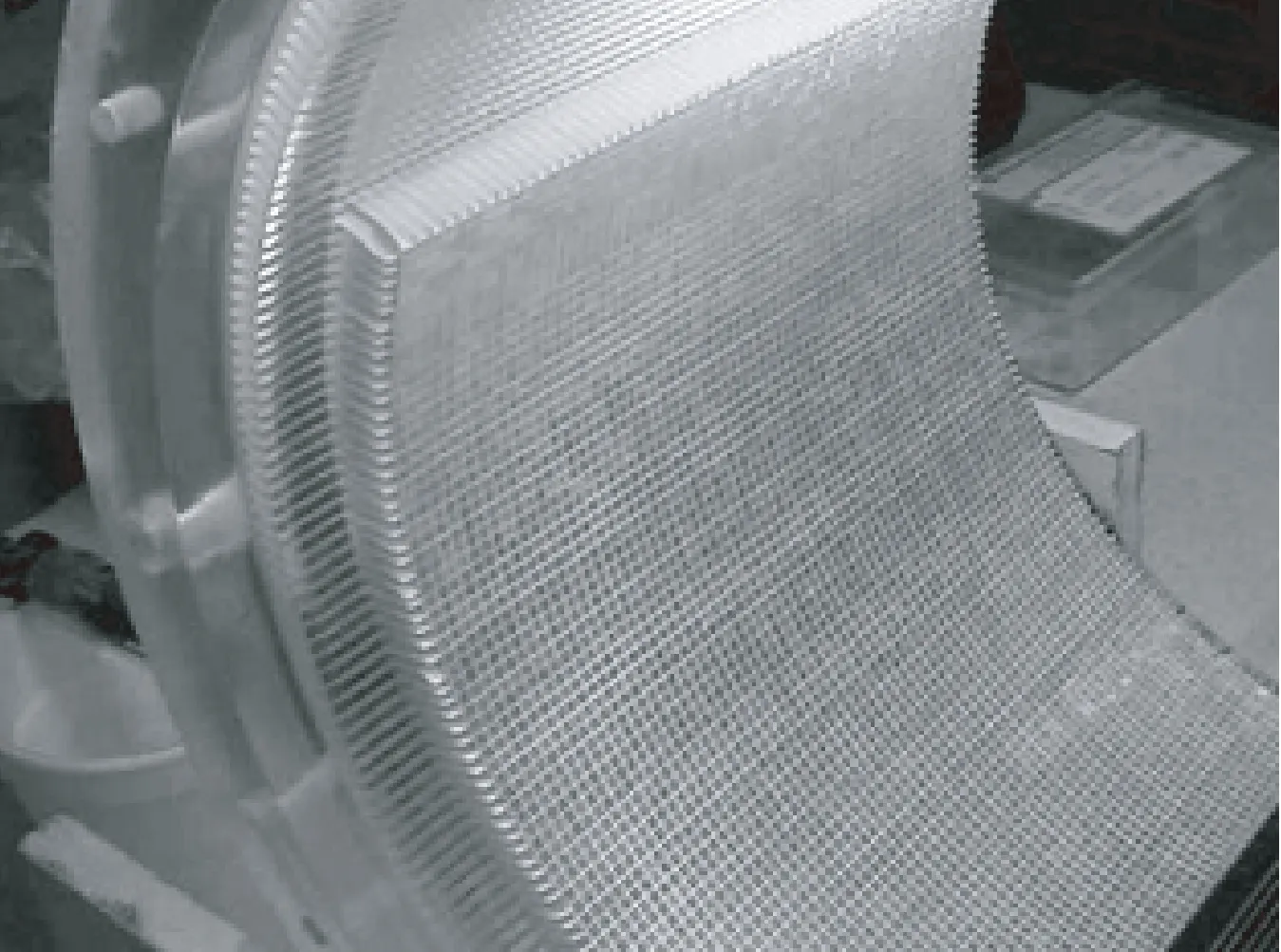
典型的环形探测器如图1所示,多个探测器单元排列成封闭的环,多个环并列构成探测器阵列,即环形探测器。环形探测器在小动物PET中应用较多,具有结构简单、灵敏度高、成像效果较好等优点,但是结构不够灵活,DOI影响较大。探测器单元中闪烁晶体的特性、尺寸、切割方式都对探测器的性能影响很大,如晶体宽度决定了分辨率极限值,同时光电倍增管的特性会影响系统空间分辨率。另外,探测器单元的排列方式也影响到PET的DOI效应大小。

图1 环形探测器Fig.1 Detector rings
CTI公司的ECAT-713[3]和Hamamastu公司的SHR-2000[4]是最早的专门用于动物PET成像的扫描仪,其空间分辨率基本与医用人体PET相同,无法满足动物研究,但是确立了动物PET的概念。
ECAT-713系统使用了与医用人体PET一样的探测器单元,BGO晶体阵列耦合到一对双头光电倍增管(photomultiplier tube,PMT)上,如图2所示。系统采用2D采集模式,噪声低,成像质量好,但会降低系统灵敏度,在小动物PET灵敏度普遍较低的情况下较少采用。

图2 ECAT-713使用的BGO耦合PMT探测器单元Fig.2 ECAT-713 detector unit: BGO coupled to PMT
SHR-2000系统使用了与医用人体PET相似的结构。使用PS-PMT(position sensitive PMT,位置灵敏光电倍增管)取代PMT。PS-PMT结构紧凑,死区较小,对晶体的定位更加准确。系统视野区域较大,可进行猴子的PET成像。SHR系列产品最新型号为SHR-41000[5]。系统采用双层LYSO晶体结构,两层晶体阵列交错半个像素,能够识别DOI信息。晶体与探测器之间加入1 mm厚的光导耦合,解决了PS-PMT边缘的死区问题。探测器环直径减小,专用于小动物的全身成像。
Sherbrooke PET第一次使用了基于雪崩二极管(avalanche photo diode,APD)的离散闪烁探测器,每个BGO晶体独立耦合到一个APD上[6]。使用APD探测器,显著提高了系统空间分辨率,适用于小动物PET。APD增益对温度非常灵敏,系统必须在恒温状态下工作。
布鲁塞尔自由大学曾开发出一套使用BaF2闪烁晶体材料的小动物PET系统[7],读出系统采用低压多级线室(low pressure multistep wire chamber)结构。BaF2晶体光输出较低,制约了系统性能。
由加州大学洛杉矶分校(University of California at Los Angeles,UCLA)研发的microPET[8-10]系统已经发展了3代,产品通过与Concord/Siemens公司合作,成功实现了商业化。
microPET是UCLA开发的第一代小动物PET型号,系统第一次将LSO晶体应用在小动物PET上,光电探测器使用MC-PMT(multi-channel PMT,多通道光电倍增管),MC-PMT具有能够分辨小晶体、增益高、串扰低、结构紧凑等特点。探测器单元使用光纤束连接晶体和探测器,主要目的是消除MC-PMT边缘死区对成像结果的影响,同时提供了PET与核磁共振成像(magnetic resonance imaging,MRI)同机融合的可能性。探测器固有分辨率为1.68 mm,是当时分辨率最高的多环PET系统,但是轴向视野小导致灵敏度不高。

表1 使用环形探测器的小动物PET系统性能比较
MicroPET II[11-13]是第二代microPET产品,其探测器单元如图3(a)所示。在晶体方面,改变了切割尺寸和安装工艺,横截面变小可以提高探测器的固有分辨率,长度增加能够改善系统灵敏度;晶体两端放置了网格矩阵,保证了晶体阵列间隔均匀;每一行晶体之间加入反光层,光学隔离相邻晶体,使光收集效率最大化(如图3(b)所示)。系统固有空间分辨率达到1.05 mm,并保持了一定的能量和时间分辨率。

图3 MicroPET II。(a)探测器单元;(b)晶体阵列Fig.3 MicroPET II. (a)Detector Unit; (b)Crystal Array
ATLAS[14](advanced technology laboratory animal scanner)是第一台具有DOI能力的小动物PET扫描仪,可与高分辨率CT图像同轴融合。采用LGSO和GSO晶体构成Phoswich(phosphor sandwich,磷三明治)结构,Phoswich像素阵列耦合PS-PMT。通过测量每一个事件的光脉冲衰减时间(LGSO:40ns;GSO:60ns),得到DOI信息。较小的探测器环能够改善系统灵敏度。
从以上机型的特点可以看出,小动物PET环形探测器的发展主要体现在晶体材料及切割尺寸、排列方式的变化,以及所选取光电倍增管性能的提升,同时晶体和光电倍增管的耦合方式也在不断改进。小动物PET经历了从单纯追求高分辨率到同时追求高分辨率和高灵敏度的转变,性能如表1所示。由于很难同时提高分辨率和灵敏度,所以最新的探测器设计均是在两者之间寻找平衡。
1.2平板型探测器在小动物PET中的应用
采用平板型探测器的小动物PET一般由相对安装在旋转机架上的平板型探测器构成,可以是双平板结构或四平板结构。如图4所示。平板PET的探测器可根据被测动物调节探测器的间距和角度,使用灵活,且其灵敏度高于传统环形PET的灵敏度。探测器间距会影响γ光子入射角,从而影响探测器分辨率和灵敏度。晶体的类型、长度同时影响探测器的分辨率和灵敏度,及光电探测器的特性,以及两者的耦合方式。

图4 平板型探测器Fig.4 Flat panel detectors
HIDAC-PET[2]系统使用了比较少见的气体探测器。探测器基于多丝正比室(multi-wire proportional chamber,MWPC)技术,可进行小动物全身成像,图像分辨率能够提高到0.7 mm,是现有小动物PET的最高值。HIDAC探测器能识别DOI信息,但散射符合较多,灵敏度低,没有能量分辨能力。

表2 使用平板型探测器的小动物PET系统性能比较
MAD-PET采用了LSO耦合APDs的结构[15]。晶体与APDs探测器像素一对一耦合。系统包含2个扇区,每个扇区有3个探测器模块,2个扇区可围绕被探测物旋转。与同样使用APDs探测器的Sherbrooke PET相比,能量分辨率好,分辨率有所提高。

图5 4个探测器的YAP-PET系统[19]Fig.5 Quad-detector YAP-PET system[19]
YAP-PET使用了YAP:Ce晶体作为闪烁体[18-19]。这种晶体具有闪烁快、光输出高、密度高、物理性能好等优点。晶体间加入反光层,晶体阵列直接与PS-PMT耦合组成探测器模块。
YAP-(S)PET是YAP-PET的升级型号,可以进行PET/SPECT数据采集[20]。系统沿用了YAP-PET的探测器结构,但是晶体长度减少了5 mm,降低了DOI效应对分辨率一致性的影响。
SmartPET(The small animal reconstruction tomograph for PET) 系统由两个相对安装在旋转机架上的HPGe(high-purity germanium,高纯度锗)平板型探测器组成,每个探测器包含一块两面切割为正交带状的平面Ge晶体[21]。SmartPET采用数字脉冲波形分析(pulse shape analysis,PSA)技术,分析探测器的响应脉冲,获得时间、位置和能量信息。
平板型探测器一般具有较高的分辨率和灵敏度,性能如表2所示。但是,平板型PET的几何结构特点导致其计数效率低于传统的环形PET,且其缺失角度较大,导致采集信息缺失,因而降低了重建图像的准确度,这使得平板型PET的发展受到限制。平板PET探测器在设计时除了要考虑到使用灵活性、高分辨率、高灵敏度外,如何提高重建图像的准确度仍将是一个重大的课题。
1.3商业化小动物PET的探测器
截止到2006年,已经商业化的小动物PET机型有5个:eXplorer Vista(General Electric Healthcare),microPET Focus(Concord Microsystems Inc.),Quad-HIDAC(Oxford Positron Systems Ltd.),MOSAIC(Philips Medical Systems),YAP-PET(I.S.E. Srl,Italy)[22]。Quad-HIDAC(4探头HIDAC系统)和YAP-PET在前文已有介绍,以下比较GE、Siemens和Philips三家公司的代表产品。
MicroPET Focus是第三代microPET系统[23]。该系统晶体横截面变大,长度变短,牺牲了一些空间分辨率,在保持系统灵敏度的同时改善了全视野分辨率的不均匀性;光纤长度减小,降低了光传输过程的损耗。
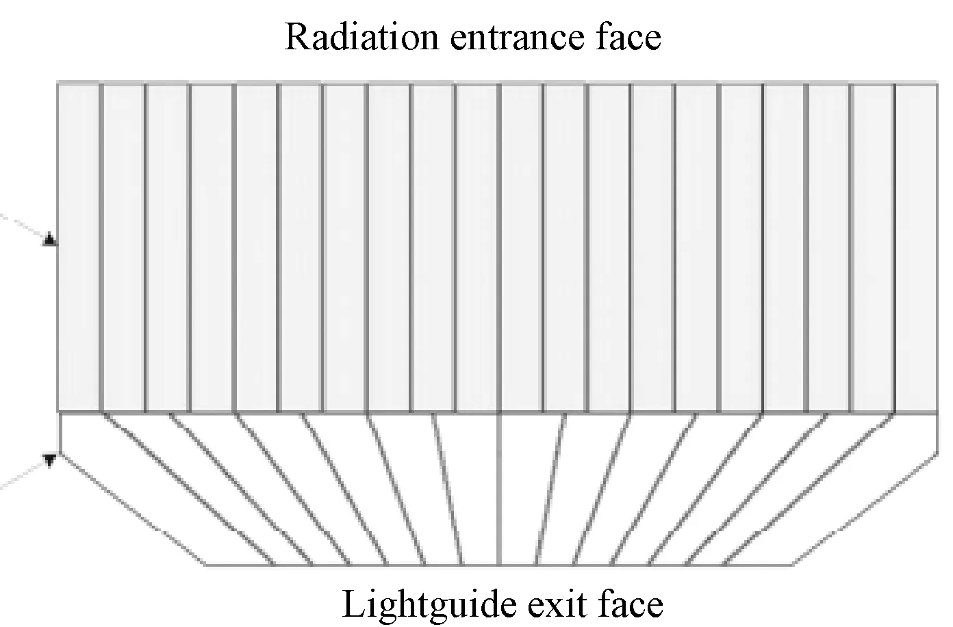
Inveon small animal PET (Siemens Medical Solutions)是microPET Focus的后续产品型号[24-25]。Inveon系统使用了与Focus系统一样的晶体,晶体阵列变大,使用锥形多像素光导耦合到PS-PMT,其探测器单元如图6所示。大面积晶体阵列耦合小面积PS-PMT能够提高光子吸收效率,降低光电探测器数量,提高轴向视野。这样,系统能够进行小动物全身成像。
CNN具有2种特性:局部连接和参数共享。CNN中相邻2层的连接方式为局部连接,当前层每个神经元的值是对上一层进行卷积操作得到的,且每次卷积的参数相同。如图1所示。相同颜色的线表示相同的参数,li+1层神经元的值依赖于上一层神经元的值, li+1 层每个神经元共享参数。

图6 使用锥形多像素光导的Inveon探测器单元[24]Fig.6 The Inveon detector unit using a tapered multiple-element lightguide[24]
eXplore VISTA Small Animal PET[26]是GE公司开发的专门用于啮齿类小动物PET的成像系统。系统使用双层晶体Phoswich结构修正DOI误差,上层晶体使用LYSO,下层晶体使用GSO,根据两种晶体的衰减时间不同来得到DOI信息。光电探测器使用PS-PMT。系统固有空间分辨率和灵敏度均达到较高水平,能量分辨率要比单层闪烁体探测器稍差。
MOSAIC small animal PET系统在空间分辨率方面进行了一定的妥协,尽量提高视野范围,其灵敏度在反映低比活度的放射性配体时表现仍可接受[27-29]。原型机使用GSO耦合PMTs的探测器结构,后使用LYSO晶体替换GSO晶体,LYSO具有更高的阻断能量和光输出。闪烁晶体和PMTs之间采用连续带沟槽的光导连接,如图7所示。连续光导能够最小化探测器死区的影响,沟槽结构是为了更好地分辨晶体。MOSAIC系统由于具有高等效噪声计数率(Noise Equivalent Count Rate,NECR)和大视野区域,可以进行高速全身小动物PET成像。

图7 MOSAIC系统连续光导和LYSO晶体[27]Fig.7 The continuous light guide and LYSO in MOSAIC[27]
商业化产品大多采取环形探测器结构,使用比较成熟的技术,在分辨率、灵敏度、成本和系统稳定性之间寻找平衡,尤其无法兼顾分辨率和灵敏度,但多以追求高的空间分辨率为主,如表3所示。

表3 部分商业化小动物PET性能比较
2 小动物PET探测器技术挑战与展望
2.1研究结构简单、成本较低的DOI修正方法
目前,已经有多种修正DOI误差的方法提出,比如前面已经提到的Phoswich结构。同样采用Phoswich结构的还有Hyun 等设计的高性能TraPET[30],其探测器模块由整块锥形LSO晶体连接LuYAP晶体阵列组成,通过分析SiPM(Si PMT,硅光电倍增管)阵列输出脉冲的波形获取DOI信息。
Nishikido 等开发出基于4层LYSO晶体阵列一对一耦合PSAPD阵列的小动物PET原型机,通过识别伽马射线穿过每层晶体的位置获取DOI信息[31]。
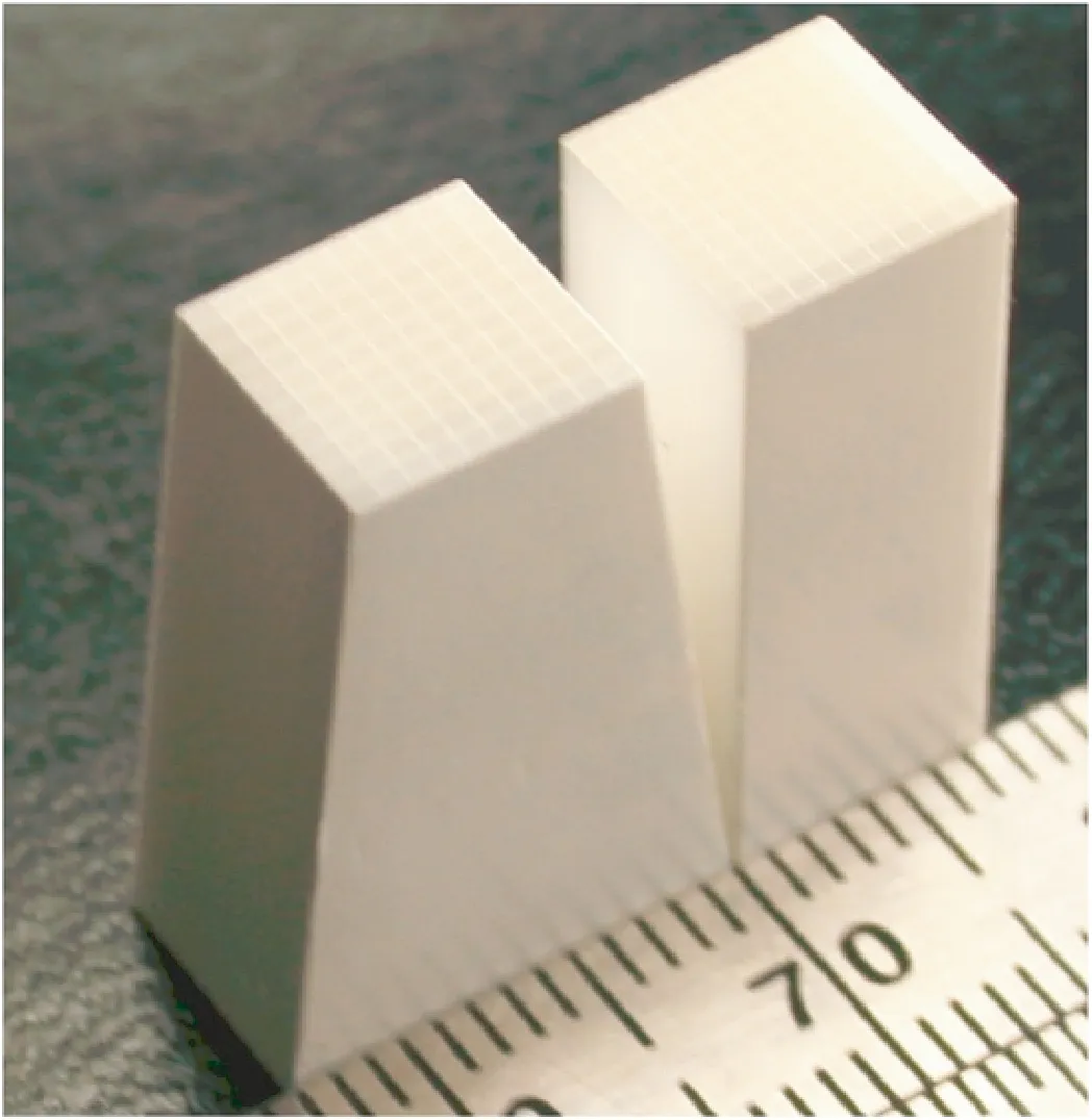
近年来,很多具有DOI能力的小动物PET采用锥形晶体阵列两端耦合PSAPD的探测器模块,锥形晶体阵列如图8所示。锥形结构可大大减小探测器间距,显著提高系统灵敏度。St James 等设计的小动物PET探测器,在通道数不变的情况下,比采用传统矩形晶体阵列的探测器灵敏度提高了64%[32]。Yang等设计的小动物PET探测器,灵敏度也实现了41%的提高,同时具有2.6mm的DOI分辨率[33]。Rodríguez-Villafuerte等提出的小鼠脑部PET,基于像素锥形晶体和PSAPDs双端读出方式,其深度编码精度达到2 mm,并且获得(0.70±0.05)mm的超高分辨率。此外,还有基于连续晶体探测器模块的DOI修正方法[34]。

图8 锥形晶体阵列和矩形晶体阵列[33]Fig.8 A tapered array together with a cuboid array[33]
这些方法从实验上证明DOI引起的误差可以进行修正,但都会导致硬件成本增加[32]。因此,研究出能够减少硬件消耗,尤其是通过软件实现的方法,是一个很有潜力的方向。
2.2开发性能更好的新型半导体探测器材料
2.2.1SiPM(silicon photomultiplier)
SiPM又称盖革模式雪崩二极管(Geiger mode avalanche photodiode,GAPD)、SSPM(solid state photomultiplier,固态光电倍增管),具有结构紧凑、增益高、响应迅速、偏压低等优点,近年来逐渐取代传统PMT,在PET上得到应用。SiPM对磁场不敏感的特性使PET可与MRI结合[35],其快速响应时间能够满足TOF-PET(time of flight,飞行时间)技术的要求[36]。
Kwon等提出LGSO晶体阵列耦合SiPM阵列的小动物PET探测器[37]。Llosá等提出基于连续LYSO晶体耦合集成SiPM阵列的PET探测器探头设计,通过算法获取DOI信息,使用5 mm厚度晶体可实现0.7 mm半高宽(full width at half maximum,FWHM)的空间分辨率[38]。Cerello 等提出基于连续LYSO晶体双面耦合SiPM阵列的PET探测器模块,该探测器可通过DOI信息去除视差,并获得各向相近的高分辨率,而且能够应用TOF技术精确地测量湮灭事件在响应线上的位置[39]。
2.2.2CdTe(碲化镉)、CdZnTe(碲锌镉)
CdTe、CdZnTe等半导体材料能够直接将伽马射线转换成电子,灵敏度高,能量分辨率好,阻止本领高,可用于制造不需闪烁晶体和光电倍增管的新型PET探测器。
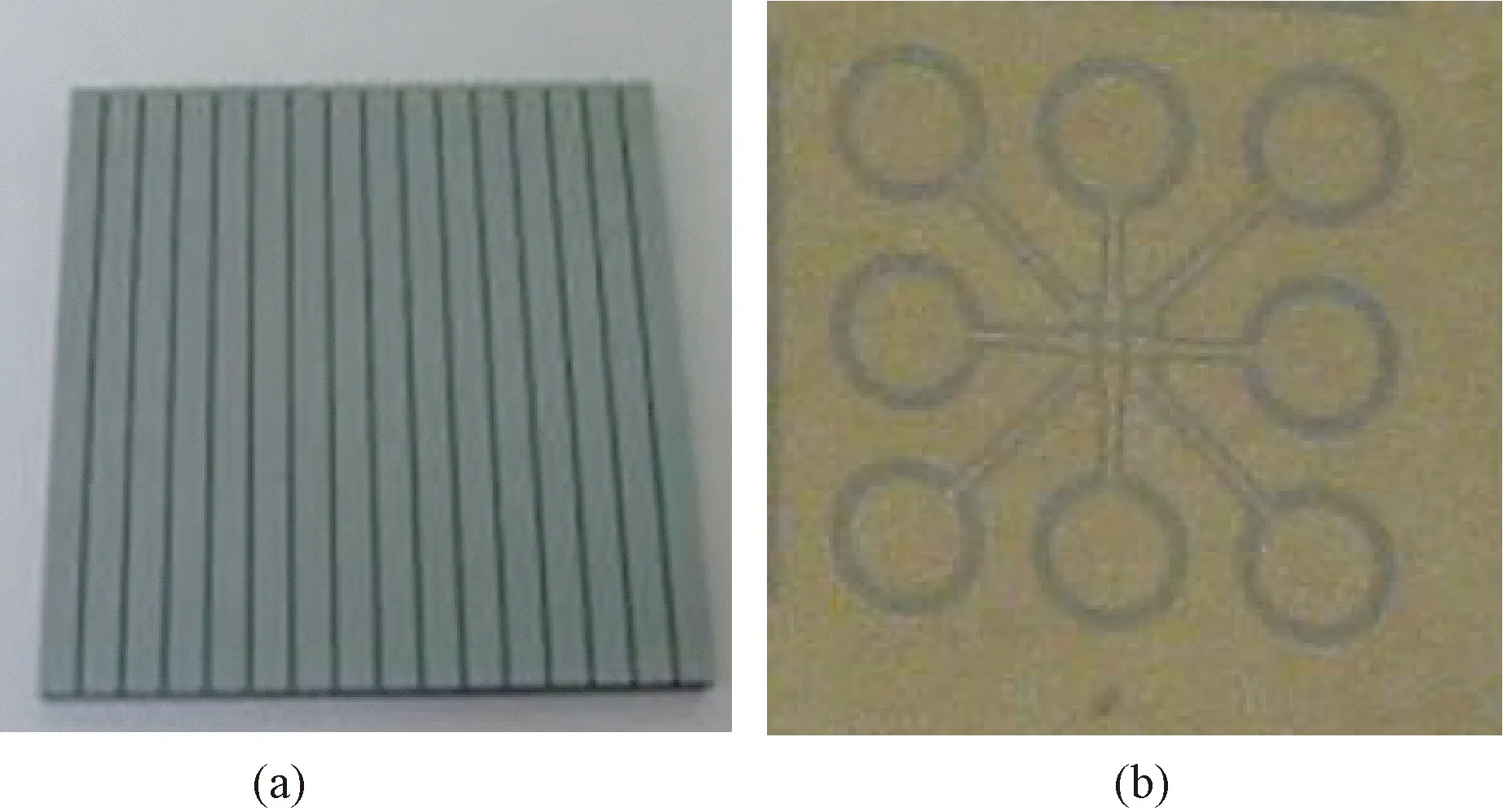
Ishii等开发的小动物PET探测器模块由双层毫米级带状CdTe探测器组成,具有DOI能力,可实现0.8 mm FWHM的FOV中心分辨率[40]。该团队又开发出基于二维位置敏感型带状CdTe探测器(见图9(a))的高分辨率PET,并提出叠加这种新型CdTe探测器可获得具有DOI能力的三维超高分辨率PET[41]。Ario等研究了一种肖特基CdTe二极管探测器,得到1.2% FWHM(511keV)的能量分辨率和6 ns FWHM(500 keV)的时间分辨率。这表明,CdTe探测器可用于开发新型PET等核医学探测器[42]。
CdZnTe探测器已实现在小动物PET中的应用[43],并且体现出高性能特点。Yin等提出的高像素(350 μm)CdZnTe小动物PET探测器(见图9(b)),可获得优于700 μm的高空间分辨率[44-45]。Gu等开发出基于CdZnTe晶体探测器的小动物PET,CZT晶体两面为交叉带状电极,使探测器具有3D位置灵敏能力,能够获得(0.44±0.07)mm的分辨率和3.06%±0.39%(511 keV)的能量分辨率[46]。Yoon等提出的CZT Compton PET探测器使用小像素CdZnTe,通过获取康普顿散射信息的方法,使能量分辨率提高了2.75倍[47]。

图9 新型CdZn/CdZnTe探测器。(a)二维位置敏感型带状CdTe探测器[41];(b)高像素CdZnTe探测器[44]Fig.9 Novel CdZn/CdZnTe detectors. (a) Two-dimensional position-sensitive strip CdTe detector[41]; (b) Highly pixelated CdZnTe detector[44]
目前,SiPM已在PET探测器上得到部分应用,但由于其造价较高等因素未广泛应用,因此开发新工艺、降低成本是其得到推广的前提。CdTe、CdZnTe探测器的性能直接受其晶体工艺技术和电子学结构的影响[48],开发出具有较高电阻率、较好完整性和较大单片面积的晶体,对设计更高性能的半导体探测器具有十分重要的意义。
2.3通用的前端电子学设计
在探测器单元中,前端电子学线路包括放大甄别电路和符合系统电路两部分,分别对光电转换部分输出的电信号进行放大、时间及能量甄别和符合判断等处理。目前开发的小动物PET系统,其探测器的前端电子学线路都是根据系统参数特殊定制的,除少数同系列产品可以通用外(microPET P4与microPET II使用了基本相同的处理电路),基本不具备跨平台移植性。无论是位置译码读出方式,还是像素独立读出方式,输出信号处理过程具有一定的相似性,理论上可以进行跨平台移植。
设计具有一定通用性的前端电子学线路能够节省大量重复工作,可将研究重心转移到探测器组态、新材料研发和改进等方面,有利于小动物PET的快速发展。目前,已经有适用于多种基于SiPM小动物PET探测器的读出电路模块设计[49]。
3 结束语
小动物PET技术已经发展了20余年,探测器也由改装人体PET探测器过渡到专门设计,性能有了很大提高,并且应用最新技术、最新材料,在科研和商用领域都取得了很大的进步。设计高性能的小动物PET探测器,最大的难点在于兼顾空间分辨率和灵敏度。未来小动物PET探测器技术的研究重点仍然在探测器单元所包含的晶体、光电探测器、前端电子学层面,对DOI方法的改进、新型半导体探测器的研发和通用电子学的设计是十分有意义的发展方向。
[1] Myers R, Hume S. Small animal PET[J]. Eur Neuropschopharmacology, 2002,12:545-555.
[2] Jeavons AP,Chandler RA, Dettmar CAR.A 3D HIDAC-PET camera with sub-millimeter resolution for imaging small animals[J]. IEEE Trans on Nucl Sci,1999,46(3):468-473.
[3] Cutler PD, Cherry SR, Hoffman EJ,etal. Design features and performance of a PET system for animal research[J]. J Nucl Med,1992,33(4):595-604.
[4] Watanabe M, Uchida H, Okada H,etal. A high resolution PET for animal studies[J]. IEEE Trans on Med Imag,1992,11(4):577-580.
[5] Yamada R, Watanabe M, Omura T,etal. Development of a small animal PET scanner using DOI detectors[J]. IEEE Trans on Nucl Sci,2008,55(3):906-911.
[6] Lecomte R, Cadorette J, Richard P,etal. Design and engineering aspects of a high resolution positron tomograph for small animal imaging[J]. IEEE Trans on Nucl Sci,1994,41(4):1446-1452.
[7] Bruydonckx P, Liu xuan, Tavernier S,etal. Performance study of a 3D small animal PET scanner based on BaF2crystals and a photo sensitive wire chamber[J]. Nucl Instr and Meth in Phys Res A,1997,392:407-413.
[8] Cherry SR, Shao Yiping, Silverman RW,etal. MicroPET: A high resolution PET scanner for imaging small animals[J]. IEEE Trans on Nucl Sci,1997,44(3):1161-1166.
[9] Slates R, Cherry SR, Boutefnouchet A,etal. Design of a small animal MR compatible PET scanner[J]. IEEE Trans on Nucl Sci,1999,46(3):565-570.
[10] Chatziioannou AF, Cherry SR, Shao Yiping,etal. Performance evaluation of microPET:A high-resolution lutetium oxyorthosilicate PET scanner for animal imaging[J]. J Nucl Med,1999,40(7):1164-1175.
[11] Tai Yuanchuan, Chatziioannou AF, Silverman RW,etal. MicroPET II: An ultra-high resolution small animal PET system[C]. IEEE Nuclear Science Symposium Conference Record.2002:1848-1852.
[12] Tai Yuanchuan, Chatziioannou AF, Yang Yongfeng,etal. MicroPET II: Design, development and initial performance of an improved microPET scanner for small-animal imaging[J]. Phys Med Biol, 2003,48:1519-1537.
[13] Yang Yongfeng, Tai Yuanchuan, Siegel S,etal. Optimization and performance evaluation of the microPET II scanner for in vivo small-animal imaging[J]. Phys Med Biol, 2004,49:2527-2545.
[14] Seidel J, Vaquero JJ, Pascau J,etal. Features of the NIH ATLAS small animal PET scanner and its use with a coaxial small animal volume CT scanner[C]. IEEE International Symposium on Biomedical Imaging.2002:545-548.
[15] Ziegler SI, Pichler BJ, Boening G,etal. A prototype high-resolution animal positron tomograph with avalanche photodiode arrays and LSO crystals[J]. Eur J Nucl Med,2001,28(2):136-143.
[16] Weber S, Terstegge A, Herzog H,etal. The design of an animal PET: flexible geometry for achieving optimal spatial resolution or high sensitivity[J]. IEEE Trans on Med Imag,1997,16(5):684-689.
[17] Weber S, Herzog H, Cremer M,etal. Evaluation of the TierPET system[J]. IEEE Trans on Nucl Sci,1999,46(4):1177-1183.
[18] Guerra AD, Domenico GD, Scandola M,etal. High spatial resolution small animal YAP-PET[J]. Nucl Instr and Meth in Phys Res A,1998,409:537-541.
[19] Guerra AD, Domenico GD, Scandola M,etal. YAP-PET: First results of a small animal positron emission tomograph based on YAP:Ce finger crystals[J]. IEEE Trans on Nucl Sci,1998,45(6):3105-3108.
[20] Guerra AD, Bartoli A, Belcari N,etal. Performance evaluation of the fully engineered YAP-(S)PET scanner for small animal imaging[J]. IEEE Trans on Nucl Sci,2006,53(3):1078-1083.
[21] Cooper RJ, Boston AJ, Boston HC,etal. Positron Emission Tomography imaging with the SmartPET system [J]. Nuclear Instruments and Methods in Physics Research Section A: Accelerators, Spectrometers, Detectors and Associated Equipment, 2009,606(3): 523-532.
[22] Larobina M, Brunetti A, Salvatore M. Small animal PET:A review of commercially available imaging systems[J]. Current Medical Imaging Reviews,2006,2(2):187-192.
[23] Tai Yuanchuan, Ruangma A, Rowland D,etal. Performance Evaluation of the microPET Focus:A Third-Generation microPET Scanner Dedicated to Animal Imaging[J]. J Nucl Med,2005,46(3):455-463.
[24] Mintzer RA, Siegel SB. Design and performance of a new pixilated LSO/PSPMT Gamma-Ray detector for high resolution PET imaging[C].//Dressendorfer PV,eds. IEEE Nuclear Science Symposium Conference Record. Honolulu:IEEE,2007: 3418-3422.
[25] Visser EP, Disselhorst JA, Brom M,etal. Spatial resolution and sensitivity of the Inveon small-animal PET scanner[J]. J Nucl Med, 2009,50(1):139-147.
[26] Wang Yuchuan, Seidel J, Tsui BMW,etal. Performance evaluation of the GE Healthcare eXplore VISTA dual-ring small-animal PET scanner[J]. J Nucl Med, 2006,47(11):1891-1900.
[27] Surti S, Karp JS, Perkins AE,etal. Design evaluation of A-PET: A high sensitivity animal PET camera[J]. IEEE Trans on Nucl Sci,2003,50(5):1357-1363.
[28] Surti S, Karp JS, Perkins AE,etal. Imaging performance of A-PET: A small animal PET camera[J]. IEEE Trans on Med Imag, 2005,24(7):844-852.
[29] Huisman MC, Reder S, Weber AW,etal. Performance evaluation of the Philips MOSAIC small animal PET scanner[J]. Eur J Med Mol Imaging, 2007,34:532-540.
[30] Hyun Chung Y, Yeon Hwang J, Baek C-H,etal. TraPET: High performance small animal PET with trapezoidal phoswich detector[J]. Nuclear Instruments and Methods in Physics Research Section A: Accelerators, Spectrometers, Detectors and Associated Equipment, 2011,652(1): 802-805.
[31] Nishikido F, Inadama N, Oda I,etal. Four-layer depth-of-interaction PET detector for high resolution PET using a multi-pixel S8550 avalanche photodiode[J]. Nucl Instr and Meth in Phys Res A, 2010,621(1-3): 570-575.
[32] St James S, Yang Y, Bowen SL,etal. Simulation study of spatial resolution and sensitivity for the tapered depth of interaction PET detectors for small animal imaging[J]. Phys Med Biol, 2010,55(2): N63-74.
[33] Yang Y, James SS, Wu Y,etal. Tapered LSO arrays for small animal PET[J]. Phys Med Biol, 2011,56(1): 139-153.
[34] Carles M, Lerche CW, Sánchez F,etal. Performance of a DOI-encoding small animal PET system with monolithic scintillators [J]. Nucl Instr and Meth in Phys Res A, 2012,69: 5317-5321.
[35] Zaidi H, Del Guerra A. An outlook on future design of hybrid PET/MRI systems [J]. Med Phys, 2011,38(10): 5667-5689.
[36] Yeom JY, Vinke R, Levin CS. Optimizing timing performance of silicon photomultiplier-based scintillation detectors[J]. Phys Med Biol, 2013,58(4): 1207-1220.
[37] Kwon SI, Lee JS, Yoon HS,etal. Development of small-animal PET prototype using silicon photomultiplier (SiPM): initial results of phantom and animal imaging studies[J]. J Nucl Med, 2011,52(4): 572-579.
[38] Llosá G, Barrillon P, Barrio J,etal. High performance detector head for PET and PET/MR with continuous crystals and SiPMs [J]. Nucl Instr and Meth in Phys Res A, 2013,702: 3-5.
[39] Cerello P, Pennazio F, Bisogni MG,etal. An innovative detector concept for hybrid 4D-PET/MRI imaging[J]. Nucl Instr and Meth in Phys Res A, 2013,702: 6-9.
[40] Ishii K, Kikuchi Y, Matsuyama S,etal. First achievement of less than 1mm FWHM resolution in practical semiconductor animal PET scanner [J]. Nucl Instr and Meth in Phys Res A, 2007,576(2-3): 435-440.
[41] Ishii K, Kikuchi Y, Takahashi K,etal. Development of a new two-dimensional position-sensitive detection based on resistive charge division and using CdTe detectors for a high-resolution semiconductor-based PET scanning [J]. Nucl Instr and Meth in Phys Res A, 2011,631(1): 138-143.
[42] Arino G, Chmeissani M, De Lorenzo G,etal. Energy and coincidence time resolution measurements of CdTe detectors for PET [J]. J Instrum, 2013,8:C02015.
[43] Drezet A, Monnet O, Mathy F,etal. CdZnTe detectors for small field of view positron emission tomographic imaging [J]. Nucl Instr and Meth in Phys Res A, 2007,571(1-2): 465-470.
[44] Yin Y, Chen X, Wu H,etal. 3-D spatial resolution of 350μm pitch pixelated CdZnTe detectors for imaging applications [J]. IEEE Trans on Nucl Sci, 2013,60(1): 9-15.
[45] Yin Y, Chen X, Komarov S,etal. Study of highly pixelated CdZnTe detector for PET applications [J]. Physics Procedia, 2012, 37: 1537-1545.
[46] Gu Y, Matteson JL, Skelton RT,etal. Study of a high-resolution, 3D positioning cadmium zinc telluride detector for PET[J]. Physics in medicine and biology, 2011,56(6): 1563-1584.
[47] Yoon C, Lee W, Lee T. Simulation for CZT Compton PET (Maximization of the efficiency for PET using Compton event) [J]. Nucl Instr and Meth in Phys Res A, 2011,652(1): 713-716.
[48] Washington AL, Teague LC, Duff MC,etal. The effect of various detector geometries on the performance of CZT using one crystal[J]. J Electron Mater, 2011,40(8): 1744-1748.
[49] Ko GB, Yoon HS, Kwon SI,etal. Development of a front-end analog circuit for multi-channel SiPM readout and performance verification for various PET detector designs [J]. Nucl Instr and Meth in Phys Res A, 2013,703: 38-44.
DevelopmentofSmallAnimalPETDetector
YANG Kun1*LI Zhen1YANG Yong-Xin1WANG Wen-Li1REN Qiu-Shi2
1(CollegeofQualityandTechnicalSupervision,HebeiUniversity,Baoding071000,China)2(CollegeofEngineering,PekingUniversity,Beijing100871,China)
The performance of PET for small animal examination is largely dependent on the performance of the detector. Therefore, the designing for detector units with high resolution and high sensitivity becomes one of crucial issues in the investigation of small animal PET. This paper reviews the characteristics and performance of ring or flat panel detectors and a number of commercial PET for small animal examination. We made analysis with and summarized the development of the PET detector technology from components of a detector unit such as crystal and PMT. The development directions of determining new DOI, exploring new semiconductor detectors and designing for universal electronics are discussed as well.
small animal PET; detector; depth of interaction (DOI) methods; semiconductor detectors; universal electronics
10.3969/j.issn.0258-8021. 2014. 02.012
2013-09-27, 录用日期:2014-03-11
国家自然科学基金(11104058);河北省自然科学基金(A2011201155);国家科技部重大科学仪器专项(2011YQ03011405)
R318.08
A
0258-8021(2014) 02-0218-09
*通信作者。E-mail: yangkun9999@hotmail.com
