Treating and management in acute Laugier's fracture: a case report
2014-03-22PredragGruborGabrieleFalzaranoAntonioMediciLuigiMeccarielloMilanGrubor
Predrag Grubor, Gabriele Falzarano, Antonio Medici, Luigi Meccariello, Milan Grubor
1Orthopaedics and Traumatology Clinic Banja Luka, Republic of Srpska, Bosnia and Herzegovina
2U.O.C. Orthopedics and Traumatology , AO G.Rummo, Benevento, Italy
3Department of Medical and Surgical Sciences and Neuroscience, Section of Orthopedics and Traumatology, University of Siena, University Hospital “Santa Maria alle Scotte”, Siena, Italy
Treating and management in acute Laugier's fracture: a case report
Predrag Grubor1*, Gabriele Falzarano2, Antonio Medici2, Luigi Meccariello3, Milan Grubor1
1Orthopaedics and Traumatology Clinic Banja Luka, Republic of Srpska, Bosnia and Herzegovina
2U.O.C. Orthopedics and Traumatology , AO G.Rummo, Benevento, Italy
3Department of Medical and Surgical Sciences and Neuroscience, Section of Orthopedics and Traumatology, University of Siena, University Hospital “Santa Maria alle Scotte”, Siena, Italy
Laugier's fractures are rare because they are located deep in the elbow joint and are thus protected from any direct trauma. Laugier's fractures have been insufficiently described in the literature. Surgical treatment does not have an alternative, and timely diagnosis and surgical and physical therapy. We presented a case report of a 23 years' old man, sustained a Laugier’s fracture in June 2012 after falling from motocycle (low energy trauma) and hit with his flexed elbow against the street.
ARTICLE INFO
Article history:
Received 8 Jan 2015
Received in revised form 11 Jan 2015
Accepted 13 Jan 2015
Available online
Laugier's fracture
1. Introduction
Isolated fractures of the humeral capitellum and trochlea (Laugier’s fracture) are not common because they are located deep in the elbow joint and are thus protected from direct injuries. These fractures most often occur in case of a direct fall on the olecranon or on the fist, with the lower arm outstretched. After a fracture, the humeral capitellum and trochlea behave like free fragments which after sustaining the fracture most often stay within the intra-articular portion of the elbow, with the joint capsule preserved. Laugier was the first to provide an original description of the fracture in 1853, and as a result it is often referred to as Laugier’s fracture[1]. Fracture of humeral trochlea is usually associated with elbow dislocations or capitellar fracture. Isolated trochlea fracture in adult is a surgical rarity as compared to its capitellar counterpart[1,2]. This fracture is not mentioned in the standard orthopedic text book[3]. The rarity of this fracture is probably as a result of its anatomical location which is sited deep within the olecranon fossa. Therefore, there is still very little known regarding the mechanism of injury and the treatment results of this injury[3]. The clinical presentation of Laugier’s fractures is non-specific. It’s not very easy to understand this injury[3]. Most frequently, the patient complains of pain, uncharacteristic swelling and a limited range of motion at the elbow. These movements can fool you by forming an isosceles triangle when flexing the elbow or the Hueter sign in elbow extension. We would like to report one case of isolated displaced trochlea fracture that was treated and managed with open reduction and internal fixation in emergency.
2. Case presentation
The patient was a 23 years’ old man, sustained a fracture in June 2012 after falling from motocycle (low energy trauma) and hit with his flexed elbow against the street. He returned at home with normal pain at the elbow Joint, Aftertwo days, he went to the family doctor who based on clinical signs, diagnosed an elbow contusion and sent her back home with prescription of resting the arm for 10 d. After 10 d, the pain worsened. He went to emergency care and he did normal XR elbow’s projection. If we had a careful inspection of the anteroposterior radiograph of the elbow, we could find the fracture (Figure 1).
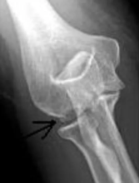
Figure 1. In posterior-anterior radiography, only a good view can show the fracture (see black arrow).
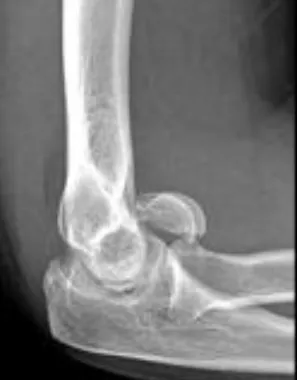
Figure 2. Lateral radiograph shows a semicircular (moon-like) dislocation line of Laugier's fracture which is above the radial head in front of the distal humerus.

Figure 3. 3D CT scan reconstruction.

Figure 4. Latero-lateral XR projection after the surgery.
The lateral radiograph shows a semicircular (moonlike) dislocation line of Laugier’s fracture which is above the radial head in front of the distal humerus (Figure 2). The rotation of the humeral capitellum and trochlea,inadequately placed elbow and radiograph can show a small portion of the subchondral bone, and thus render making a diagnosis and choice of treatment more difficult. We used 3D CT to indicate the size and position of the fracture (Figure 3). Having understood the severity of the injury, we subjected the patient at MRI scan of the elbow to understand the ligaments’ injures. We decide to operate in emergency because the Laugier’s fracture was old for 10 d. The surgery was performed applying an Esmarch bandage. The posterolateral Campbell approach was used; the ulnar nerve freed and mobilised for a distance of 12 cm. A tenotomy of the triceps brachii muscle and a posterior capsulectomy of the elbow joint were performed. After repositioning and temporarily fixing the humeral trochlea and head, we performed a permanent fixation using two screws (3.2 mm). The screws were inserted from the back, through the posterolateral and posteromedial pillars of the humeral condyle, to the front and medially. The tip of the screw was kept subchondrally in the bone of the humeral head,i.e.trochlea so that it did not go through the cartilage (Figures 4 and 5). The stitches of the wound were done by layers, and prior to that a transposition of the ulnar nerve was performed medially. A plaster cast was worn on the upper arm for three weeks, with mandatory prophylaxis of periarticular ossification by taking Celecoxib 200 mg twice a day for three weeks[4]. Upon removing the plaster cast, the patient underwent outpatient physical therapy, followed by inpatient physical therapy with full weight-bearing.

Figure 5. Posterior-anterior XR projection after the surgery.
3. Discussion
The actual mechanism of injury of this condition is still uncertain. There are numerous theories concerning the mechanism of injury for this fracture. The unique articular relation of distal humerus, with proximal ulna and radius results indifferent action of force at various angles of flexion[3]. The usual anterosuperior dislodgement of the fractured fragment into the coronoid fossae suggests anaxial loading force against the trochlea. The intact capitellum suggests that it is either spared or sub-jected to a lesser force[3]. A humeral capitellum fracture may be connected with soft tissue injuries: medial collateral ligament or lateral ligament complex[5]. There are many classifications about elbow’s fracture like Bryan and Morrey[1], McKee[6], AO classification[7] and Ring D[8]. The clinical symptomatology is not enough to understand the severity of the injury and the medical malpractice can be very common. For a correct diagnosis, we need the radiological imaging. XR of the isolated humeral trochlea fracture shows a half moon-shape osteochondral fragment in the lateral view, which sometimes can be mistaken for capitellum fracture[9]. Therefore, the antero-posterior radiograph which shows an irregularity of the trochleaolecranon articulation is crucial in differentiating these fractures. Correlation of the clinical findings always provides clinicians with more information[9]. If in doubt, a computer tomography scan can be helpful as 3D CT reconstructions[3]. We need also to understand the ligaments injuries with MRI[10]. Non-operative treatment has been recommended for undisplaced humeral trochlea fracture[6-11]. Unsatisfactory outcomes following conservative treatment were observed: dislocation of the trochlea, subluxation, luxation, contracture and elbow anchylosis. The autopsy established that the humeral capitellum was dislocated in a superior position in the anterior aspect of the humerus and it caused a limited elbow flexion[12]. The advantage of surgical treatment by stabilizing humeral capitellum and trochlea fractures with osteosynthetic material compared to conservative treatment was described in the late 1970s[12]. Nowadays, surgical stabilization or removal of the fractured fragment is universally acceptable if the repositioning by surgery is not satisfactory[13]. The small fragments can seriously damage the elbow joint if in a closed procedure, as there is an excessive pressure[14]. Early mobilization of the elbow is desirable and it depends on the quality of stabilisation and fixation of fragments, and the existence or lack of intraoperational iatrogenous injuries[13]. Mobilization is the best way to prevent elbow inflexibility and disability, which may be caused by a long plaster cast immobilization[13]. The patient regained 12°-155° of elbow motions 24 months post-operatively (Figure 6). InEnglish literature, we found only four case reports about an isolated displaced humeral trochlea fracture[3,11,15,16]. Three of these treated with screws and one with K wires[3,11,15,16]. From the results of our case, we notice that the early diagnosis and the early fixation in emergency of isolated displaced fracture of humeral trochlea using the screws can provide an adequate stable fixation for early mobilization. Functional assessment performed showed a good recovery of elbow ranges of motion with excellent results.
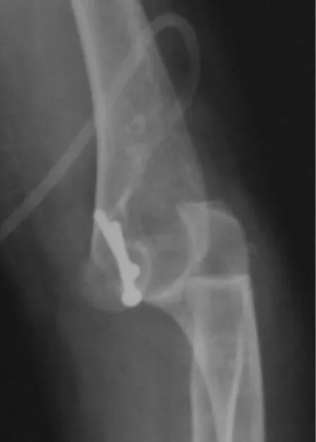
Figure 6. 24 months post-operatively, the patient regained a good extension of the elbow.
4. Conclusion
In summary, the mechanism of this rare injury is likely due to the direct impact on the flexed elbow. A good choice of radiological imaging permits to find and characterize this injure. Open reduction and internal fixation with screws not only restore the articular congruity but also provide a stable fixation which allows early mobilization and subsequently, a good functional result.
Conflict of interest statement
The authors report no conflict of interest.
[1] Dubberley JH, Faber KJ, Macdermid JC, Patterson SD, King GJ. Outcome after open reduction and internal fixation of capitellar and trochlear fractures. J Bone Joint Surg Am 2006; 88(1): 46-54.
[2] Ruszkowski I, Pečina M, i Muftić O. Temelji funkcionalne anatomije i biomehanike lakatnog zgloba. Arh Zast Majke i Djeteta 1978; 22: 181-198.
[3] Kwan MK , Khoo EH, Chua YP, Manso A. Isolated displaced fracture of humeral trochlea: A report of two rare cases. Injury Extra 2007; 38: 461-465.
[4] Saudan M, Saudan P, Perneger T, Riand N, Keller A, Hoffmeyer P. Celecoxib versus ibuprofen in the prevention of heterotopic ossification following total hip replacement: a prospective randomised trial. J Bone Joint Surg Br 2007; 89(2): 155-159.
[5] Dushuttle RP, Coyle MP, Zawadsky JP, Bloom H. Fractures of the capitellum. J Trauma 1985; 25(4): 317-321.
[6] McKee MD, Jupiter JB, Bamberger HB. Coronal shear fractures of the distal end of the humerus. J Bone Joint Surg Am 1996; 78(1): 49-54.
[7] Mueller ME, Nezarian S, Koch P, Schatzker J. The comprehensive clasification of fractures of long bones. Berlin: Springer - Verlag; 1990.
[8] Ring D, Jupiter J, Gulotta L. Articular fractures of the distal part of the humerus. J Bone Surg Am 2003; 85-A(2): 232-238.
[9] Yiannakopoulos CK, Vraggalas V, Darmanis S. Synchronous fractures of the trochlea and the radial neck without elbow dislocation. J Trauma 2002; 53(1): 125-130.
[10] Mak S, Beltran LS, Bencardino J, Orr J, Jazrawi L, Cerezal L, et al. MRI of the annular ligament of the elbow: review of anatomic considerations and pathologic findings in patients with posterolateral elbow instability. AJR Am J Roentgenol 2014; 203(6): 1272-1279.
[11] Foulk DA, Robertson PA, Timmerman LA. Fracture of the trochlea. J Orthopaed Trauma 1995; 9: 530-532,
[12] Wong AS, Baratz ME. Elbow fractures: distal humerus. J Hand Surg Am 2009; 34(1): 176-190.
[13] Poynton AR, Kelly IP, O’Rourke SK. Fractures of the capitellum - a comparison of two fixation methods. Injury 1998; 29(5): 341-343.
[14] Morrey BF. Current concepts in the treatment of fractures of the radial head, the olecranon and the coronoid. Instr Course Lect 1995; 44: 175-185.
[15] Nakatani T, Sawamura S, Imaizumi Y. Isolated fracture of the trochlea: a case report. J Shoulder Elb Surg 2005; 14(3): 340-343.
[16] Worrell RV. Isolated, displaced fracture of the trochlea. NY State J Med 1971; 71: 2314-2315.
ment heading
10.1016/S2221-6189(14)60070-6
*Corresponding author: Prof. Predrag Grubor, Ul.Aleja Svetog Save 20/24, 78 000 Banja Luka, Republic of Srpska, Bosnia and Herzegovina.
Tel: 00 387 65 513 115
E-mail: predraggrubor@gmail.com
Elbow joint trauma
Acute management
杂志排行
Journal of Acute Disease的其它文章
- The acute effect of the antioxidant drug “U-74389G” on red blood cells levels during hypoxia reoxygenation injury in rats
- Different profiles of mental and physical health and stress hormone response in women victims of intimate partner violence
- Time-critical AMI Detection: A novel and fast technique using the 12-lead ECG
- Epidemiological survey on scorpionism in Gotvand County, Southwestern Iran: an analysis of 1 067 patients
- Successful treatment of lower urinary tract obstruction with peritonealamniotic and vesicoamniotic shunting
- Simvastatin-induced Toxic Epidermal Necrolysis