Comparative evaluation of surgical alternatives in the treatment of acute cervical myelopathy and in the decompression of cervical spinal canal
2014-03-22borCziglczkiZoltPappCsabaPadnyiterBanczerowski
Gábor Czigléczki, Zoltán Papp, Csaba Padányi, Péter Banczerowski,*
1Department of Neurosurgery, Faculty of Medicine, Semmelweis University, Budapest, Hungary
2National Institute of Neurosurgery, Budapest, Hungary
Comparative evaluation of surgical alternatives in the treatment of acute cervical myelopathy and in the decompression of cervical spinal canal
Gábor Czigléczki1, Zoltán Papp2, Csaba Padányi2, Péter Banczerowski1,2*
1Department of Neurosurgery, Faculty of Medicine, Semmelweis University, Budapest, Hungary
2National Institute of Neurosurgery, Budapest, Hungary
Symptoms of cervical myelopathy are caused by the compression of the cervical spinal cord in the narrowed spinal canal. Several techniques including less invasive and minimally invasive methods have been developed with the aim of decompressing the cervical spinal canal, preserving posterior motion segments and paraspinal muscles as much as possible, reducing iatrogenic consequences and promoting faster recoveries of patients. The purpose of this article is to summarize these procedures and evaluate their efficacy with comparing them to each other. The applicable methods are presented shortly but the differences between them are discussed in details. Comprehensive examination did not reveal the proven superiority of any techniques and in most cases the less invasive or minimally invasive treatment choices should be individually determined, considering the location and extension of pathology and the familiarity of surgeon with techniques.
ARTICLE INFO
Article history:
Received 15 January 2015
Received in revised form 16 January 2015
Accepted 17 January 2015
Available online 18 January 2015
Acute cervical myelopathy
1. Introduction
Cervical myelopathy (CM) is a common disorder that affects primarily the middle-aged people, but as an acute disease can evolve at any age of patients. The natural history of CM has not explored entirely but multiple factors include static, dynamic and biomolecular factors[1-4]. All static factors such as spondylosis, degenerative disc disease, congenital stenosis and ossification of the posterior longitudinal ligament (OPLL) or ligamentum flavum are able to cause local ischemia, neurological injury and dysfunction by mechanical compression in the narrowed spinal canal[1]. Dynamic factors consist of dynamical changes in neck movements that cause repetitive axonal injuries by putting increased biomechanical forces on the spinal cord and narrowing the space within the spinal canal[1]. Biomolecular factors can be the causes of CM as well as the consequences of the aforementioned factors and include ischemic injury, excitotoxicity and neuronal apoptosis[1]. Symptoms of CM are diverse and can be divided in two main groups. Segmental symptoms involve radiating pain and neural deficit in the supply area of a nerve root. Long-tract symptoms include pathological reflexes, quadriparesis-plegia, sensory loss mainly on extremities and bladder-bowel dysfunction[2].
From multiple factors, dynamic factors especially in traumatic cases may be the main reasons of an acute disease with rapidly evolving symptoms. It is accepted that poor outcome may eventuate with delay in surgical intervention[5-7], and in traumatic cases the rate of complications can be decreased if the operation is carried out within 24 h[8]. Prognostic factors of surgical outcome are also divisive and contain the duration of symptoms before surgery[6] or a high-signal area on T2-weighted MR scans[9,10].
In general, an ideal surgical procedure for decompression of CM should be individualized to patients, should minimize the damage of normal structures and should be highly effective. Minimally invasive spine surgery techniques (MISSTs) have been developed with the goal to achieve better clinical outcomes than traditional procedures may offer. MISSTs aim to preserve posterior motion segmentsand paraspinal muscles as much as possible, reduce iatrogenic consequences and promote faster recovery with allowing patients to resume normal daily activities sooner. In addition, some less invasive surgical approaches have been introduced that have advantages over other traditional techniquese.g.in posterior muscle function preservation or in the rate of complications. These procedures are important steps in the evolution of minimal invasiveness but they are not strictly considered to be MISSTs.
The purpose of this article is to summarize and evaluate traditional techniques and MISSTs that may be applied in the treatment of acute CM. We aimed to present shortly the applicable methods and discuss in details the differences between these procedures.
2. Anterior approaches
Anterior approaches are the most appropriate ways of achieving decompression in anteriorly localized compressive factors and when the posterior approaches are contraindicated. In selected cases, they may perform better early postoperative clinical outcomes and may preserve posterior muscle functions[11].
2.1. Anterior cervical discectomy and fusion (ACDF)
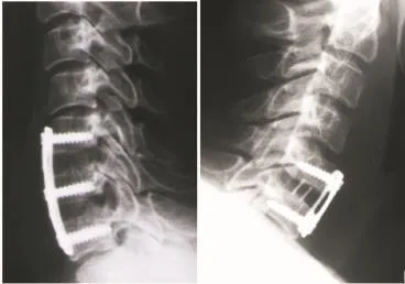
The surgery is performed with operative microscope that allows adequate visualization of the disc space, uncovertebral joints, nerve roots and anterior duralsac. Discectomy can be supplemented with additional nerve root decompression if both canal and foraminal decompressions are required[12]. In case of OPLL, the compressive agent is the ligament itself thus the resection of hypertrophied and ossified ligament may also be crucial during the procedure. In the classical Smith Robinson fashion, iliac crest autograft is used for grafting, but other alternatives can also be applied such as spacers (Figure 1)[13].

Figure 1. Postoperative X-ray photographs show the decompression and fusion after multilevel (left picture) and single-level ACDF (right picture).
2.2. Anterior cervical corpectomy and fusion (ACCF)
Stenotic levels are exposed under microscopic visualization. Resection of the vertebral body allows extensive decompression of the spinal cord and can be supplemented with removal of osteophytes or the resection of hypertrophied ligament. Vertebral body screws and typically iliac crest autografts are used to achieve sufficient fusion, but other alternative spacers also exist (Figure 2). Preventing graft dislodgement and restoring lordosis, constrained, semi-constrained or dynamic anterior plates may be utilized[12,13].

Figure 2. Left picture: Intraoperative photograph shows single-level corpectomy and decompression of the spinal cord; Right picture: Postoperative axial CT scan demonstrates ACCF with application of spacer.
Recently, hybrid procedures have been under investigation that address to strengthen the stability of grafts. The combination of a single-level ACDF with adjacent level corpectomy may be applied for treatment of multilevel diseases. The technique enables greater postoperative stability of spine but needs more operating time and graft contouring[12]. Beside sufficient early results, no long-term examinations have been conducted about its efficacy[12].
Matzet al.[14] used an evidence-based approach to examine the outcomes of surgical intervention between mild and severe CM. In case of mild CM, Class II results showed equivalency between surgical and non-operative therapy in the short term (3 years). Only in case of severe CM, surgery seemed to be superior to non-operative therapy according to Class III evidences. They emphasized these evidences based on the inadequacy of underlying studies. Despite these results, Coricet al.[15] proved the long-term efficacy of ACDF in a prospective randomized study and Gaoet al.[16] also reported good long-term clinical outcomes in the setting of ACCF according to a retrospective study.
3. Posterior approaches
3.1. Modifications of laminectomies
Laminectomy alone is one of the oldest techniques in the treatment of CM with extended devastation of posterior elements and some drawbacks. Common negativeconsequences are the postlaminectomy kyphosis that may lead to the recurrence of CM, spine deformity and neck pain[17-19]. Various modifications have been developed to avoid the disadvantages of laminectomies and fulfil the requirements of less invasiveness.
3.2. Laminectomy with fusion
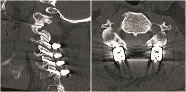
Figure 3 shows laminectomy with fusion. The main indication for laminectomy with fusion consists of kyphotic alignment and presence of instability which is determined by the measurements of subluxation and angulations on static and dynamic views. Under the condition of instability, the possibility of postoperative progressive deformity is highly increased without fusion[20,21]. Lateral mass fixation is regularly performed with lateral mass screws and rods but additional bony grafts can be used to increase the fusion rates[17]. Andersonet al.[18] performed an evidence-based approach to evaluate the effectiveness of laminectomy with fusion. They found Class III evidences that showed neurological improvement after surgery with the rate of 70%-95%. The reported complications included screw dislodging, hardware failure with loss of alignment and radiculopathy. Improved postoperative neck pain, prevented postlaminectomy kyphosis and instability may prove the efficiency of this modification[18].

Figure 3. Multilevel laminectomy with fusion on sagittal (left) and axial (right) postoperative CT scans in case of a patient with myelopathy, significant preoperative neck pain and kyphotic alignment.
3.3. Skip laminectomy
With limiting the destruction of posterior spinal elements, postoperative kyphosis and neck pain, skip laminectomy may fulfill the requirements of less invasiveness. By removing alternated laminae and preserving the posterior arches of interval vertebrae, skip laminectomy leaves intact muscle attachments. In other words, standard laminectomies alternate with partial laminectomies of the lower adjacent vertebra where the muscular attachments of the skipped vertebrae remain intact. Imaging studies determine how many laminae need to be removed but its number should be small to prevent the stability of spine. Shiraishi[22] performed successfully this surgery on 24 patients and measured an average recovery rate of 61%. No neurological deteriorations or recurrences of stenosis were evident in the follow-up periods. A 2-year follow-up study reported nearly similar average recovery rate (59.2%) and found skip laminectomy effective in avoiding postoperative complications that are frequent after laminectomy alone[23].
3.4. Laminoplasty techniques
Laminoplasty techniques have been developed as MISSTs with the goal of preserving dorsal elements and cervical motions as much as possible. Various types of laminoplasties have been described and all of them aim to widen the spinal canal with preventing postsurgical cervical spine instability. Multilevel cervical spondylosis (three or more levels), OPLL and spinal cord tumors are the main indications for laminoplasty[24]. Contraindications consist of kyphotic cervical disorders and less than three-level diseases. Many studies aimed to summarize laminoplasty techniques and described technical features thus we confine ourselves to indicate the main differences between methods.
3.5. Open-door laminoplasty (Hirabayashi laminoplasty) [24-26]
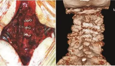
After dissecting the paraspinal muscles, the required cervical level is exposed and a bony trough is created at the medial one third of the lateral mass on the hinge (“closed”) side of the laminae. On the aperture (“open”) side, the lamina is drilled away entirely and the door can be opened with gently force to avoid the fracture of the laminae. Suture should be applied on the hinge side to maintain sufficient decompression or autograft can also be placed to achieve this goal (Figures 4 and 5). The method may also be repeated on other cervical levels accordingly to the expansion of the pathological process.

Figure 4. Left picture: Intraoperative photograph demonstrates open-door laminoplasty with autograft spacers and miniplates; Right picture: The 3D CT reconstruction image shows postoperative situation after modified open-door laminoplasty.
3.6. French-door laminoplasty[24,26]
In this technique, the spinous processes are splitted in the midline then bilateral troughs are made as similar as in the open-door method. The integrities of laminae remain intacton both sides. The split laminae are opened with gently forces in sequence as are French doors. Various spacers can be used to maintain sufficient decompression including bony autografts or synthetic spacers. Two types of modification of this method have been reported in the literature. Kurokawa modification[24,27] includes removing the posterior aspect of spinous processes and using them as spacers. Tomita modification (or T-saw laminoplasty)[24,28] involves using of a wire-saw to split the spinous processes.

Figure 5. Left picture: Sagittal pre-operative T2-weighted MR scan shows stenosis of the spinal canal and signs of myelopathy in the spinal cord; Right picture: Sagittal T2-weighted post-operative MR scan demonstrates the decompression of spinal cord with open-door laminoplasty.
3.7. Z-laminoplasty[24,29]
The method requires the removal of spinous processes. After thinning the laminae, a “Z” shapes are cut in the laminae so the sides of the laminar opening are alternated. Separating the sections enables the enlargement of spinal canal and it can be maintained by securing the laminae with sutures or wires.
Complications of laminoplasty techniques consists of nerve root palsy (mainly C5 root palsy), closure of opened laminae, axial pain, infections, dural tears, pseudomyelomeningocele and decrease in lordosis[26]. The last one highlights lordotic patients are the best candidates for laminoplasties. Additional consequence contains some loss of cervical range of motion but not as much as can be noticed in fusion techniques. This alteration in movements is controversial[30] because it can decrease the possibility of cord injury via dynamic components[31] but the stiffness increases the evolution rate of adjacent-segment disease[32].
Matzet al.[30] used an evidence-based approach to evaluate the efficacy of laminoplasty techniques. Class III evidences anticipated a recovery rate of 55-60%. Class II evidences revealed duration of symptoms, poorly controlled diabetes, severity of myelopathy and stenosis are associated with poorer clinical outcomes. A few studies proved the long-term effectiveness (over 10 years) of laminoplasty techniques. Chibaet al.[33] reported satisfactory long-term neurological results after open-door laminoplasty but they noticed some deterioration in patients who presented with OPLL that may refer to the natural progression of disease. Seichiet al.[34] found similar long-term results after 10 years using double-door laminoplasty. In summary, improvements are evident after laminoplasties even in long-term followup examinations[24].
A newly developed variation of laminoplasty techniques is the cervical microendoscopic laminoplasty (C-MEL). This endoscopic version has been developed as a MISST to minimize the destructive components of laminoplasties. Good clinical outcomes have been reported but the number of cases involved in the study was small to draw conclusions about the long-term efficacy and safety[35].
3.8. Split laminotomy and the“archbone”technique
The multilevel spinous process splitting and distracting laminotomy was primarily developed for adults to explore intramedullary spinal pathologies with the aim of preservation the anatomical integrity of posterior structures and spine stability[36,37]. Although, split laminotomy may also be an applicable procedure by decompression and moderate enlargement of the spinal canal in case of CMs. Leaving the muscle attachments intact and reducing postoperative complications, split laminotomy fulfils the requirements of MISSTs.
In midline posterior approach, the interspinous ligaments are dissected longitudinally and the ligamentum flavum is removed at the middle part. The spinous processes are split in the midline with an oscillating saw or craniotome then are separated and distracted with Cloward-type retractors. Preventing the fracture of the spinous process, gently forces may be applied for the retraction. Grafts or spacers can be placed between the bony parts of spinous processes facing each other to decompress and enlarge moderately the spinal canal (Figure 6). The method is similar to the placement of an ”archstone” into the arch of a vault in architecture, this theme was borrowed and modified to ”archbone” for surgery.

Figure 6. Intraoperative photographs show the separation and distraction of spinous processes with Cloward-type retractors (left) and the situation after placing spacers between the splitted spinous processes for moderate enlargement of the spinal canal (right).
The authors recommend split laminotomy and the“archbone” technique in cases which requires moderate enlargement of the spinal canal. The technique is an effective method for creating extra intraspinal space and decompression without signs of postoperative spineinstability or deformation but further studies are required to evaluate the limitations, long-term effectiveness and safety in the treatment of CM.
3.9. Microendoscopic stenosis decompression (MEDS)
The technique is a newly developed modification of the dorsal laminoforaminotomy and an alternative posterior MISST for bony decompression caused by spinal stenosis. Reducing the dissection of muscles and preserving the stability of spine may offer better clinical outcomes and decrease the rate of postoperative pain and time of hospital stay. Postoperative kyphosis which is a common complication of traditional procedures can be prevented by avoiding the destruction of midline dorsal cervical tension band, muscular attachments and facet complexes. The authors recommended kyphotic deformities as contraindications of this method[38,39].
After positioning patients sitting, a guide wire is gently docked onto the bone on the required stenotic level under fluoroscopic guidance to avoid inadequate positioning. Placing initial dilators down to the lamina under fluroscopy is required to dilate the paraspinal musculature. The working channel is placed over the final dilator that is fixed with a table-mounted flexible arm. After removing the dilators, the endoscope is inserted into the working channel. The lateral aspect of the lamina and the base of spinous process are drilled away to perform foraminotomy and achieve adequate decompression. To visualize the contralateral part of the spinal canal, resection of the ligamentum flavum may be a helpful surgical step. Additional decompression can be performed on other levels with angulating the working channel cranially or caudally[38,39].
Dahdalehet al. recommended MEDS as an alternative technique to open laminectomy and laminoplasty in acceptable selected patients, but they emphasized more comparative studies should be carried out to evaluate the efficacy and complications[39].
4. Discussion
Controversial results exist about which technique may be the most effective and we aimed to discuss and possibly answer this question.
In general, examining the question anterior or posterior surgical approach is the better choice for multilevel CM[11], better clinical outcomes and more complications have been observed after anterior surgery only in the early postoperative stage (<5 years). More than 5-year follow-up examination has revealed similar outcomes and complication rates between the two approaches. Only in case of OPLL patients with more than 60% occupying ratio of OPLL, the superiority of anterior surgery has been proven in clinical outcomes[11]. On the other side, posterior approaches may be technically easier with fewer perioperative complications and shorter operative times[20]. However, some limitations of posterior approaches are kyphotic disorders that were marked as absolute contraindications. In case of more than three level stenotic CM and post-traumatic or postlaminectomy kyphosis, a combined anteroposterior approach may be recommended but no controlled studies have been carried out to examine the clinical outcomes[11,40].
Comparison of anterior procedures has revealed ACCF diminishes the number of graft-bone surfaces that are needed for fusion in multilevel stenosis and decreases the risk of pseudoarthrosis more than multilevel ACDF[12]. Significant differences between ACDF and ACCF results in regard to sagittal alignment, cervical lordosis, graft subsidence, and adjacent-level ossification have not been notified[41,42]. Comparing ACCF to laminoplasty, higher rate of complications including adjacent segment degeneration and higher amount of pain medication have been reported in the ACCF group[43]. Moreover, disadvantages of ACCF and ACDF consist of longer surgical time, more blood loss, and potentially more complications[44,45].
Laminectomy with fusion compared to anterior approaches enables better neurological results, although, only Class III evidences prove its efficacy[18,46]. Contrasting laminectomy with fusion to laminoplasty, no significant differences in neurological recovery and in the rate of postoperative axial pain have been revealed[18]. Other studies have noticed better clinical outcomes, lower complication rates, lower implant costs and rates of reoperations in laminoplasty groups[47,48]. Although, laminectomy with fusion may be recommended in case of significant preoperative neck pain by reducing more efficiently the rate of pain[48] and in kyphotic disorders by preserving sagittal alignment better than laminoplasty[49].
Skip laminectomy may provide better clinical outcomes in respect to complication rate, surgical trauma, preserved posterior segments and range of motion than laminoplasty[23,50]. However, the superiority of skip laminectomy cannot be emphasized because only limited studies exist that examine the differences in efficacy between these procedures[23,51].
Considering laminoplasty techniques, none of them has been proven superior to each other in outcome, cervical alignment or preserving range of motions[24]. Minor difference may be that open-door laminoplasty expands the spinal canal asymmetrically by using one side as hinge point, whereas the French-door laminoplasty opens the spinal canal symmetrically in the midline[24,52]. Comparisons of laminoplasties to other methods have been discussed above in details.
The effectiveness of MEDS and split laminotomy has been proven in decompressing the spinal canal sufficiently. Clinical outcomes are promising but no comparative studies have been conducted to contrast them to other treatment options of CM.
The purpose of this review was to summarize traditional techniques and MISSTs that may be applied in the treatment of acute CM and evaluating their efficacy with comparing them to each other. None of them has been proven to be a superior technique to each other, so we conclude the choice of treatment option of CM should be individually determined considering the location and extension of pathology, the familiarity of surgeon with techniques and as far as possible requirements of minimally or less invasiveness.
5. Conclusion
Significant development of less invasive techniques and MISSTs has been witnessed in spine surgery recently to prevent negative biomechanical consequences that were noticed following some traditional methods. The general demand for developing and performing these procedures led to the report of many less invasive and MISSTs that may also be applied in the treatment of acute CM. The summarization and comparison of these methods did not reveal superiority of any procedures and individual decisions may be required in most cases.
Conflict of interest statement
The authors report no conflict of interest.
[1] Baptiste DC, Fehlings MG. Pathophysiology of cervical myelopathy. Spine J 2006; 6(6 Suppl):190S-197S.
[2] LaRocca H. Cervical spondylotic myelopathy: natural history. Spine 1988; 13: 854-855.
[3] Matz PG, Anderson PA, Holly LT, Groff MW, Heary RF, Kaiser MG, et al. The natural history of cervical spondylotic myelopathy. J Neurosurg Spine 2009; 11(2): 104-111.
[4] Yonenobu K. Cervical radiculopathy and myelopathy: when and what can surgery contribute to treatment? Eur Spine J 2000; 9(1): 1-7.
[5] Fujiwara K, Yonenobu K, Ebara S, Yamashita K, Ono K. The prognosis of surgery for cervical compression myelopathy. An analysis of the factors involved. J Bone Joint Surg Br 1989; 71: 393-398.
[6] Koyanagi T, Hirabayashi K, Satomi K, Toyama Y, Fujimura Y. Predictability of operative results of cervical compression myelopathy based on preoperative computed tomographic myelography. Spine 1993; 14: 1958-1963.
[7] Yonenobu K, Fuji T, Ono K, Okada K, Yamamoto T, Harada N. Choice of surgical treatment for multisegmental cervical spondylotic myelopathy. Spine 1985; 10: 710-716.
[8] Bourassa-Moreau É, Mac-Thiong JM, Ehrmann Feldman D, Thompson C, Parent S. Complications in acute phase hospitalization of traumatic spinal cord injury: does surgical timing matter? J Trauma Acute Care Surg 2013; 74(3): 849-854.
[9] Okada Y, Ikata T, Yamada H, Sakamoto R, Katoh S. Magnetic resonance imaging study on the results for surgery for cervical compression myelopathy. Spine 1993; 18: 2024-2029.
[10] Bucciero A, Vizioli L, Carangelo B, Tedeschi G. MR signal enhancement in cervical spondylotic myelopathy. Correlation with surgical results in 35 cases. J Neurosurg Sci 1993; 37: 217-222.
[11] Liu T, Xu W, Cheng T, Yang HL. Anterior versus posterior surgery for multilevel cervical myelopathy, which one is better? A systematic review. Eur Spine J 2011; 20(2): 224-235.
[12] Klineberg E, McLain RF, Bell GR. Cervical spondylotic myelopathy: anterior approach: multilevel anterior cervical discectomy and fusion versus corpectomy. Semin Spine Surg 2007; 19: 27-34.
[13] Epstein N. Anterior approaches to cervical spondylosis and ossification of the posterior longitudinal ligament: review of operative technique and assessment of 65 multilevel circumferential procedures. Surg Neurol 2001; 55(6): 313-324.
[14] Matz PG, Holly LT, Mummaneni PV, Anderson PA, Groff MW, Heary RF, et al. Anterior cervical surgery for the treatment of cervical degenerative myelopathy. J Neurosurg Spine 2009; 11(2): 170-173.
[15] Coric D, Kim PK, Clemente JD, Boltes MO, Nussbaum M, James S. Prospective randomized study of cervical arthroplasty and anterior cervical discectomy and fusion with long-term follow-up: results in 74 patients from a single site. J Neurosurg Spine 2013; 18(1): 36-42.
[16] Gao R, Yang L, Chen H, Liu Y, Liang L, Yuan W. Long term results of anterior corpectomy and fusion for cervical spondylotic myelopathy. PLoS One 2012; 7(4): e34811.
[17] Ryken TC, Heary RF, Matz PG, Anderson PA, Groff MW, Holly LT, et al. Cervical laminectomy for the treatment of cervical degenerative myelopathy. J Neurosurg Spine 2009; 11(2): 142-149.
[18] Anderson PA, Matz PG, Groff MW, Heary RF, Holly LT, Kaiser MG. Laminectomy and fusion for the treatment of cervical degenerative myelopathy. J Neurosurg Spine 2009; 11(2): 150-156.
[19] Epstein NE. Laminectomy for cervical myelopathy. Spinal Cord 2003; 41(6): 317-327.
[20] Komotar RJ, Mocco J, Kaiser MG. Surgical management of cervical myelopathy: indications and techniques for laminectomy and fusion. Spine J 2006; 6(6 Suppl): 252S-267S.
[21] Epstein N, Epstein JA. Treatment of cervical myelopathy: Part A. Laminectomy. In: TCSR Society, editor. The cervical spine. Philadelphia: Lippincott Williams & Wilkins; 2005, p. 1043-1056.
[22] Shiraishi T. Skip laminectomy-a new treatment for cervical spondylotic myelopathy, preserving bilateral muscular attachments to the spinous processes: a preliminary report. Spine J 2002; 2(2): 108-115.
[23] Shiraishi T, Fukuda K, Yato Y, Nakamura M, Ikegami T. Results of skip laminectomy-minimum 2-year follow-up studycompared with open-door laminoplasty. Spine 2003; 28(24): 2667-2772.
[24] Steinmetz MP, Resnick DK. Cervical laminoplasty. Spine J 2006; 6(6 Suppl): 274S-281S.
[25] Hirabayashi K, Miyagawa J, Satomi K, Maruyama T, Wakano K. Operative results and postoperative progression of ossification among patients with ossification of cervical posterior longitudinal ligament. Spine 1981; 6: 354-364.
[26] Patel CK, Cunningham BJ, Herkowitz HN. Techniques in cervical laminoplasty. Spine J 2002; 2(6): 450-455.
[27] Kurokawa T, Tsuyama N, Tanaka H. Enlargement of spinal canal by the sagittal splitting of the spinous process. Bessatusu Seikeigeka 1982; 2: 234-240.
[28] Tomita K, Kawahara N, Toribatake Y, Heller JG. Expansive midline T-saw laminoplasty (modified spinous processsplitting) for the management of cervical myelopathy. Spine (Phila Pa 1976) 1998; 23: 32-37.
[29] Oyama M, Hattori S, Moriwaki N. [A new method of posterior decompression]. Chubuseisaisi 1973; 16: 792. Japanese.
[30] Matz PG, Anderson PA, Groff MW, Heary RF, Holly LT, Kaiser MG. Cervical laminoplasty for the treatment of cervical degenerative myelopathy. J Neurosurg Spine 2009; 11(2): 157-169.
[31] Kimura I, Oh-Hama M, Shingu H. Cervical myelopathy treated by canal-expansive laminoplasty. Computed tomographic and myelographic findings. J Bone Joint Surg Am 1995; 66: 914-920.
[32] Shaffrey CI, Wiggins GC, Piccirilli CB, Young JN, Lovell LR. Modified open-door laminoplasty for treatment of neurological deficits in younger patients with congenital spinal stenosis: analysis of clinical and radiographic data. J Neurosurg (Spine) 1999; 90: 170-177.
[33] Chiba K, Ogawa Y, Ishii K, Takaishi H, Nakamura M, Maruiwa H, et al. Long-term results of expansive opendoor laminoplasty for cervical myelopathy--average 14-year follow-up study. Spine 2006; 31(26): 2998-3005.
[34] Seichi A, Takeshita K, Ohishi I, Kawaguchi H, Akune T, Anamizu Y, et al. Long-term results of double-door laminoplasty for cervical stenotic myelopathy. Spine 2001; 26(5): 479-487.
[35] Minamide A, Yoshida M, Yamada H, Nakagawa Y, Maio K, Kawai M, et al. Clinical outcomes of microendoscopic decompression surgery for cervical myelopathy. Eur Spine J 2010; 19(3): 487-493.
[36] Banczerowski P, Vajda J, Veres R. Exploration and decompression of the spinal canal using split laminotomy and its modification, the “archbone” technique. Neurosurgery 2008; doi: 10.1227/01.neu.0000326031.31843.99.
[37] Banczerowski P, Bognár L, Rappaport ZH, Veres R, Vajda J. Novel surgical approach in the management of longitudinal pathologies within the spinal canal: the split laminotomy and“archbone” technique: alternative to multilevel laminectomy or laminotomy. Adv Tech Stand Neurosurg 2014; 41: 47-70.
[38] Santiago P, Fessler RG. Minimally invasive surgery for the management of cervical spondylosis. Neurosurgery 2007; 60(1 Supp1 1): S160-S165.
[39] Dahdaleh NS, Wong AP, Smith ZA, Wong RH, Lam SK, Fessler RG. Microendoscopic decompression for cervical spondylotic myelopathy. Neurosurg Focus 2013; doi: 10.3171/2013.3.FOCUS135.
[40] Chin KR, Ozuna R. Options in the surgical treatment of cervical spondylotic myelopathy. Curr Opin Orthop 2000; 11: 151-157.
[41] Park Y, Maeda T, Cho W, Riew KD. Comparison of anterior cervical fusion after two-level discectomy or single-level corpectomy: sagittal alignment, cervical lordosis, graft collapse, and adjacent-level ossification. Spine J 2010; 10(3): 193-199.
[42] Wang JC, McDonough PW, Endow KK, Delamarter RB. A comparison of fusion rates between single-level cervical corpectomy and two-level discectomy and fusion. J Spinal Disord 2001; 14(3): 222-225.
[43] Edwards CC 2nd, Heller JG, Murakami H. Corpectomy versus laminoplasty for multilevel cervical myelopathy: an independent matched-cohort analysis. Spine 2002; 27(11): 1168-1175.
[44] Fang Z, Tian R, Sun TW, Yadav SK, Hu W, Xie SQ. Expansion open-door laminoplasty with foraminotomy versus anterior cervical discectomy and fusion for coexisting multilevel cervical myelopathy and unilateral radiculopathy. J Spinal Disord Tech 2013; doi: 10.1097/BSD.0000000000000074.
[45] Wada E, Suzuki S, Kanazawa A, Matsuoka T, Miyamoto S, Yonenobu K. Subtotal corpectomy versus laminoplasty for multilevel cervical spondylotic myelopathy: a long-term follow-up study over 10 years. Spine 2001; 26(13): 1443-1447; discussion 1448.
[46] González-Feria L, Peraita-Peraita P. Cervical spondylotic myelopathy: a cooperative study. Clin Neurol Neurosurg 1975; 78(1): 19-33.
[47] Heller JG, Edwards CC 2nd, Murakami H, Rodts GE. Laminoplasty versus laminectomy and fusion for multilevel cervical myelopathy: an independent matched cohort analysis. Spine 2001; 26(12): 1330-1336.
[48] Highsmith JM, Dhall SS, Haid RW Jr, Rodts GE Jr, Mummaneni PV. Treatment of cervical stenotic myelopathy: a cost and outcome comparison of laminoplasty versus laminectomy and lateral mass fusion. J Neurosurg Spine 2011; 14(5): 619-625.
[49] Rhee JM, Basra S. Posterior surgery for cervical myelopathy: laminectomy, laminectomy with fusion, and laminoplasty. Asian Spine J 2008; 2(2): 114-126.
[50] Sivaraman A, Bhadra AK, Altaf F, Singh A, Rai A, Casey AT, et al. Skip laminectomy and laminoplasty for cervical spondylotic myelopathy: a prospective study of clinical and radiologic outcomes. J Spinal Disord Tech 2010; 23: 96-100.
[51] Yuan W, Zhu Y, Liu X, Zhou X, Cui C. Laminoplasty versus skip laminectomy for the treatment of multilevel cervical spondylotic myelopathy: a systematic review. Arch Orthop Trauma Surg 2014; 134(1): 1-7.
[52] Hukuda S, Mochizuki T, Ogata M, Shichikawa K, Shimomura Y. Operations for cervical spondylotic myelopathy. A comparison of the results of anterior and posterior procedures. J Bone Joint Surg Br 1985; 67(4): 609-615.
ment heading
10.1016/S2221-6189(14)60059-7
*Corresponding author: Péter Banczerowski M.D., Ph.D., National Institute of Neurosurgery &Semmelweis University, Faculty of Medicine, Department of Neurosurgery, Amerikai út 57.Budapest, Hungary, 1145
Tel: +36 1 2512999
Fax: +36 1 2515678
E-mail: bancpet@gmail.com, banczerowski.peter@med.semmelweis-univ.hu
Minimal invasive spine surgery techniques
Traditional techniques
杂志排行
Journal of Acute Disease的其它文章
- Acute and sub-acute toxicity study of Clerodendrum inerme, Jasminum mesnyi Hance and Callistemon citrinus
- Time-critical AMI Detection: A novel and fast technique using the 12-lead ECG
- Epidemiological survey on scorpionism in Gotvand County, Southwestern Iran: an analysis of 1 067 patients
- The acute effect of the antioxidant drug “U-74389G” on red blood cells levels during hypoxia reoxygenation injury in rats
- Successful treatment of lower urinary tract obstruction with peritonealamniotic and vesicoamniotic shunting
- Simvastatin-induced Toxic Epidermal Necrolysis
