肢端恶性黑色素瘤前哨淋巴结活检 35 例报告
2014-02-13杨发军刘巍峰孙扬丁易牛晓辉
杨发军 刘巍峰 孙扬 丁易 牛晓辉
肢端恶性黑色素瘤前哨淋巴结活检 35 例报告
杨发军 刘巍峰 孙扬 丁易 牛晓辉
目的探索99Tcm标记的右旋糖苷结合 γ 射线探测仪在淋巴结显像中的用药规律和前哨淋巴结活检的准确性;探讨前哨淋巴结活检在恶性黑色素瘤早期转移诊治中的临床意义。方法2012 年 3 月至2013 年 5 月,我科收治 35 例肢端恶性黑色素瘤患者,其中手部 8 例、足部 27 例。所有病灶 Breslow 厚度均>1 mm,且无临床及影像学淋巴结转移的证据。除外淋巴结已有转移的患者。术前 4~6 h 在病灶周围注射1~2 mCi 的99Tcm标记的右旋糖苷,注射后 30 min、2、4 h 行核素显像,获得前哨淋巴结的显像图。然后在麻醉下切取前哨淋巴结,术中用 γ 射线探测仪帮助定位和切取前哨淋巴结。术后淋巴结行 3 mm 一层的切片,行HE 染色和 HMB-45,S-100,Melan-A 免疫组化染色。前哨淋巴结活检结果阳性的行局部淋巴结清扫。结果核素注射后 4 h 前哨淋巴结显像稳定。27 例足部黑色素瘤患者前哨淋巴结中有 7 例窝及腹股沟均显像,其余20 例仅腹股沟淋巴结显像;8 例手部黑色瘤患者中有 2 例滑车上及腋窝淋巴结均显像,其余 6 例仅腋窝淋巴结显像。35 例均检出前哨淋巴结,前哨淋巴结检出率为 100%。前哨淋巴结的个数为 1~3 个。5 例患者的前哨淋巴结病理检查发现有转移,阳性率为 14.3%。此 5 例均行淋巴结清扫术。结论用99Tcm标记的右旋糖苷作为显像剂、术中应用 γ 射线探测仪的前哨淋巴结活检技术在肢端黑色素瘤中是一种可靠的技术。
黑色素瘤;前哨淋巴结活组织检查;放射性核素淋巴显像;γ 射线
黑色素瘤是临床上较为常见的恶性肿瘤之一,也是发病率增长较快的恶性肿瘤之一。虽然黑色素瘤在我国发病率较低,但近年来成倍增长,每年新发病例约 2 万例。黑色素的早期诊断和正确治疗非常重要,早期患者可以治愈。因此黑色素瘤的分期诊断至关重要。黑色素瘤通过淋巴系统远处转移,淋巴结转移对分期诊断、治疗、预后的判断有重要的影响。因此前哨淋巴结的活检具有重要的临床意义。我国黑色素瘤的发病特点之一为肢端黑色素瘤多。2012 年 3 月至 2013 年 5 月,我科收治 35 例肢端黑色素瘤患者,并进行了前哨淋巴结活检,现报告如下。
临床资料
本组 35 例,男 19 例,女 16 例。中位年龄为 58 ( 29~89 ) 岁。原发病灶:手部 8 例,足部27 例。35 例中有 24 例原发病灶有溃疡。35 例Breslow 厚度均>1 mm,区域淋巴结均行 B 超、MRI、或全身的 PET-CT 检查除外了局部有淋巴结转移,因此 35 例均行前哨淋巴结活检术。
一、前哨淋巴结核素扫描
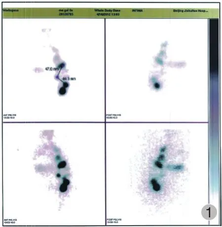
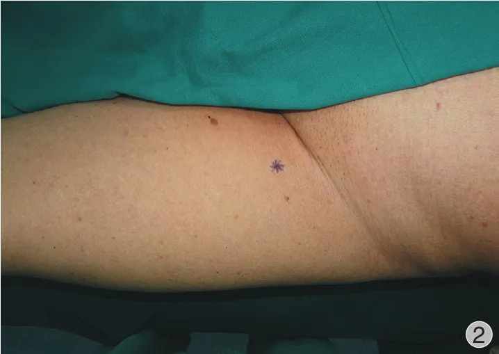
我院使用的前哨淋巴结显像剂为99Tcm标记的右旋糖苷 ( 北京森科医药有限公司提供 )。在术前4~6 h 在病灶周围行 4 点注射,剂量为 2 mCi。注射后 30 min、2、4 h 行全身扫描,确定前哨淋巴结的位置,并行体表标记 (图1~2 )。

图1 腹股沟前哨淋巴结核素显像 Fig.1 The radionuclide images of the SLN in the groin

图2 根据前哨淋巴结核素扫描的体表定位 Fig.2 Surface localization of the SLN according to the radionuclide images
二、前哨淋巴结的切取
在全麻或椎管内麻醉下切取前哨淋巴结,术者均为有丰富淋巴结活检经验的骨肿瘤科医师。消毒前先用 Neoprobe 2000 γ 射线探测仪 ( 美国强生公司生产 ) 再次确定前哨淋巴结的位置。然后消毒铺单。术中在 γ 射线探测仪的帮助下切取前哨淋巴结。
三、前哨淋巴结的病理检查
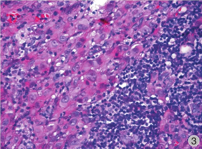
淋巴结每 3 mm 一层的切片,行 HE 染色和HMB-45,S-100,Melan-A 免疫组化染色 (图3 )。前哨淋巴结活检结果阳性的行腹股沟淋巴结清扫术或腋窝淋巴结清扫术。
结 果
一、核素扫描结果
核素扫描在注射99Tcm后 30 min 可发现,核素自原发灶沿淋巴管向区域淋巴结扩散;注射后 4 h 前哨淋巴结显像稳定。35 例核素扫描均发现 1~3 枚淋巴结。8 例手部黑色瘤中 2 例滑车上淋巴结和腋窝淋巴结同时显像,其余 6 例仅腋窝淋巴结显像。27 例足部黑色素瘤患者,5 例窝和腹股沟淋巴结同时显像,其余 22 例仅腹股沟部淋巴结显像。
二、手术及病理检查结果
所有显像的前哨淋巴结均切取出淋巴结,淋巴结个数为 1~3 个。前哨淋巴结检出率为 100%。本组 35 例中有 5 例足部黑色素瘤患者,其腹股沟前哨淋巴结病理检查可发现转移,阳性率为 14.3% (图3 )。此 5 例均行腹股沟淋巴结清扫术。1 例术后伤口出现皮瓣坏死,1 例出现伤口感染,经再次手术后痊愈。

图3 淋巴细胞周围可见恶性黑色素瘤细胞 ( HE ×20 ) Fig.3 Malignant melanoma cells were seen around the lymphocytes ( HE ×20 )
讨 论
1977 年 Cabanas 为阴茎癌患者进行阴茎背侧淋巴管造影时发现了一组“特殊”的淋巴结,临床上尚未发现淋巴结转移却常常发现这组淋巴结受累的现象。Cabanas 将这组淋巴结命名为前哨淋巴结( sentinel lymph node,SLN ),并定义为是最先接受肿瘤淋巴引流,最早发生淋巴结转移的淋巴结[1]。目前前哨淋巴结活检技术已广泛应用于乳腺癌和皮肤黑色素瘤的临床上。
Morton[2]在 1992 年首先应用99Tcm标记的硫胶体在前哨淋巴结活检术中。目前,多数文献报道前哨淋巴结活检的示踪剂为:硫胶体。但目前国内没有药厂生产和进口的硫胶体药品,因此硫胶体多为自己单位加工[3]。国外有些文献报道:99Tcm标记的右旋糖苷是一种可靠的前哨淋巴结显像剂[4]。而且国内有市售的99Tcm标记的右旋糖苷,因此我院用99Tcm标记的右旋糖苷做为前哨淋巴结活检的显像剂。
本组 8 例手部黑色瘤患者中有 2 例滑车上及腋窝淋巴结均显像,其余 6 例仅腋窝淋巴结显像。其原因与上肢淋巴回流途径相关,即:手部尺侧和前臂尺侧皮肤淋巴引流先至肘窝淋巴结,再到腋窝淋巴结;其余部位直接至腋窝淋巴结。本组 27 例足部黑色素瘤患者,前哨淋巴结中有 7 例窝及腹股沟均显像,其余 20 例仅腹股沟淋巴结显像。其原因与下肢淋巴回流途径相关,即:足底外侧及小腿后侧皮肤先引流至窝淋巴结,然后再到腹股沟淋巴结;其余部位直接引流到腹股沟淋巴结。本组 35 例前哨淋巴结活检的结果完全符合肢端淋巴引流的解剖规律。
2012 年美国 ASCO 和 SSO ( society of surgical oncology ) 推荐的前哨淋巴结活检的指南如下:( 1 )中等厚度任何位置的黑色素瘤 Breslow 厚度 1~4 mm被推荐行前哨淋巴结活检;( 2 ) 厚黑色素瘤 ( Breslow厚度>4 mm ) 可能被推荐行前哨淋巴结活检;( 3 ) 对于薄黑色素瘤 Breslow 厚度<1mm,且有溃疡和有丝分裂率≥1 / mm2推荐行 SLN 活检[5]。本组所有病灶Breslow 厚度均>1 mm,且无临床及影像学淋巴结转移的证据。完全符合前哨淋巴结活检的适应证。
有学者对 282 例 Breslow 厚度>1 mm 的黑色素瘤患者行前哨淋巴结活检,结果 47 例前哨淋巴结呈阳性,阳性率为 16%[6]。本组 35 例黑色素瘤患者,Breslow 厚度均>1 mm,5 例前哨淋巴结阳性,阳性率为 14.3%。本组前哨淋巴结的转移阳性率与文献报道基本一致。
总之,用99Tcm标记的右旋糖苷作为显像剂,术中用 γ 射线探测仪帮助定位和切取前哨淋巴结,是黑色素瘤前哨淋巴结活检中一种可靠的方法。
[1] Cabanas RM. An approach for the treatment of penis carcinoma. Cancer, 1977, 39(2):456-466.
[2] Morton DL, Wen DR, Wong JH, et al. Technical details of intraoperative lymphatic mapping for early stage melanoma. Arch Surg, 1992, 127(4):392-399.
[3] 徐宇, 朱蕙燕, 吴江宏, 等. 皮肤恶性黑色瘤的前哨淋巴结活检技术. 肿瘤, 2011, 31(1)64-68.
[4] Masiero PR, Xavier NL, Spiro BL, et al. Scintigraphic sentinel node detection in breast cancer patients: paired and blindedcomparison of99mTc dextran 500 and99mTc phytate. Nucl Med Commun, 2005, 26(12):1087-1091.
[5] Wong SL, Balch CM, Hurley P, et al. Sentinel lymph node biopsy for melanoma: American Society of Clinical Oncology and Society of Surgical Oncology joint clinical practice guideline. Ann Surg Oncol, 2012, 19(11):3313-3324.
[6] Ellis MC, Weerasinghe R, Corless CL, et al. Sentinel lymph node staging of cutaneous melanoma: predictors and outcomes. Am J Surg, 2010, 199(5):663-668.
( 本文编辑:李贵存 )
Sentinel lymph node biopsy in the extremity with malignant melanoma: 35 cases report
YANG Fa-jun, LIU Wei- feng, SUN Yang, DING Yi, NIU Xiao-hui. Department of Orthopedic Oncology, Beijing Jishuitan Hospital, Beijing, 100035, PRC
ObjectiveTo explore how to use the technetium-99m(99Tcm) dextran and the gamma-ray detector in the sentinel lymph node mapping ( SLNM ) and the accuracy of SLN biopsy, and to investigate the clinical value of SLN biopsy in the early diagnosis and treatment of malignant melanoma metastasis.MethodsFrom March 2012 to May 2013, 35 patients with malignant melanoma in the extremities were admitted. There were 8 cases in the hands and 27 cases in the feet. The Breslow thickness of all primary lesions were more than 1 mm, and no lymph node metastasis was found clinically and radiographically. The patients with lymph node metastasis were ruled out. All patients received intradermal injection of 1-2 millicurie ( mCi ) of99Tcmdextran around the lesions at 4-6 hours before the operation. Radionuclide imaging was performed at 30, 120, 240 minutes after the injection to achieve the SLN images. And the SLN was excised under anesthesia. During the operation, the gamma-ray detector was used to help identify and excise the SLN. After the operation, a piece of 3 mm SLN was sliced, and then the HE dying, Melanoma Marker ( HMB-45 ), S-100 and Melan-A immunohistochemical staining were performed. The patients with the positive SLN accepted lymph node dissection locally.ResultsThe SLN images were clear at 4 hours after the injection. Seven patients had popliteal and inguinal images among the 27 patients with melanoma in the feet, and the remaining 20 patients had inguinal images. Two out of 8 patients with melanoma in the hands had epitrochlear and axillary lymph node images, and the remaining 6 patients only had axillary lymph node images. The SLN was detected in all 35 patients, and the rate was 100%. The number of SLN was 1-3. The pathologic examination revealed that 5 patients had SLN metastasis, and the positive rate was 14.3%. All 5 patients underwent lymphadenectomy.ConclusionsThe SLN biopsy is a reliable technique for the patients with melanoma in the extremity with the99Tcmdextran as the imaging agent, and the gamma-ray detector is used during the operation.
Melanoma; Sentinel lymph lode ( SLN ) biopsy; Lymphoscintigraphy; Gamma-ray
10.3969/j.issn.2095-252X.2014.02.005
R739.5
100035北京积水潭医院骨肿瘤科
牛晓辉,Email: niuxiaohui@263.net
32013-12-23 )
