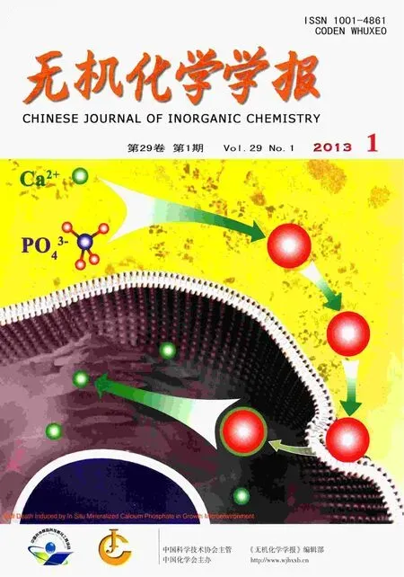
生长微环境下原位矿化磷酸钙诱导的细胞死亡
2013-08-20王广川徐旭荣唐睿康
刘 朋 陈 燕 王广川 王 本 徐旭荣 唐睿康
(浙江大学理学院化学系,杭州 310027)
Calcium phosphate nanoparticles (CaP-NPS) have received considerable interest in an interdisciplinary study involving chemistry, materials, biology and medicine[1-4], particularly in biomedical field. Calcium phosphate, the major inorganic composition of natural hard tissues (bone, dentin and enamel) is extensively applied in biomedical applications due to its excellent biocompatibility, bioactivity and osteo-conductivity characteristics[5-9]. Calcium phosphate based composite nanoparticles is also suggested as an attractive candidate for bioimaging and therapeutic delivery applications[10]. Unfortunately, large amounts of CaPNPS can also induce cell death, but this side effect has not been broadly investigated. Cell death includes two typical forms, necrosis and apoptosis. Necrosis is a degenerative phenomenon that follows irreversible injury and apoptosis appears to be an active process requiring protein synthesis for its execution[11]. In nature, a phenomenon is that almost all cell death induced by phosphate is relied on the presence of calcium, which implies that the cell death can be induced by the co-existence of calcium and phosphate in the growth microenvironment[12]. Both calcium and phosphate are the essential elements in biological fluids and generally, their co-existence is inert on cell death. Therefore, the amount control may be the key to the insecurity of calcium phosphate materials.
Previous studies have suggested that it is the CaP-NPS rather than the free calcium or phosphate ions in biological solutions that induce the cell death[11-12]. However, these studies about the lethal inducing-effect are limited to the influences of CaPNPS sizes or crystallities[13-15]. In order to understand the roles of calcium ion, phosphate ion and CaP-NPS in the cell death quantitatively, we investigate their effects on MG-63 cells derived from osteosarcoma, a common malignant tumor of bone, in different culture media[16]. By adding calcium into calcium-free culture media, CaP-NPS can be in situ synthesized in the culture media. We reveal that the cell viability is directly relevant to the concentration of CaP-NPS in the culture media. An interesting finding is there exsits a critical point of 240 μg·mL-1CaP-NPS to ensure the cell viability. Less than this value, the influence of CaP-NPS on cell can be ignored whereas the cell death is induced remarkably when the CaPNPS amount in media exceeded this critical point. We suggest that a possible reason may be the endocytosis of CaP-NPS. The finding can provide a guidance on the appropriate amount of CaP-NPS in medical bioengineering such as drug delivery, bioimaging, and gene transfection etc.
1 Experimental
1.1 Preparation of calcium phosphate nanoparticles
The chemicals are analysis-grade, purchased from Sinopharm Chemical Reagent Co., Ltd (China)without additional purification prior to their usage. All water in the experiments was triply distilled.
The cell culture media without calcium or phosphate were prepared as described everywhere[17].Calcium phosphate nanoparticles were in situ prepared in the culture media by adding 10 mmol·L-1calcium into the calcium free solution.
1.2 Characteristics
Transmission electron microscopy (TEM, JEM-200CX, Japan) and scanning electron microscopy(SEM, S-4800, Hitachi, Japan) were used to investigate CaP-NPS. X-ray diffraction (XRD, D/max-2550pc, Rigaku, Japan) was employed to identify the structure. The concentrations of calcium and phosphate ions were determined by ultraviolet-visible examination (UV-Vis, T6, New century, China)according to the literatures[18-20].
1.3 Cell culture
Human osteoblast-like MG-63 cell line(American Type Culture Collection, USA) was used.Cells were seeded at a density of 8×10³per well in 24-well plates. Incubation was performed in Dulbeccos modified Eagles media (DMEM, Genom Biomed Technology Inc., China) containing 10% fetal bovine serum (FBS, Hangzhou Sijiqing Biological Engineering Materials Co. Ltd., China). When the cells in plate reached monolayer confluence, the culture media were changed to calcium and phosphate free culture media with different quantity of calcium phosphate (0, 120, 240, 360, 480 and 600 μg·mL-1,respectively) and the incubation continued for 24 h.
1.4 MTT assay
Cell viability was measured using MTT (3-[4,5-dimetylthiazole-2-yl]-2,5-diphenyl tetrazolium bromide)assay. The MTT reagent was enzymatically converted by living cells into a blue/purple formazan product.Since the intensity of color produced was directly related to the number of viable cells, it reflected cell viability in vitro. MTT reagent was added to each sample and incubated at 37 ℃for 4 h in a 24-well plate. The blue formazan product was dissolved using dimethylsulfoxide and the liquid of each sample was removed to a 96-well plate for assay. Absorbance was read on a microplate reader at 490 nm (Bio-Tek Instruments).
1.5 Ultra thin section observation
The cells cultured for 24 h were detached by 0.25% trypsin-EDTA, and fixed with 2.5% glutaraldehyde. After a treatment with 1% osmium tetroxide,the samples were dehydrated with alcohols and infiltrated with epoxy resin. The resin sample block was trimmed, thin-sectioned and collected on formvarcoated copper grids. The obtained ultra thin sections were stained using uranylacetate and lead citrate for the TEM examination.
1.6 Statistics
Data shown are means±SD. Comparisons between two means were performed using the Student t test[13].
2 Results
2.1 Characteristics of CaP-NPS
The CaP-NPS formed in the culture media were examined by TEM (Fig.1A) and SEM (Fig.1B). The in situ biomineralized CaP-NPS were grain-like and consisted of numerous flake-like nanoparticles with the diameter of about 50 nm. The formed CaP-NPS with the diameter of 200 nm were well dispersed in the culture media. The XRD pattern of CaP-NPS is shown by Fig.1C; all the diffraction peaks could be assigned using the standard hydroxyapatite XRD pattern (PDF No.09-0432). Therefore, the precipitated nano solid phase was hydroxyapatite, the most thermostable phase of calcium phosphate phase under physiological conditions[21].

Fig.1 Characterizations of in situ mineralized CaP-NPS formed in the culture media
2.2 Cell viability with different quantity CaPNPS

Fig.2 Viability of MG-63 cells in the culture media with different concentrations of calcium phosphate
The effect of calcium phosphate quantity on the viability of MG-63 cells was also evaluated (Fig.2).The MTT results showed that the co-cultured cell viability was gradually decreased with the growing amount of CaP-NPS added. Notably, more than 90%cells were still viable when the concentration of CaPNPS was less than 240 μg·mL-1; the cell viability decreased rapidly when the concentration was larger than 240 μg·mL-1. These results indicated that 240 μg·mL-1of CaP-NPS was a critical point for cell viability.
2.3 Concentration of calcium and phosphate

Fig.3 Concentrations of free calcium and free phosphate ions in the culture media with different CaP-NPS amounts
To confirm that the death of co-cultured cells was caused by the CaP-NPS, the concentrations of free calcium and free phosphate ions in cell cocultured media with different amounts of calcium phosphate phase at 37 ℃for 24 h were examined by UV-Vis. As shown in Fig.3, the concentrations of calcium ion and phosphate ion were kept as the constants of 1.2~1.5 mmol·L-1and 1.2~1.6 mmol·L-1,respectively. The increase of CaP-NPS quantity did not obviously change the concentrations of calcium and phosphate ions, indicating that the decrease of cell viability was not caused by the calcium ion or phosphate ion generated by the mineral dissolution.More importantly, the concentrations of calcium and phosphate ions in the CaP-NPS containing media were similar with those in the normal culture media,implying that the influence of calcium ion and phosphate ion on cell viability can be excluded.
2.4 Ultra thin section
To determine the influence of CaP-NPS on cell inner structures and the presence of CaP-NPS in the inner cell, the ultra-thin sectional samples were prepared and examined by TEM. Under biological TEM, no calcium phosphate particle was observed inside cells when CaP-NPS concentration was less than 240 μg·mL-1(Fig.4A), however, lots of CaP-NPS were observed in the cytoplasm of MG-63 cells when the concentration exceeded 240 μg·mL-1(Fig.4B).More importantly, the morphology and size of CaPNPS in the cytoplasm were consistent with the in situ mineralized particles in culture media (Fig.4C),indicating that the cell death might be induced by the endocytic CaP-NPS. However, such a induced death was only observed when CaP-NPS was more than 240 μg·mL-1in the culture media. Under a condition of<240 μg·mL-1, we suggest that the particles could be dissolved and cleared by cells so that their presences could not be detected at large scale.
3 Discussion

Fig.4 TEM images of ultra thin section of MG-63 cells
We investigated the cell death inducing-effect of CaP-NPS depending on their quantity with the influence of CaP-NPS size and crystallite excluded.The stable CaP-NPS in situ prepared in a biocompatible/biologically friendly way provided an opportunity to investigate the relationship between CaP-NPS and their biological effects. Previous reports demonstrated that the physical characteristics including size, morphology and crystallinity of the ex situ prepared can affect cell behaviors signficantly[22-24].Here, a uniform CaP-NPS with the same size and crystallity was in situ prepared in cell culture media,so that the quantity effect of CaP-NPS can be exactly examined. We observed that the cell toxicities of CaPNPS on MG-63 cells were in accordance with previous reports on other cells, such as liver, colon, and stomach cancer cells[25-26]and the effect is strengthened with increasing particles concentration. When the value is higher than 240 μg·mL-1, CaP-NPS in cell culture microenvironment can significantly impair cell viability of MG-63. It follows a quantity effect of the interaction of CaP-NPS and living cells, which provides a guide for application of composite hybrid material for biomedical goals.
As the death-inducing effect of calcium or phosphate ions on cells has been reported, we examined whether the concentration of calcium ion or phosphate ion contributed to this death inducement.The final concentration of calcium or phosphate ion in culture media was measured and their influence could be excluded because the concentration of calcium ion or phosphate ion in culture media containing CaPNPS was similar with that in normal culture media[16].In addition, the ultra-thin TEM results revealed that lots of CaP-NPS were observed in cytoplasm of cocultured cells when media CaP-NPS was >240 μg·mL-1. However, no calcium phosphate nanoparticle was found in cytoplasm when media CaP-NPS below the critical point.
The mechanism of cell death induced by CaPNPS may be explained by the increased intracellular calcium concentration. The endocytic CaP-NPS are rapidly degraded in lysosomes and subsequent acidification leads to the release of calcium ions into the cell and the released calcium is initially sequestered by calcium stores or pumped out of the cell. However, at high CaP-NPS concentration, the large amount of endocytic CaP-NPS result in a severe increasing of calcium ions in cytoplasm, which exceeds the self-handling ability of cell and causes the loss of function of membrane pumps.[12]In contrast,at low concentration, the impairment from the endocytic CaP-NPS can be eliminated by cell selfhandling. The biomineral phase is degraded in lysosomes and the acidification leads to the release of calcium ions which are subsequently sequestered by calcium stores or pumped out of the cell.
We notice that, in the clinic applications of CaPNPS, especially gene transfection and drug delivery,CaP-NPS are expected to enter into cells to improve the efficiencies of gene transfection and drug delivery,but exceeded CaP-NPS can also severely induce the death of cells. Therefore, the quantity control of CaPNPS should be considered as a vital factor before clinic applications are carried out. Herein, we clearly demonstrate that 240 μg·mL-1is a critical point for the in situ mineralized calcium phosphate in biological growth microenvironment.
4 Conclusions
In this study, the CaP-NPS are in situ synthesized in calcium and phosphate free culture media. The correlation of their quantity and lethal effect on MG-63 cells is evaluated. Our results demonstrate that cell death induction effect is related to the concentration of CaP-NPS and 240 μg·mL-1is a critical limitation to keep cell viability. This work provides an instruction for the applications of calcium phosphate nanoparticles to ensure biosecurity in biomedical applications.
[1] Dorozhkin S. Materials, 2009,2(2):399-498
[2] Tao J H, Jiang W G, Pan H H, et al. J. Cryst. Growth, 2007,308(1):151-158
[3] Koutsopoulos S. J. Biomed. Mater. Res., 2002,62(4):600-612
[4] Lemos A, Rebelo A, Rocha J, et al. Key Eng. Mater., 2005,284-286(17):67-70
[5] Wang M. Biomaterials, 2003,24(13):2133-2151
[6] Epple M, Ganesan K, Heumann R, et al. J. Mater. Chem.,2010,20(1):18-23
[7] Nishimura N, Yamamuro T, Taguchi Y, et al. J. Appl.Biomater., 1991,2(4):219-229
[8] Venugopal J, Low S, Choon A, et al. J. Mater. Sci.-Mater.Med., 2008,19(5):2039-2046
[9] LIANG Wei(梁伟),XU Jian(徐建), GE Shu-Hua(葛淑华),et al. Chinese J. Inorg. Chem.(Wuji Huaxue Xuebao), 2012,28(7):1397-1402
[11]Walker N, Harmon B, Gobé G, et al. Methods and Achievements in Experimental Pathology: Vol.13. Jasmin G Ed.,New York: Karger, 1988:18-54
[12]Bourgine A, Beck L, Khoshniat S, et al. Arch. Oral Biol.,2011,56(10):977-983
[13]Ewence A E, Bootman M, Roderick H L, et al. Circ. Res.,2008,103(5):E28-E34
[14]Shi Z L, Huang X, Cai Y R, et al. Acta Biomater., 2009,5(1):338-345
[15]Shi Z L, Huang X, Liu B, et al. J. Biomater. Appl., 2010,25(1):19-37
[16]Bramwell V H. Curr. Opin. Oncol., 2000,12(4):330-336
[17]Liu Y K, Lu Q Z, Pei R, et al. J. Biomed. Mater. Res.,2009,4(2):025004(8pp)
[18]Qiu X, Zhang Y, Zhu Y. Analyst, 1983,108(1287):754-757
[19]Christoffersen J, Christoffersen M R, Kibalczyc W, et al. J.Cryst. Growth, 1989,94(3):767-777
[20]Drummond L, Maher W. Anal. Anal. Acta, 1995,302(1):69-74
[21]Dorozhkin S V, Epple M. Angew. Chem. Int. Edit., 2002,41(17):3130-3146
[22]Cai Y R, Liu Y K, Yan W Q, et al. J. Mater. Chem., 2007,17(36):3780-3787
[23]Hu Q H, Tan Z, Liu Y K, et al. J. Mater. Chem., 2007,17(44):4690-4698
[24]CAO Jun(曹 俊),CAI Yu-Rong(蔡 玉 荣), MA Yin-Sun(马 寅孙), et al. Chinese J. Tissue Eng. Res.(Zhongguo Zuzhi Gongcheng Yanjiu), 2012,16(29):5341-5344
[25]Bauer I, Li S P, Han Y C, et al. J. Mater. Sci.-Mater. Med.,2008,19(3):1091-1095
[26]Chen X, Deng C, Tang S, et al. Biol. Pharm. Bull., 2007,30(1):128-132
