LONG-TERM TREND OF BONE DEVELOPMENT IN THE CONTEMPORARY TEENAGERS OF CHINESE HAN NATIONALITY
2012-11-18WANGYahuiYINGChongliangWANLeiZHUGuangyou
WANG Ya-hui,YING Chong-liang,WAN Lei,ZHU Guang-you
(Shanghai Key Laboratory of Forensic Medicine,Institute of Forensic Science,Ministry of Justice,P.R.China, Shanghai 200063,China)
LONG-TERM TREND OF BONE DEVELOPMENT IN THE CONTEMPORARY TEENAGERS OF CHINESE HAN NATIONALITY
WANG Ya-hui,YING Chong-liang,WAN Lei,ZHU Guang-you
(Shanghai Key Laboratory of Forensic Medicine,Institute of Forensic Science,Ministry of Justice,P.R.China, Shanghai 200063,China)
Objective To further improve the accuracy of bone age identification using the time of secondary ossification center appearance and epiphyseal fusion of 7 joints to estimate the age of living individuals.Methods DR films were taken from 7 parts including sternal end of clavical and the left side of shoulder,elbow,carpal,hip,knee and ankle joints of 1 709 individuals who came from eastern China, central China and southern China,whose ages were between 11.0 and 20.0 years.From those 7 joints 24 osteal loci were selected as bone age indexes,which could better reflect age growth of teenagers.The characteristics of secondary ossification center appearance and epiphyseal fusion were observed,and the mean and age range of secondary ossification center appearance and epiphyseal fusion were calculated. Results The fusion time of the 24 epiphyses were advanced at different degrees,the most obvious epiphyses the sternal end of clavicle,scapular acromial end,distal end of the radius,distal end of the ulna, iliac crest,ischial tuberosity,the upper and lower end of tibia and fibula.The appearance time of sternal end of clavicle,scapular acromial end,iliac crest and ischial tuberosity epiphyses were all found to be after the age of 12,and the female’s age,approximately 1 year ahead of schedule in comparison with the male’s.Conclusion The relevant forensic information and data for bone age identification should be updated every 10-15 years so as to provide accurate and objective evidence for court testimony,conviction and sentencing.
forensic anthropology;X-ray film;age determination by skeleton;epiphyses;adolescent;Han nationality
1 Introduction
Secular changing trend of body growth certainly correlates with bone development[1],which is easy to be influenced by such factors as nutritional state, physical environment and genetic gene[2].With the rapid speed of Chinese economy in the 21st century,especially in the recent years,the living standard has been unprecedentedly improved.Therefore,the time of bone development has been constantly advanced. There has been much literature on bone development in Chinese teenagers.However,few reportshave made on the proceeding or time of limb joints in Chinese Han teenagers.
In the course of forensic bone age identification in living subjects and the study on the rules of bone development,incorrect results can be obtained based on the past norms or data.Quibus conducted a research on secondary ossification center appearance and epiphyseal fusion state of teenagers aged 11-20 living in eastern,central and southern China using digital X-ray camera technique according to the investigations on constitution and health in Chinese teenagers in several districts.The documents of bone age research in living subjects for forensic anthropology were accumulated.And the data,employed to estimate bone age by the time of punctum. Moreover,secondary ossification centerappearance and epiphyseal fusion should be updated in time as scientific evidence forage estimation in Chinese teenagers.
2 Materials and methods
2.1 Collection of subjects
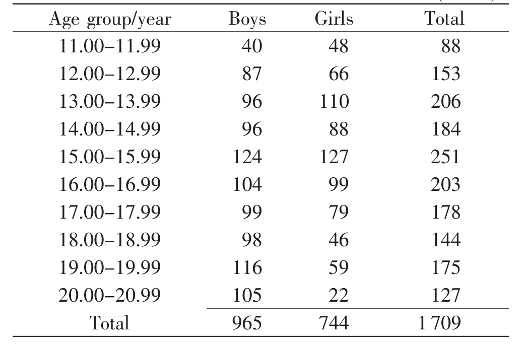
From Eastern,Central and Southern China,1 709 healthy teenagers aged 11-20 for the current study were collected,via stratified sampling method,which composed of 744 girls and 965 boys,respectively. The true data of their births were confirmed by hospital birth certificates.The average age of menarche was 12.9 inquired among the female subjects.On the basis of obtaining volunteers’informed consent, these samples were divided into ten groups every one year in terms of age and sex distribution(Table 1).

Table 1 Age and gender distribution(cases)
The height and weight of each subject were measured.The sampleentrance criterion included normal height and body mass according to the national criteria[3],i.e.,physical wellbeing and good nutritional status.Those were excluded who had received special recreation and sports training,taken the medications which can influence bone development,or had a history of bone injury or bone development disorder.
2.2 Photographs of X-ray
Considering that the images of sternal end of clavical,manubrium of sternum,lungs and ribs were overlapped in X-ray films,it was difficult from a single side to confirm whether the secondary ossification center of sternal end of clavical had appeared.Thus,two sides of sternal end of clavical and the left side of shoulder,elbow,wrist,hip,knee and ankle joints were photographed.Antero-posterior and lateral X-ray projection were taken for each joint by X-ray apparatus with tube current from 200 to 500 mA,voltage from 80 to 100 kV.Consequently, 11963 valid radiographs were obtained.
2.3 Bone age indexes
As bone age indexes,24 osteal loci of the joints were selected.In accordance with those selected by previous researches[4-8],it was difficult to observe the distinctive changes of 8 carpals,since the carpals of boys and girls almost matured after the age of 12. Therefore,only 7 indexes from hand and wrist joints were chosen,which were of significance to bone development in teenagers.The collected X-ray films observed,17 indexes from other 6 joints in all were found to reflect bone development(Table 2).

Table 2 The osteal loci(bone age indexes)and variables
2.4 Film reading
According to the radiological grading method of adolescent bone development,which was developed by Zhu et al.[9],all the X-ray films were read using MIWORKS 5.0.0.6 PACS radiograph reader software by two radiology experts and two post-graduates.Only the time of secondary ossification center appearance and epiphyseal fusion investigated,since both are recognized to be conspicuous identification markers of the development.All the data were recorded in Epidata 2.0 database and examined twice before being transformed into SPSS data sets.
2.5 Statistics
Skewness and kurtosis were used to exam the age distribution about appearance and fusion of each index as Gaussian distribution or skewness distribution.Mean(x),standard deviation(s)and method of percentileswereused toestimatereferencevalue scope(RVS)and confidence interval(CI)of the time about secondary ossification center appearance and epiphyseal fusion.
If the curves were presented as Gaussian distribution,was used to estimate 95%RVS(μ=1.96),but if as skewness distribution,percentiles were employed to estimate 95%RVS and 95%CI(μ=1.96)[10].
All statistic procedures were done by SPSS 14.0 software package with a significant level at 0.05 for both sides[11].
3 Results
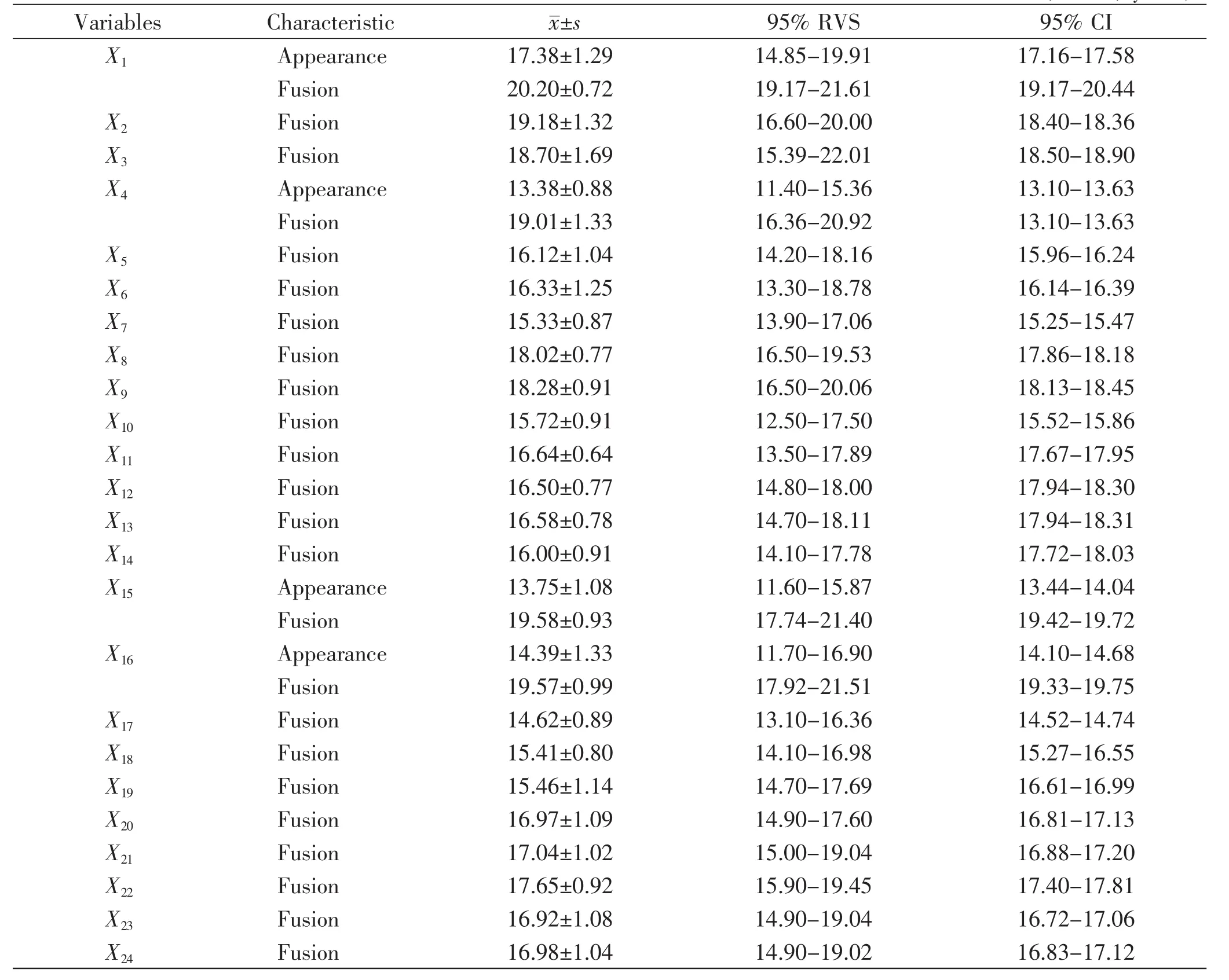
In the teenagers of Chinese Han nationality,the age intervals of the secondary ossification center appearance and epiphyseal fusion with±s,95%RVS and 95%CI were detailed in Table 3 and 4.
In addition,it was found that four secondary ossification centers(X1,X4,X15,X16)appeared in both the boys and girls after the age of 12,its mean age 17.38,13.38,13.75 and 14.39 in the former,and 16.15,12.64,12.88,13.34 in the latter,respectively. The time of those four indexes was found to have been advanced by about 1 year in the girls when compared with that in the boys of the teenagers of Chinese Han nationality.

Table 3 Age interval of secondary ossification center appearance and epiphyseal fusion in boys(n=965,years)

Table 4 Age interval of secondary ossification center appearance and epiphyseal fusion in girls(n=744,years)
4 Discussion
4.1 Several rules of secondary ossification center appearance and epiphyseal fusion
The latest scientific method available for age estimation is the radiological approach to bone development.In the past few decades,the investigations on constitution and health have revealed that growth and development of Chinese Han teenagers have presented a trend of evident acceleration.Therefore,the time of secondary ossification center appearance and epiphyseal fusion of current Chinese Han teenagers has been advanced.The two physical features,which are of distinctive marks in the long period ofbonegrowth,areeasy tobeobserved through X-ray films.There are normal ranges of the time of every secondary ossification center appearance and epiphyseal fusion,but they are disparate in different joints thanks to secondary ossification center or epiphysis.
In the past few years,the correlative studies have indicated that the floating interval of those normal ranges could be changed within several years[5]. When compared with the state of bone development in 14-year-old teenagers,it probably displays the same characteristics as 12 years,even as 11 years. In the current study,we found that the time of secondary ossification center appearance and the advance of epiphyseal fusion in the girls were much earlier than that in the boys.
4.2 The secular trends of bone development in Chinese Han teenagers
Currently,the widely used forensic approach to age estimation of living subjects in China judges the time of secondary ossification center appearance and epiphyseal fusion because of its convenience and facility.The method is based on the data collected by Li et al.[5,12]in 1960s,and sometimes even on the foreign data.However,the samples of these data are neither adequate nor integrate,so their reliability and accuracy can be questioned.It has been recently proved that bone development is differed to region and climate,except to the influence of heredity and living habit.With health status and living condition improved,the time of skeletal development has been advanced,suggesting that the data used to estimate age of living subjects should be updated every 10-15 years,and that the results of different countries or races can be only used in the corresponding areas.
The studies of Gu et al.[12-13]found the time of epiphyseal fusion of X10,X11,X12,X13,X14to be at the age of 18 in both boys and girls,and the time of secondary ossification center appearance of X1and epiphyseal fusion of X15to be at the age of 18-20 years and 20-25 years in boys and girls,respectively. However,our data demonstrated that the time of epiphyseal fusion of X10,X11,X12,X13,X14approximately advanced to the age of 16 and 15 in the boys and girls,respectively;that the time of secondary ossification center appearance of X1,to the age of 17 and 16 in the boys and girls,respectively;and that the age of epiphyseal fusion of X15were both approximately at the age of 19 in the boys and girls. The study of Xi[14]in 1997 reported that the time of epiphyseal fusion of knee joint were at the age of 18-21 and 17-19 in the boys and girls,respectively.In the current study,however,the results indicated the age of 17 and 16 in the boys and girls,respectively.In comparison with thepreviousdata, therefore,it’s obvious that the development age has been made earlier by 2-3 years,which confirmed the study of Gu’s et al.[15]It can be concluded that the time of epiphyseal fusion of X4,X15,X16and secondary ossification center appearance ofX10,X17were nearly to the average age of menarche(Table 4). Therefore,these five indexes could better forecast the time of menarche.
4.3 The implication of the new data
In most European countries,the legally relevant key age limit ranges are 14-22 years[16].Economic globalization,European integration,cross-border guilt and the current armed conflicts have led to a rise in cross-border migration in Europe,which in turn have resulted in a steady increase of the foreign population in many European countries[17].This development has produced a growing demand for age estimation for those involved in criminal proceedings. Therefore,the radiological study of bone development,especially the studies on the development of secondary ossification center appearance and epiphyseal fusion are becoming globally urgent in forensic anthropology.
In China,forensic experts are often required to report the probable age in various civil and criminal cases when the age ranges are 12-18 years.For decades,the investigation on the time of secondary ossification center appearance and epiphyseal fusion has been considered as a reasonable scientific and acceptable method for age estimation by court of law in China because it helps enhance legal certainty by ensuring equal treatment of persons with or without valid identity documents.Such investigationshelp prevent perpetrators from wrongfully benefiting from false claims to be younger than they really are,and also supply exonerating evidence for those who are erroneously suspected ofmaking false statements about their ages[18].
However,the data in this area need to be updated in accordance with the advancement of bone development.In order to gain relatively accurate age estimation in living subjects,much more bone age indexes should be taken into account.In the current study,24 indexes of 7 joints,selected from articulatio sternoclavicularis,joints of upper extremity and lower extremity,can be used as new data to reflect the accelerated trends of bone development in the contemporary Chinese teenagers,and to contribute to the literature of bone age researches on living subjectsfrom a perspectiveofforensicanthropology. The new results can have far-reaching values in formulating bone norms in the future,and also in conducting clinical researches such as forecasting the time of menarche,diagnosing teenagers’growth retardation and choosing the materials for athletes.
Acknowledgements
This study was funded by the Ministry of Science and Technology of the People’s Republic of China(No.2004DEA70970)and Science and Technology Commission ofShanghaiMunicipality(No. 042512036).We are grateful to all the boys and girls as the subjects of the current study for their cooperation.We are also thankful to the Department of Radiology and Statistics for their technical support.
[1] Zhang YB,Li TM,Wang SQ,et al.A comparative study on bone growth for Chinese in the past 30 years[J]. JournalofTianjin Institute ofPhysicalEducation, 1997,12(1):28-31.
[2] Banerjee KK,Agarwal BB.Estimation of age from epiphyseal union at the wrist and ankle joints in the capital city of India[J].Forensic Sci Int,1998,98(1-2): 31-39.
[3] 中国学生体质与健康研究组.改革开放20年中国汉族学生体质状况的动态分析[M]//中国学生体质与健康调研报告.北京:高等教育出版社,2002:54-93.
[4] Tanner JM,Healy MJR,Goldstein H,et al.Assessmentofskeletalmaturity and prediction ofadult height(TW3 Method)[M].Oxford,UK:Blackwell Scientific Publications,2001.
[5] 李果珍,张德苓,高润泉.中国人的骨发育研究:上肢骨发育的初步研究[J].中华放射学杂志,1979,13(1):19-23.
[6] 贾静涛.法医人类学[M].沈阳:辽宁科学技术出版社,1993:171-312.
[7] 叶义言.中国儿童骨龄评分法[M].北京:人民卫生出版社,2005:83-151.
[8] Zhang SY,Liu LJ,Wu ZL,et al.The skeletal development standards of hand and wrist for Chinese children—China 05Ⅰ.TW3-C RUS,TW3-C Carpal,and RUS-CHN Methods[J].Chin J Sports Med,2006,25(5): 509-516.
[9] Zhu GY,Fan LH,Zhang GZ,et al.Staging methods of skeletal growth by X-ray in teenagers[J].Fa Yi Xue Za Zhi,2008,24(1):18-24.
[10]孙振球.医学统计学[M].北京:人民卫生出版社,2002:9-83,140-318,427-477.
[11]余建英,何旭宏.数据统计分析与SPSS应用[M].北京:人民邮电出版社,2003:141-291,365-459.
[12]Gu GN,Wu XZ.Ossification and maturation of bones of hand and wrist among the Chinese[J].Acta Anatomica Sinica,1962,5(2):173-183.
[13]Wu RK,Bo HY.Attrition of molar teeth in relation to age in Northern Chinese skulls[J].Vertebrate PalAsiatica,1965,9(2):217-222.
[14]席焕久.人的骨骼年龄[M].沈阳:辽宁民族出版社,1997:24-43.
[15]顾光官,朱海贤,邹新农,等.国人标准骨龄的研究及应用[J].苏州医学院学报,1998,18(8):877-878.
[16]Schmeling A,Geserick G,Reisinger W,et al.Age estimation[J].Forensic Sci Int,2007,165(2-3):178-181.
[17]Angenendt S.Asylum and migration policies in the European Union[M].Europa Union Verlag,Bonn,1999.
[18]Schmeling A,Reisinger W,Geserick G,et al.Age estimation of unaccompanied minors.PartⅠ.General considerations[J].Forensic Sci Int,2006,159(S1):S61-S64.
date:2012-05-07)
(Editor:LIANG Lu)
DF795.1
A
10.3969/j.issn.1004-5619.2012.04.007
Article IC:1004-5619(2012)04-0269-06
Author:WANG Ya-hui(1982—),male,assistant research fellow, MD in forensic medicine;E-mail:wangyh@ssfjd.cn
ZHU Guang-you,male,research fellow, master tutor in forensic clinic and research;E-mail:zhugy@ssfjd.cn
