三种胰腺癌细胞株Mesothelin的表达及成瘤速度的比较
2012-11-07潘璜吴洪玉陈士跃刘敬禹邵成伟田建明
潘璜 吴洪玉 陈士跃 刘敬禹 邵成伟 田建明
·论著·
三种胰腺癌细胞株Mesothelin的表达及成瘤速度的比较
潘璜 吴洪玉 陈士跃 刘敬禹 邵成伟 田建明
目的比较3种人胰腺癌细胞株在体外和裸鼠体内Mesothelin的表达及裸鼠皮下种植瘤生长速度的差异。方法培养人胰腺癌细胞株SW1990、BxPC3、PANC1,取对数生长期分别注射于裸鼠左腋窝皮下,每周测量裸鼠皮下种植瘤的长、短径,计算体积,SW1990、BxPC3组裸鼠观察3周,PANC1组裸鼠观察5周;用蛋白质印迹法分别检测3种细胞及裸鼠皮下种植瘤组织中的Mesothelin表达,用免疫组化染色检测3种裸鼠皮下种植瘤组织中Mesothelin表达。结果人胰腺癌细胞株体外Mesothelin表达的强弱顺序是BxPC3>PANC1>SW1990,体内种植瘤组织表达的强弱顺序是SW1990>BxPC3>PANC1。3种人胰腺癌细胞种植于裸鼠皮下的成瘤率均为100%,各组肿瘤的生长速度顺序为SW1990>BxPC3>PANC1。结论不同人胰腺癌细胞株Mesothelin的表达量不同,裸鼠皮下种植瘤的生长速度亦不同,两者无相关性。
胰腺肿瘤; 细胞系,肿瘤; Mesothelin蛋白; 小鼠,裸; 印迹法,蛋白质; 免疫组织化学
近来研究发现,Mesothelin蛋白表达于胰腺癌细胞膜上,而正常胰腺细胞及其他原发性胰腺疾病时则不表达,因而对于胰腺癌具有较高的特异性,成为研究早期诊断、靶向治疗胰腺癌的分子标靶之一。由于各实验所使用的细胞株多不相同,使得结果在一定程度上缺乏统一性。因此本研究采用常用的3种胰腺癌细胞株,对各细胞株Mesothelin表达及其在裸鼠体内的成瘤情况进行比较,探讨它们之间的关联,希望能为基于胰腺癌细胞Mesothelin表达的实验研究提供有价值的参考信息。
资料与方法
一、细胞株及实验动物
人胰腺癌细胞株SW1990、BxPC3、PANC1由第二军医大学长海医院消化内科实验室馈赠,常规体外培养、传代。BALB/C裸小鼠购自第二军医大学实验动物中心,雌性,4~5周龄,体重(18±2)g,SPF级,饲养环境恒温25~27℃,恒湿45%~50%。
二、裸鼠皮下成瘤实验
取3株对数期细胞,分别用PBS调整细胞悬液密度至2×10⁷/ml。将18只裸鼠按完全随机法分为3组,每组6只,分别将SW1990、BxPC3及PANC1细胞悬液0.2 ml种植于左腋窝皮下,接种后分笼饲养。每周用游标卡尺测量各肿瘤长(a)、短(b)径,按瘤体积=a×b2×0.4的公式计算[1],并绘制生长曲线。根据肿瘤生长速度,SW1990、BxPC3组裸鼠饲养3周,PANC1组裸鼠饲养5周后处死,摘取皮下种植瘤。
三、Mesothelin表达的检测
取对数生长期细胞,提取细胞总蛋白,常规行蛋白质印迹法检测细胞mesothelin蛋白表达。鼠抗人Mesothelin一抗购自美国SANTA CRUZ生物技术公司,工作浓度1∶200。
取皮下种植瘤组织,采用蛋白质印迹法和免疫组化法检测Mesothelin蛋白表达。蛋白质印迹法所用一抗同上,免疫组化所用一抗为美国R&D公司产品,工作浓度1∶100。所得图像用Imagel软件作半定量分析,以目的条带与内参条带灰度值比作为蛋白表达量。
四、统计学处理
3组裸鼠皮下肿瘤第3周的体积均数采用ANOVA检验,3组间的差异采用SNK检验。P<0.05认为差异具有统计学意义。
结 果
一、种植瘤的生长情况
裸鼠成瘤率100%,病理检查均证实为胰腺癌(图1)。各组种植瘤平均体积及生长曲线见表1、图2。
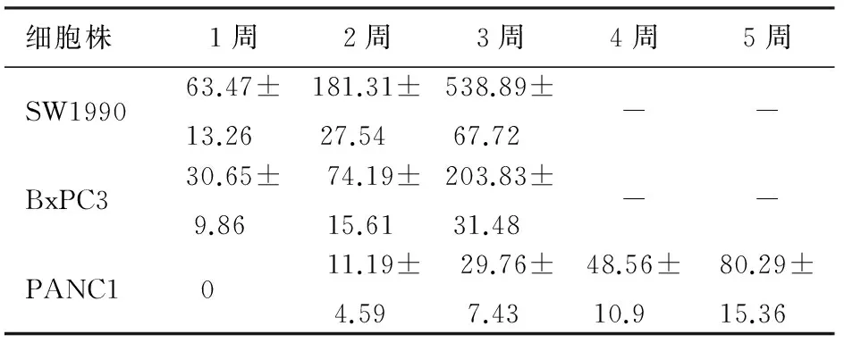
表1 裸鼠皮下种植瘤各周平均体积(mm3)
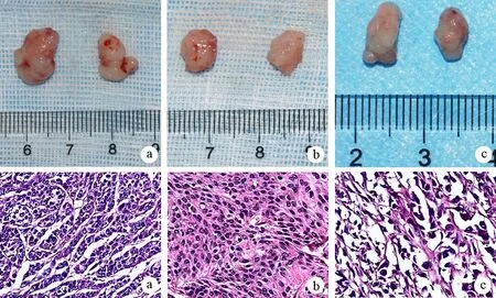
图1SW1990(a)、BxPC3(b)、PANC1(c)细胞的种植瘤(上)及病理(下)改变(HE ×100)
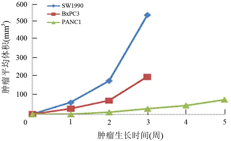
图2 3组种植瘤生长曲线
二、Mesothelin表达
SW1990、BxPC3、PANC1细胞株均有Mesothelin蛋白表达(图3a),表达量分别为0.76±0.03、1.14±0.08、1.02±0.05,以SW1990表达最低,它们的种植瘤组织Mesothelin表达量分别为2.26±0.17、0.20±0.03、0.06±0.01,以SW1990细胞种植瘤表达最高,PANC1最低(图3b)。SW1990细胞种植瘤呈强阳性染色,BxPC3及PANC1种植瘤仅见少数细胞弱阳性染色(图4)。
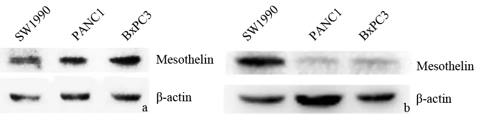
图3胰腺癌细胞株(a)和种植瘤组织(b)的mesothelin表达(蛋白质印迹法)

图4SW1990(a)、PANC1(b)及BxPC3(c)细胞种植瘤组织Mesothelin表达(免疫组化染色 ×400)
讨 论
Mesothelin是一种分化抗原,在正常人体组织中只有胸膜、心包和腹膜上的间皮细胞表达[2-3]。既往报道,某些人类肿瘤,如间皮瘤、卵巢癌等Mesothelin异常高表达。2001年Argani等[4]首次报道胰腺导管腺癌高表达Mesothelin mRNA及蛋白,而正常胰腺组织无表达。随后得到许多研究的证实[5-8]。
本研究结果显示,3株人胰腺癌细胞均有Mesothelin蛋白表达,但表达的量有差异。它们的种植瘤亦均有Mesothelin表达,但表达量与体外实验结果不同。体外实验以SW1990表达量最低,而体内种植瘤则以SW1990表达量最高,这一方面反映机体对肿瘤细胞基因表达具有调控作用,另一方面表明不同的肿瘤细胞对机体的反应也不一致。
裸鼠种植瘤模型是研究人体肿瘤的一种较为理想的模型,在临床研究中广泛应用,是最能反映人类胰腺癌生物学特性的“活试管”[9-10]。应用直接皮下细胞注射法制作裸鼠种植瘤的成功率高达80%~
100%[11]。本研究结果显示,裸鼠成瘤率达100%。但3种不同细胞株的瘤体积差异具有统计学意义,SW1990细胞种植瘤平均体积较PANC1细胞种植瘤约大17倍,BxPC3种植瘤平均体积也较PANC1种植瘤约大6倍;即使5周后PANC1种植瘤体积仍然小于仅3周的SW1990及BxPC3种植瘤。
SW1990细胞种植瘤组织的Mesothelin表达量较其他两株细胞的种植瘤显著上调,提示Mesothelin参与体内癌细胞的增殖,其机制有待进一步研究。
[1] Hassan R, Williams-Gould J, Steinberg SM, et al. Tumor-Directed Radiation and the Immunotoxin SS1P in the Treatment of Mesothelin-Expressing Tumor Xenografts. Clin Cancer Res, 2006,12:4983-4988.
[2] Chang K, Pastan I. Molecular cloning of mesothelin, a differentiation antigen present on mesothelium, mesotheliomas, and ovarian cancers. Proc Natl Acad Sci USA, 1996,93:136-140.
[3] Chang K, Pastan I, Willingham MC. Isolation and characterization of a monoclonal antibody, K1, reactive with ovarian cancers and normal mesothelium. Int J Cancer, 1992,50:373-381.
[4] Argani P, Iacobuzio-Donahue C, Ryu B, et al. Mesothelin is overexpressed in the vast majority of ductal adenocarcinomas of the pancreas: identification of a new pancreatic cancer marker by serial analysis of gene expression (SAGE). Clin Cancer Res, 2001,7:3862-3868.
[5] Hassan R, Laszik ZG, Lerner M, et al. Mesothelin is overexpressed in pancreaticobiliary adenocarcinomas but not in normal pancreas and chronic pancreatitis. Am J Clin Pathol, 2005,124:838-845.
[6] Ordonez NG. Application of mesothelin immunostaining in tumor diagnosis. Am J Surg Pathol, 2003,27:1418-1428.
[7] Li M, Bharadwaj U, Zhang R, et al. Mesothelin is a malignant factor and therapeutic vaccine target for pancreatic cancer. Mol Cancer Ther, 2008,7:286-296.
[8] 朱庆云,屠振兴,李兆申,等. Mesothelin在胰腺癌组织的表达及其临床意义. 胰腺病学, 2005,5:17-20.
[9] 叶燕丽,周昕熙,王莲桂. 裸小鼠的繁殖及在肿瘤学中的应用. 实验动物科学与管理, 2005,22:6-8.
[10] Celinski SA, Fisher WE, Amaya F, et al. Somatostatin receptor gene transfer inhibits established pancreatic cancer xenografts. J Surg Res, 2003,115:41-47.
[11] 王凤力,李德春,张子祥,等. 胰腺癌裸鼠皮下瘤模型建立的比较研究. 苏州大学学报(医学版), 2007,27:359-376.
Comparisonofexpressionofmesothelinamongthreekindsofpancreaticcancercelllinesanddevelopmentspeedintheirnudemousemodels
PANHuang,WUHong-yu,CHENShi-yue,LIUJing-yu,SHAOCheng-wei,TIANJian-ming.
DepartmentofRadiology,LushanSanatoriumofPLA,Jiujiang332000,ChinaCorrespondingauthors:SHAOCheng-wei,Email:cwshao@sina.com;TIANJian-ming,Email:tianjianming1952@hotmail.com
ObjectiveTo compare the mesothelin expressions in 3 human pancreatic cancer cell lines between in vitro and in vivo and the developing speed among the subcutaneous tumors implanted with the 3 human pancreatic cancer cell lines in nude mice.MethodsThe human pancreatic cancer cell lines (SW1990, BxPC3 and PANC1) were cultured and then were implanted subcutaneously into left axillas of nude mice. The volumes of these subcutaneous tumors were recorded every week to estimate their developing speed. The mice implanted with SW1990 and BxPC3 cells were observed for three weeks, while the mice implanted with PANC1 cell were observed for five weeks. The Western blot method was used to measure the expressions of mesothelin in the 3 kinds of cells and subcutaneous tumors, while immunohistochemical staining was applied to determine the expressions of mesothelin in 3 kinds of subcutaneous tumors.ResultsThe sequence of quantities of expressions of mesothelin in these cell lines in vitro were BxPC3>PANC1>SW1990, and the sequence of quantities of expressions in vivo were SW1990>BxPC3>PANC1. One handrued percent of the tumors grew out successfully, and the sequence of speeds of their growth was SW1990>BxPC3>PANC1.ConclusionsThe mesothelin expressions among 3 kinds of pancreatic cancer cell line are different. The developing speeds of tumors originated from different subcutaneous tumors in nude mice are also different, and there is no association between them.
Pancreatic neoplasms; Cell line, tumor; Mesothelin protein; Mice, nude; Blotting, western; Immunohistochemistry
10.3760/cma.j.issn.1674-1935.2012.05.011
国家自然科学基金(30970801)
332000 江西九江,解放军庐山疗养院放射科(潘璜);第二军医大学长海医院消化内科(吴洪玉),放射科(潘璜、陈士跃、刘敬禹、田建明、邵成伟)
邵成伟,Email:cwshao@sina.com;田建明,Email:tianjianming1952@hotmail.com
2012-08-07)
(本文编辑:屠振兴)