小鼠胃和阑尾组织中h-CD降解规律与死亡时间的相关性研究
2011-11-10梁新华
刘 嘉,梁新华,焦 炎
(山西医科大学法医学院,山西太原 030001)
小鼠胃和阑尾组织中h-CD降解规律与死亡时间的相关性研究
刘 嘉,梁新华,焦 炎
(山西医科大学法医学院,山西太原 030001)
目的:观察小鼠死亡后不同时间胃和阑尾组织中h-CD含量的变化,探讨不同温度条件下h-CD的降解规律与死亡时间的关系及其组织差异性。方法:选取72只健康成年昆明小鼠,采用机械性窒息致死后,随机分为两组,分别置于4℃冰箱和25℃人工气候箱中,并于死后0、24、48、72、96、120 h提取胃及阑尾组织,采用免疫印记及计算机图像分析技术检测上述组织中h-CD的含量,应用SPSS 16.0软件对数据进行统计学分析。结果:①h-CD的含量随死亡时间的延长而降低,与死亡时间具有相关性;②不同温度条件下,胃和阑尾组织中h-CD的降解速度均存在明显差异(P<0.05),h-CD在25℃组降解速度快于4℃组(P<0.05);③h-CD的降解速度在胃和阑尾组织中存在组织差异性,在阑尾组织中的降解速度较快(P<0.05)。结论:h-CD的含量随死亡时间延长而降低,其降解速度受环境温度影响,温度升高,降解速度加快,且胃和阑尾组织中h-CD的降解速度具有组织差异性,其在阑尾组织中的降解速度快于胃组织中的降解速度。
法医病理学;重型钙调蛋白结合蛋白;死亡时间;免疫印记
机体死亡后,发挥重要生命功能的蛋白质也会随之发生降解。蛋白质的降解不仅受蛋白酶的作用,而且受尸体所处环境温度的影响[1]。本实验应用免疫印记法,观察小鼠死亡后不同温度条件下胃和阑尾组织中重型钙调蛋白结合蛋白(h-CD)的降解规律,为死亡时间的推断提供有效的法医学依据。
1 材料与方法
1.1 实验动物分组及制备
本研究选用实验动物已获得本校伦理委员会的同意。选取72只健康成年昆明小鼠,雌雄不限,体重(30±5)g(由山西医科大学实验动物中心提供)。采用机械性窒息方式致死后随机分为低温组和高温组,每组36只,分别放置于4℃冰箱和25℃人工气候箱中,再依据死亡时间将两组分别再分为对照组、死后 24、48、72、96、120 h 组 6 个亚组,每组 6 只。
1.2 免疫印记检测
分别称取100 mg胃和阑尾组织,加入1 ml裂解液(RIAP)和10 μmol/L蛋白酶抑制剂(PMSF)。加入适量液氮,在研钵中充分研磨。 将组织匀浆后抽取500 μl,4℃ 12 000×g离心30 min,移取上清液测蛋白浓度后置于-80℃冰箱中备用。取20 μg蛋白样品,在10%变性聚丙烯酰胺凝胶中垂直板电泳,80 V,20 min浓缩胶,120 V,70 min分离胶。用水平电泳槽将凝胶进行电转移100 V,50 min,将蛋白转移至硝酸纤维素膜(购自武汉博士德生物工程有限公司)上。将含有蛋白质的硝酸纤维膜用TBST脱脂奶粉封闭,平缓摇动过夜,按1∶300加入caldesmon单克隆抗体(购自武汉博士德生物工程有限公司),室温下 1 h,TBST 洗膜 10 min×3 次,按 1∶400 分别加入二抗,室温下1 h,TBST洗膜10 min×3次。然后分别加入等体积发光剂(购自北京普利来基因有限公司)和增强剂反应1 min,分别对二者进行检测后,柯达(KODAK Image station 440)全自动显影系统显像。免疫印迹法结果进行图像扫描后采用Image proplus 5.0测出各条带的积分光密度值(integrate optical density,IOD)。 实验数据用均数±标准差(±s)表示。应用SPSS 16.0软件对数据进行t检验。以P<0.05为差异有统计学意义。
2 结果
Western-blot检测可见,存放于4℃和25℃温度条件下的小鼠胃、阑尾组织中的h-CD的含量均随死亡时间的延长而逐渐减少(图1、2),且变化趋势表现出一定的规律性,四组条带颜色均随死亡时间的延长而逐渐变浅,同一时间点处,25℃条件下,胃和阑尾中h-CD的含量明显少于4℃条件时h-CD的含量,而在温度相同的条件下可见,h-CD在阑尾组织中的降解速度快于其在胃组织中的速度。
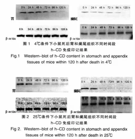
IOD值分析结果显示:在α=0.05检验水准下,4℃条件时,对胃组织在各时间点时h-CD的含量与相邻上一时间点时 h-CD的含量进行检验,t值依次 为 45.205、57.429、62.799、110.229、78.282,可得 P 值均小于 0.05,具有统计学意义;对阑尾组织进行相同的检验,t值依次为43.171、51.382、275.094、121.302、60.061,P 均<0.05, 差异具有统计学意义。25℃条件时,对胃组织进行相同检验,t值依次为9.132、15.684、59.941、63.392、51.878,P 均<0.05, 差异有统计学意义;对阑尾组织进行相同的检验,t值依次为115.429、187.980、178.497、83.328、55.579,P 均<0.05,差异具有统计学意义。由上述分析可知,各组中h-CD的含量均随时间的延长而减少。见表1。各时间点h-CD含量的相对变化以死后不同时间点h-CD含量占死亡即刻含量的百分数来反应,在α=0.05检验水准下,4℃条件时,对胃和阑尾组织中h-CD含量百分数进行检验,t值依次为 6.231、13.115、70.779、65.209、31.674,P均<0.05,差异具有统计学意义;在25℃条件时检验 ,t 值 依 次 为 4.430、45.632、62.525、47.156、22.517,P 均 <0.05,差异具有统计学意义。对胃组织4℃组和25℃组的数据进行检验,t值依次为 1.173、30.356、77.813、105.650、85.777,可知除死后24 h 4℃组和25℃组数据无统计学意义 (P=0.294)外,其余P均<0.05,差异具有统计学意义;对阑尾组织中各数据进行上述检验,t值依次为15.570、210.634、85.413、137.843、126.440,P 均<0.05,差异具有统计学意义。分析数据可知25℃组胃和阑尾组织h-CD降解速度均高于4℃组,且在同温度比较中阑尾组织中h-CD降解速度均高于胃组织的降解速度。见表2。
表1 小鼠死后不同时间不同温度h-CD的IOD值(±s)Tab.1 The IOD values of h-CD of mouse in the different time and temperature(±s)
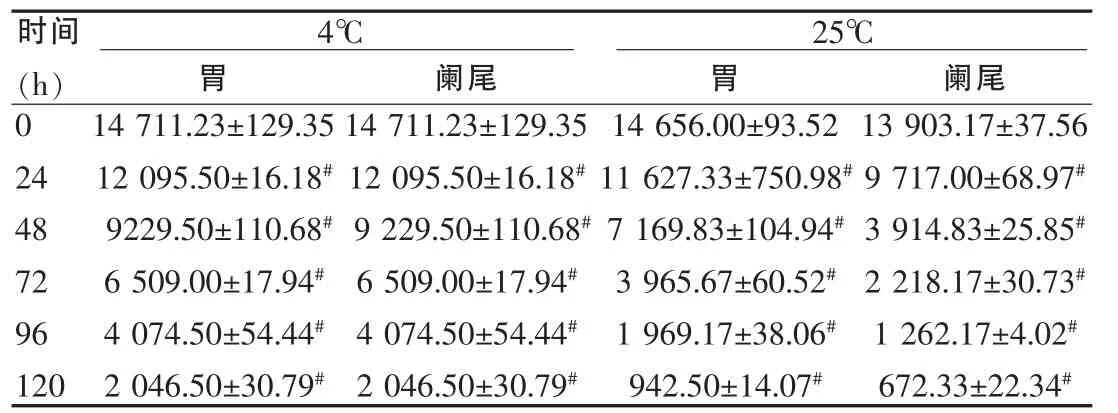
表1 小鼠死后不同时间不同温度h-CD的IOD值(±s)Tab.1 The IOD values of h-CD of mouse in the different time and temperature(±s)
与上邻组比较,#P<0.05Compared with up group in the same temperature and tissue,#P<0.05
时间(h)4℃胃阑尾25℃胃阑尾0 24 48 72 96 120 14 711.23±129.35 12 095.50±16.18#9229.50±110.68#6 509.00±17.94#4 074.50±54.44#2 046.50±30.79#14 711.23±129.35 12 095.50±16.18#9 229.50±110.68#6 509.00±17.94#4 074.50±54.44#2 046.50±30.79#14 656.00±93.52 11 627.33±750.98#7 169.83±104.94#3 965.67±60.52#1 969.17±38.06#942.50±14.07#13 903.17±37.56 9 717.00±68.97#3 914.83±25.85#2 218.17±30.73#1 262.17±4.02#672.33±22.34#
表2 小鼠死后不同时间段h-CD含量的相对变化(±s,%)Tab.2 The relative content changes of h-CD of mouse in different time and temperature(±s,%)

表2 小鼠死后不同时间段h-CD含量的相对变化(±s,%)Tab.2 The relative content changes of h-CD of mouse in different time and temperature(±s,%)
同温度不同组织间比较,*P<0.05;同组织不同温度间比较,#P<0.05Comparedwithdifferent tissuesinthe same temperature,*P<0.05;compared with different temperature in the same tissue,#P<0.05
时间(h)4℃胃阑尾25℃胃阑尾0 24 48 72 96 120 100 82.2%±0.8 62.7%±0.6 44.3%±0.4 27.7%±0.4 13.9%±0.2 100%78.6%±1.2*58.6%±0.2*29.5%±0.2*17.4%±0.2*12.2%±0.1*100%79.3%±5.5 48.9%±1.0#27.0%±0.4#13.4%±0.3#6.3%±0.1#100%69.9%±0.6*#28.2%±0.2*#15.9%±0.2*#9.1%±0.1*#4.8%±0.2*#
3 讨论
h-CD存在于正常平滑肌细胞和软组织平滑肌肿瘤[2]及多种骨肿瘤中[3],它可以起到抑制肌球蛋白Mg-ATP酶活性的作用,还可以抑制平滑肌磷酸化肌球蛋白所引发的肌动蛋白丝的移动[4-6],是一种重要的调节蛋白。有研究表明,机体死亡后96 h内h-CD的含量仍保持相对稳定[7],因此可选其作为早期死亡时间推断的测量指标。
从本实验可以看出在胃和阑尾组织中h-CD的含量随死亡时间的延长而降低,与死亡时间具有相关性;在不同温度条件下,h-CD 的含量变化存在差异(P<0.05),h-CD 在25℃组中的降解速度快于4℃组;且h-CD的降解速度在胃和阑尾组织中也存在差异(P<0.05),在阑尾组织中的降解速度较快。这主要是由于在机体死亡后,由于细胞膜渗透能力的变化,使得离子能自由的出入细胞。从细胞外环境、内质网、肌质网释放出来的游离Ca2+进入了细胞液是死后发生细胞或分子水平发生变化的重要原因之一[8-9]。Ca2+进入细胞引起一系列后果:①激活钙调蛋白。随着死亡时间的逐渐延长,大量钙调蛋白被进入细胞液的游离Ca2+激活,从而使作为靶蛋白的钙调蛋白结合蛋白由于与被活化的钙调蛋白结合[10],其含量则逐渐下降。②激活依赖Ca2+的蛋白激酶,例如钙蛋白酶[11],它作用于钙调蛋白结合蛋白后,可以根据酶解物的特异性确定钙调蛋白结合蛋白的亚型。由于死后大量的钙蛋白酶被激活,导致大量的钙调蛋白结合蛋白被酶解,因此在一定时间内,随着死亡时间的延长,由于依赖Ca2+的蛋白激酶被大量活化,钙调蛋白结合蛋白的含量逐渐下降。以上说明,死后细胞液游离Ca2+的增加导致了钙调蛋白和依赖Ca2+的蛋白激酶的激活,最终引起h-CD发生降解而含量减少,且随着死亡时间的延长,机体组织的pH值逐渐降低[12],钙蛋白酶活性下降而溶酶体释放的蛋白水解酶(最适pH值3~7)活性增强[13]。由于钙蛋白酶和死亡后期溶酶体释放的蛋白水解酶的激活,使得钙调蛋白结合蛋白发生了蛋白水解作用而降解。因此在本实验中,通过免疫印记法检测可以看到随着时间的延长,h-CD的含量逐渐降低。
在4℃组和25℃组的比较中还可发现,高温组中h-CD的降解程度高于低温组,这说明高温可以加快蛋白质的分解而低温则可起到延迟分解的作用[14-15]。这主要是因为机体死后,组织、细胞的自溶主要是在其自身固有的各种酶的作用下进行的,而酶活性的高低很大程度上受温度的调控。因此在高温环境下,组织、细胞结构破坏、溶解要早于低温环境。在相同温度条件下,肠组织自溶发生早于胃组织,其中以回盲部附近为最早,而胃体部自溶较晚[16]。从本实验也可发现,在同组比较中,h-CD在阑尾组织中的降解速度快于在胃组织的降解速度,这与尸体腐败的一般顺序相一致。此外,影响蛋白质分解的另一重要因素是腐败菌的作用。由于其在胃中分布很少,故而在相同温度条件下,阑尾组织中h-CD降解速度更快。以上的实验结果证实了h-CD的讲解规律与死亡时间及温度存在有相关性;但由于人与动物间存在种属差异以及h-CD降解过程中受多重内、外因的影响,因此要明确h-CD在人体中的降解规律则有待于今后大量人体实验的研究。此研究成果将对法医学鉴定中死亡时间的认定有很大帮助。
[1]Archer MS.Rainfall and temperature effects on the decomposition rate of exposed neonatal remains[J].Sci Justice,2004,44(1):35-41.
[2]Watanabe K,Kusakabe T,Hoshi N,et al.h-Caldesmon in leiomyosarcoma and tumors with smooth muscle cell-like differentiation:its specific expression in the smooth muscle cell tumor[J].Hum Pathol,1999,30:392-396.
[3]Kazuo Watanabe,Takahiro Tajino,Miho Sekiguchi,et al.H-Caldesmon as a specific maker for smooth muscle tumors comparison with other smooth muscle makers in bone tumors [J].Am J Clin Pathol,2000,113:663-668.
[4]Gerthoffer WT,Pohl J.Caldesmon and calponin phosphorylation in regulation of smooth muscle contraction[J].Can J Physiol Pharmacol,1994,72:1410-1415.
[5] Phillips SV,Sconwoo GC,Walsh MP,et al.Comparison of the caldesmon content of cardiac and smooth muscle[J].J Mol Cell Cardiol,1999,31:1413-1417.
[6]Savineau JP,Marthan R.Modulation of the calcium sensiuvity of the smooth muscle contractile apparatus;molecular mechanisms,pharmacological and pathophysiological implication [J].Fundam Clin Pharmacol,1997,11:289-299.
[7]Kang S,Kassam N,Gauthier ML,et al.Post-mortem changes in calmodulin binding proteins in muscle and lung [J].Forensic Sci Int,2003,131(2-3):140-147.
[8]Lametsch R,Roepstorff P,Bendixen E.Identification of protein degradation during post-mortem storage of pig meat[J].J Agric Food Chem,2002,50(20):5508-5512.
[9]Kent MP,Spencer MJ,Koohmaraie M.Postmortem proteolysis is reduced in transgenic mice overexpressing calpastatin [J].J Anim Sci,2004,82(3):794-801.
[10]Obata K,Nagata K.Overexpression of calmodulin induces cardiac hypertrophy by a calcineurin-dependent pathway[J].Biochem Biophys Res Commun,2005,26:1-18.
[11]Wang KK,Villalobo A,Roufogalis BD.Calmodulin-binding proteins as calpain substrates[J].Biochem,1989,262(3):693-706.
[12]党永辉,王振原,张联合,等.测量大鼠死后骨骼肌pH值推断早期死亡时间实验研究[J].中国法医学杂志,2005,20(4):202-205.
[13]Takeichi S,Tokunaga I.Mechanism of postmortem autolysis of skeletal muscle[J].Biochem Med,1984,32(3):341-348.
[14]Prieto JI,Magana C,et al.Interpretation of postmortem change in cadavers in Spain[J].J Forensic Sci,2004,49(5):918-923.
[15]Archer MS.Rainfall and temperature effects on the decomposition rate of exposed neonatal remains[J].Sci Justice,2004,44(1):35-41.
[16]陈新山.死后变化[A].赵子琴.法医病理学[M].3版.北京:人民卫生出版社,2004:48-49.
Associativity study between h-CD degradation regularity and the estimation of postmortem interval in stomach and appendix of mice
LIU Jia,LIANG Xinhua,JIAO Yan
(Faculty of Forensic Medicine of Shanxi Medical University,Taiyuan 030001,China)
Objective:To observe the postmortem degradation of high molecular-weight caldesmon(h-CD)in stomach and appendix smooth muscle cells of mice,study the effects of postmortem intervals,and ambient temperature on the h-CD degradation of mice,and the variability of the h-CD degradation in stomach smooth muscle cells and appendix smooth muscle cells in different ambient temperature.Methods:72 healthy adult Kunming mice were killed by asphyxia,and put them in 4℃ refrigerator and 25℃ artificial climate incubator respectively,then extractd the stomach and appendix tissues at 0,24,48,72,96,120 h after dead.The h-CD contents in the above tissues were quantitated by western-blot assay and collected by Image Pro Plus 5.0 image analysis system,then the datas were statistically analyzed with the SPSS 16.0 software.Results:①h-CD content in stomach and appendix tissues decreased with the postmortem intervals,there was correlation between the postmortem degradation of h-CD and the postmortem intervals;②The differences of the postmortem degradation of h-CD in different ambient temperature were significant(P<0.05),the degradation in 25℃ faster than that in 4℃;③The differences of the postmortem degradation of h-CD between stomach and appendix tissues were significant(P<0.05),the degradation in the appendix more faster.Conclusion:h-CD content in stomach and appendix tissues decreased gradually with the postmortem intervals,the postmortem degradation of h-CD is influenced by ambient temperature,and the degradation rate tends to be raised with higher temperature,and the differences of the postmortem degradation of h-CD between stomach and appendix tissues are significant,the degradation in the appendix more faster.
Forensic pathology;High molecular-weight caldesmon(h-CD);Postmortem intervals(PMI);Western-blot
D919.4
A
1673-7210(2011)02(c)-017-03
2010-12-16)
