lcariin accelerates bone regeneration by inducing osteogenesisangiogenesis coupling in rats with type 1 diabetes mellitus
2024-04-22ShengZhengGuanYuHuJunHuaLiJiaZhengYiKaiLi
Sheng Zheng,Guan-Yu Hu,Jun-Hua Li,Jia Zheng,Yi-Kai Li
Abstract BACKGROUND Icariin (ICA),a natural flavonoid compound monomer,has multiple pharmacological activities.However,its effect on bone defect in the context of type 1 diabetes mellitus (T1DM) has not yet been examined.AIM To explore the role and potential mechanism of ICA on bone defect in the context of T1DM.METHODS The effects of ICA on osteogenesis and angiogenesis were evaluated by alkaline phosphatase staining,alizarin red S staining,quantitative real-time polymerase chain reaction,Western blot,and immunofluorescence.Angiogenesis-related assays were conducted to investigate the relationship between osteogenesis and angiogenesis.A bone defect model was established in T1DM rats.The model rats were then treated with ICA or placebo and micron-scale computed tomography,histomorphometry,histology,and sequential fluorescent labeling were used to evaluate the effect of ICA on bone formation in the defect area.RESULTS ICA promoted bone marrow mesenchymal stem cell (BMSC) proliferation and osteogenic differentiation.The ICA treated-BMSCs showed higher expression levels of osteogenesis-related markers (alkaline phosphatase and osteocalcin) and angiogenesis-related markers (vascular endothelial growth factor A and platelet endothelial cell adhesion molecule 1) compared to the untreated group.ICA was also found to induce osteogenesis-angiogenesis coupling of BMSCs.In the bone defect model T1DM rats,ICA facilitated bone formation and CD31hiEMCNhi type H-positive capillary formation.Lastly,ICA effectively accelerated the rate of bone formation in the defect area.CONCLUSION ICA was able to accelerate bone regeneration in a T1DM rat model by inducing osteogenesis-angiogenesis coupling of BMSCs.
Key Words: Icariin;Osteogenesis-angiogenesis coupling;Type 1 diabetes mellitus;Bone defect;Bone regeneration
INTRODUCTION
Diabetes mellitus (DM) is a growing epidemic globally[1].Worldwide,about 463 million adults aged 20 years to 79 years are suffering from diabetes,with a prevalence rate of 9.3%.It is estimated that by 2030 there will be 578 million (10.2%) people living with DM[2].DM places a significant strain on medical resources and patient quality of life,which in turn places heavy burdens on society and the patient’s family[3].DM is typically categorized as type 1 DM (T1DM) and type 2 DM based on the etiology.Patients with T1DM require exogenous insulin to lower blood glucose.Otherwise,chronic high blood glucose can lead to damage in the heart,blood vessels,eyes,kidneys and nerves[4].A growing body of research has shown T1DM affects bone metabolism[5-7];however,the underlying mechanism has not yet been fully elucidated.
Although the bone has a propensity for repairing itself,cases arise that are beyond the self-repairing capacity of the bone[8].One of these cases is a bone defect in a patient with T1DM[9].Bone metabolism is disordered in T1DM,which increases the challenges for the treatment of bone defects[10-12].The current standard of treatment are allografts and autografts.However,patients find these therapies to be unsatisfactory[13].Studies have suggested that the pathogenesis of disordered bone metabolism caused by T1DM is closely related to the imbalance between bone resorption and bone formation,and a high glucose environment could significantly inhibit osteoblast-mediated bone formation and promote osteoclast-mediated bone absorption[14-16].
Icariin (ICA) is a natural flavonol glycoside primarily extracted fromHerbaEpimedii.It has multiple pharmacological activities,including anti-inflammatory,anti-rheumatic,anti-diabetic nephropathy,anti-apoptotic,and anti-oxidative properties[17-21].ICA can also promote osteogenesis and play an anti-osteoporotic role[22-24].Huangetal[25] showed that ICA promoted osteogenic differentiation through upregulation of BMAL1,and Chengetal[26] demonstrated that ICA attenuated thioacetamide-induced bone loss through downregulation of the RANKL-p38/ERK-NFAT pathway.Haoetal[27] observed that ICA accelerated bone regeneration in the defect area of rabbit skulls.Several other studies have demonstrated the protective effects of ICA in T1DM models[28-30].Considering these findings collectively,we hypothesize that ICA has therapeutic potential for bone defect repair in T1DM.
Both osteogenesis and angiogenesis exert vital roles during the bone regeneration process[28-31].Several studies have consistently demonstrated the ability of ICA to promote osteogenesis[32-34].Interestingly,ICA has been shown to play different roles in angiogenesis depending on the circumstances.Yuetal[35] reported that ICA promoted angiogenesis in glucocorticoid-induced osteonecrosis of femoral heads,and Huangetal[36] demonstated ICA inhibition of angiogenesisviaregulation of the TDP-43 signaling pathway.It is unknown how ICA affects angiogenesis during the bone regeneration process in the context of T1DM.Therefore,this study investigated the effects and potential mechanisms of ICA on bone defect repair in the context of T1DM.
MATERlALS AND METHODS
Chemicals and reagents
ICA (Cat.No.M211098) was ordered from Mreda Technology Co.,Ltd.(Beijing,China).Monoclonal antibodies against CD29 (Cat.No.11-0291-82),CD90 (Cat.No.11-0900-81),CD105 (Cat.No.MA1-19594),CD34 (Cat.No.11-0341-85),and CD45 (Cat.No.11-0461-82) were purchased from eBioscience (San Diego,CA,United States).Primary antibodies against alkaline phosphatase (ALP;Cat.No.DF6225),osteocalcin (OCN;Cat.No.DF12303),vascular endothelial growth factor A (VEGFA;Cat.No.AF5131),and platelet endothelial cell adhesion molecule 1 (CD31;Cat.No.AF6191) were obtained from Affinity Biosciences (Cincinnati,OH,United States).The primary antibody against endomucin (EMCN;Cat.No.sc-65495) was obtained from Santa Cruz Biotechnology (Dallas,TX,United States).
Cell proliferation assay
Rat bone marrow mesenchymal stem cells (BMSCs) were isolated and cultured as previously described[37].For phenotypic analysis,expression of CD29,CD90,CD105,CD34,and CD45 was evaluated.Cell counting kit-8 (CCK-8;Cat.No.C0039,Beyotime,Beijing,China) was used to evaluate the effect of ICA on BMSC proliferation.BMSCs were treated with different concentrations of ICA (0 μM,1 μM,10 μM,and 100 μM) for 1 d,3 d,5 d,and 7 d.The untreated group served as control (CON).
Colony-forming unit assay
BMSCs were seeded into 6-well plates (1 × 10³cells/well) and incubated in the presence or absence of ICA for 1 wk.Then,the clones were fixed with 4% paraformaldehyde and stained with 0.1% crystal violet for 20 min.Colonies containing 50 or more cells were quantified by ImageJ software (Release 1.51,National Institutes of Health,Bethesda,MD,United States).
Osteogenic differentiation assays
When BMSC confluency reached 80%,the medium was replaced with osteogenic medium.Varying concentrations of ICA were added.Total cellular proteins were extracted,and the supernatant liquid was collected for downstream assays.ALP activity was measured with an ALP Staining Kit (Beyotime) after 1 wk.Calcium mineralization was detected after 3 wkviaalizarin red S (ARS) solution (Beyotime).
Quantitative real-time polymerase chain reaction
Total RNA was extracted with an RNA Purification Kit (Thermo Fisher Scientific,Waltham,MA,United States),and reverse transcription was performed with a cDNA Reverse Transcription Kit (Thermo Fisher Scientific).Quantitative realtime polymerase chain reaction (qRT-PCR) was performed using SYBR Green qPCR Master Mix (Thermo Fisher Scientific).Relative gene expression was calculated using the 2-ΔΔCTmethod.The primer sequences are listed in Table 1.
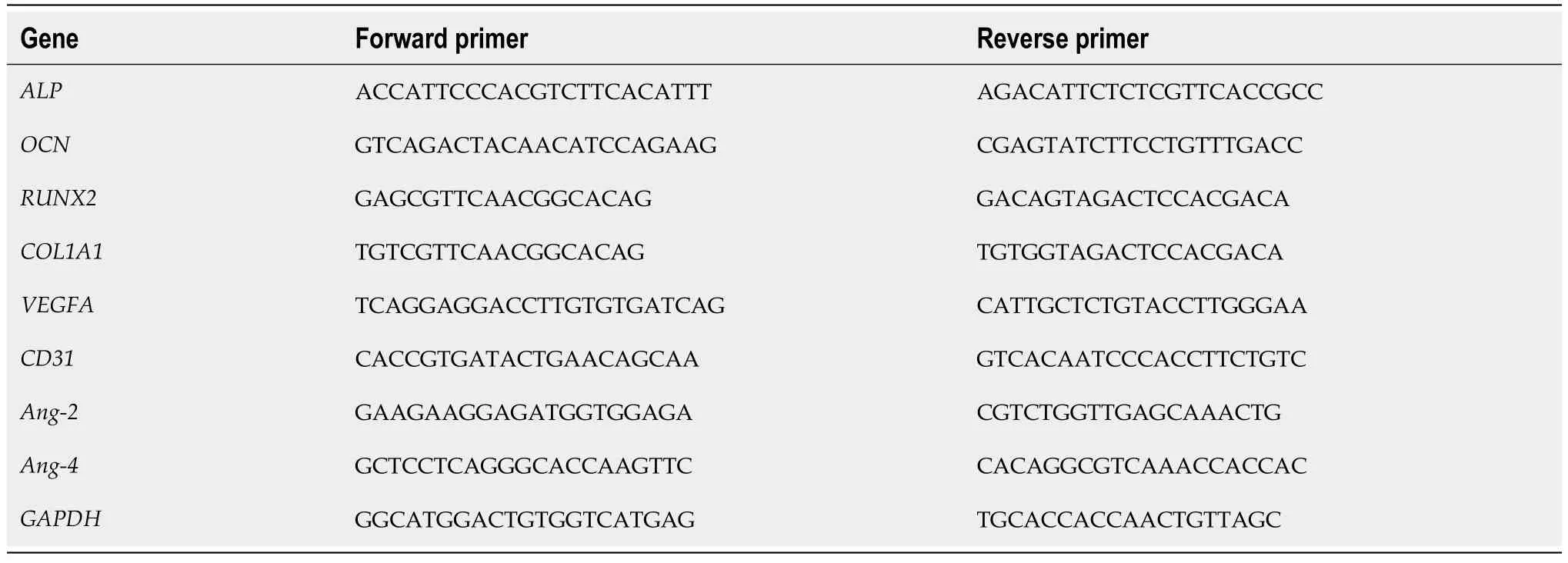
Table 1 Real-time polymerase chain reaction primer sequences
Western blot
Total protein was extracted using RIPA lysis buffer with protease inhibitors and protein phosphatase inhibitors (Beyotime) on ice.The cell lysates were collected and ultrasonicated for 10 min.After 15 min of centrifugation (4 °C,12000 rpm),the protein concentration was measured with a BCA Protein Assay Kit (Beyotime).Equal amounts of protein (30 μg) were subjected to 10% SDS-PAGE and transferred to PVDF membranes (Serva Electrophoresis GmbH,Heidelberg,Germany).The membranes were incubated with primary antibodies against ALP (1:1000 dilution),OCN (1:1000),VEGFA (1:1000),CD31 (1:2000),and β-actin (1:5000).The proteins were visualized by autoradiography and analyzed with ImageJ software.
Immunofluorescence
BMSCs were fixed with 4% paraformaldehyde for 30 min,permeabilized with 0.5% Triton X-100 for 15 min,and blocked with 1% bovine serum albumin for 30 min.The cells were incubated with primary antibodies overnight at 4 °C.Subsequently,BMSCs were washed thrice with PBS and incubated with fluorescence-conjugated secondary antibodies for 2 h.Fluorescence images were obtained with a BX63 fluorescence microscope (Olympus,Tokyo,Japan).
Angiogenesis-related assays
To further evaluate the proangiogenesis ability of ICA,BMSCs were treated with 10 μM ICA for 1 wk,and the conditioned medium (CM) was harvested.Human umbilical vein endothelial cells (HUVECs) were also cultured under different conditions: (1) Fresh medium (FM) without ICA (FM+CON group);(2) FM with 1 μM ICA (FM+ICA group);(3) CM without ICA (CM+CON group);and (4) CM with 1 μM ICA (CM+ICA group).HUVECs (Cat.No.iCell-h110) were obtained from iCell Bioscience Inc (Shanghai,China).
The scratch wound assay was conducted with HUVECs seeded into 12-well plates at 2 × 10⁵cells/well.After confluence,the cells were scratched with a sterile yellow pipette tip and then cultured in the conditions listed above.Images of the wound were taken immediately and 24 h later.Images were analyzed by ImageJ software.
The transwell migration assay was conducted with HUVECs seeded into the upper chamber of 24-well transwell plates (BD Biosciences,Franklin Lakes,NJ,United States) at 3 × 10⁵cells/well.The culture conditions listed above were added to the lower chamber.After 12 h,the cells that migrated to the lower chamber surface were stained with 0.1% crystal violet for 30 min and measured upon visualization with an inverted microscope (IX73,Olympus).The tube formation assay was conducted with HUVECs seeded into matrigel precoated 96-well plates at 1.5 × 10⁴cells/well.After 8 h of culture,tube formation was observedviaan inverted microscope.
Surgery and treatment
Male Wistar rats (280 g ± 15 g;8-wk-old;Laboratory Animal Center of Southern Medical University,Guangzhou,China) were used in this study.All animal experiments were reviewed and approved by the Animal Ethics Committee of Southern Medical University (Approval No.SMUL2022023,Date of approval: 16 November 2022,Guangzhou,China).All rats were housed in 55% humidity and 22 °C constant temperature under 12-h dark/light cycles.After 1 wk of adaptation,rats in the model group received intraperitoneal injections of streptozotocin (65 mg/kg) as previously described[38] to induce T1DM.Rats in the CON group received vehicle injections.
Blood glucose concentrations were evaluated after 3 d and 7 d.If the blood glucose concentration was higher than 16.7 mmol/L,the rats were diagnosed with T1DM and selected for further studies.Then,the T1DM rats and CON rats were weighed and anesthetized with pentobarbital sodium.The longitudinal approach was used to expose the right proximal tibial metaphysis.A standardized drill hole defect (4 mm diameter and 5 mm deep) was used to create a monocortical defect.
After surgery,according to the random number table method,the T1DM rats were classified into the following two groups: T1DM group (n=32) and ICA group (n=32).Rats in the control group were regarded as the CON group (n=32).The ICA group was treated with ICA (100 mg/kg/d) by gavage for 4 wk,and the rats in the CON group and the T1DM group were treated with equal amounts of normal saline for 4 wk.The therapeutic dose of ICA was determined based on previous experiments where ICA showed protective effects in T1DM rats[39].
Micron-scale computed tomography
Bone repair was evaluated by micron-scale computed tomography (micro-CT;Bruker,Kontich,Belgium).The region of interest was first defined as the bone defect area.After three-dimensional reconstruction,the parameters of bone mineral density,bone volume/tissue volume,trabecular number,and trabecular separation were measured by micro-CT.
Histology staining
After micro-CT imaging,the tibias were decalcified in 10% EDTA for 21 d for subsequent histological analysis.Hematoxylin and eosin staining and Masson’s trichrome staining were performed on 5 μm-thick sections.For immunohistochemical staining,6 μm-thick sections were incubated with primary antibodies against OCN (1:100),VEGFA (1:200),and CD31 (1:200).For immunofluorescence staining,4 μm-thick sections were incubated with primary antibodies against CD31 (1:500) and EMCN (1:100).Staining was visualized using the BX63 fluorescence microscope.
Sequential fluorescent labeling
All rats were injected subcutaneously with 10 mg/kg calcein (Sigma-Aldrich,St Louis,MO,United States) at 10 d and 3 d before sacrifice[40].Fluorescent agents were freshly prepared before injection and filtered through a 0.45-μm filter.Tibia samples from each group were collected for hard-tissue slicing and imaged by laser confocal microscopy (FV3000,Olympus).
Statistical analysis
Statistical significance was assessed using two-tailed Student’st-test or analysis of variance.All statistical analyses were performed using SPSS software version 26.0 (IBM Corp.,Armonk,NY,United States).Differences were considered statistically significant when thePvalue was < 0.05.Data were summarized as mean ± SEM.
RESULTS
ICA promoted BMSC proliferation and osteogenic differentiation
Flow cytometry of BMSCs confirmed their identityviapositive expression for CD29 (99.70%),CD90 (99.51%),and CD105 (99.29%) and negative expression for CD34 (0.94%) and CD45 (0.78%) (Figure 1A).The CCK-8 assay indicated that ICA (Figure 1B) promoted BMSC proliferation at certain concentrations;however,the high concentration (100 μM) showed an inhibitory effect on BMSC proliferation (Figure 1C).The proliferation-promoting concentrations (1 μM and 10 μM) were confirmed by colony forming unit assay (Figure 1D and E).Next,we evaluated the osteogenic ability of ICAviaALP and ARS staining.The groups treated with ICA had higher ALP activity,and the optimal concentration was 10 μM (Figure 1F and G).The ICA-treated BMSCs showed more calcium nodule deposits,and the optimal concentration was 10 μM (Figure 1H and I).
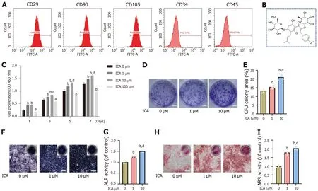
Figure 1 lcariin promoted bone marrow mesenchymal stem cell proliferation and osteogenic differentiation. A: Bone marrow mesenchymal stem cell (BMSC) surface markers were detected by flow cytometry;B: Icariin (ICA) chemical structure;C: The effect of ICA on BMSC proliferation was measured by the cell counting kit-8 assay;D: Representative images of the colony-forming unit (CFU) assay to determine the effect of ICA on BMSC proliferation;E: Quantification of the CFU assay;F: Representative images of alkaline phosphatase (ALP) staining (scale bar: 250 μm);G: ALP activity detection;H: Representative images of alizarin red S (ARS) staining (scale bar: 250 μm);I: Semi-quantitative result of ARS staining.Data are mean ± SEM (n=5).aP < 0.05 and bP < 0.01 vs control group;cP < 0.05 and dP < 0.01 vs 1 μM ICA group.ICA: Icariin.
ICA enhanced expression of osteogenesis-related and angiogenesis-related markers
After 3 d of incubation in ICA,the gene expression levels of osteogenesis-related markers were evaluated by qRT-PCR.The expression levels ofALPandOCNwere elevated in the ICA-treated groups,but the expression levels of runt-related transcription factor 2 and collagen type I alpha 1 were not significantly different from those in the CON group (Figure 2A).After 1 wk of incubation,the protein levels of ALP and OCN were also elevated in the ICA-treated groups (Figure 2B).Furthermore,immunofluorescence staining of ALP and OCN confirmed these findings (Figure 2C and D).The optimal concentration of ICA was 10 μM,which was consistent with qRT-PCR and western blot.These results suggested that ICA enhanced the expression of osteogenesis-related markers.
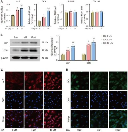
Figure 2 lcariin enhanced the expression of osteogenesis-related markers. A: Expression levels of osteogenesis-related genes [alkaline phosphatase (ALP),osteocalcin (OCN),runt-related transcription factor 2 (RUNX2),and collagen type I alpha 1 (COL1A1)] in bone marrow mesenchymal stem cells (BMSCs) following treatment with icariin (ICA);B: Protein expression levels of ALP and OCN by Western blot;C: Expression of ALP by immunofluorescence (scale bar: 100 μm);D: Expression of OCN by immunofluorescence (scale bar: 100 μm).Data are mean ± SEM (n=5).bP < 0.01 vs control group;cP < 0.05 and dP < 0.01 vs 1 μM ICA group.ICA: Icariin;ALP: Alkaline phosphatase;OCN: Osteocalcin;RUNX2: Runt-related transcription factor 2;COL1A1: Collagen type I alpha 1;qRT-PCR: Quantitative real-time polymerase chain reaction.
After 3 d of incubation in ICA,the gene expression levels of angiogenesis-related markers were evaluated by qRT-PCR.The expression levels ofVEGFAandCD31were elevated in the ICA-treated groups.However,the expression levels of angiopoietin-2 and angiopoietin-4 detected in the ICA-treated groups were not significantly different from those in the CON group (Figure 3A).After 1 wk of incubation,the protein expression levels of VEGFA and CD31 were also elevated in the ICA-treated groups (Figure 3B).Immunofluorescence staining of VEGFA and CD31 confirmed that ICA enhanced the expression of VEGFA and CD31 (Figure 3C and D).Significantly,the optimal concentration of ICA was also 10 μM.This indicates that the optimal concentration of ICA for promoting osteogenesis and angiogenesis was the same.

Figure 3 lcariin enhanced the expression of angiogenesis-related markers. A: Expression levels of angiogenesis-related genes [vascular endothelial growth factor A (VEGFA),platelet endothelial cell adhesion molecule 1 (CD31),angiopoietin-2 (Ang-2),and angiopoietin-4 (Ang-4)] in bone marrow mesenchymal stem cells (BMSCs) following treatment with icariin (ICA);B: Protein expression levels of VEGFA and CD31 by Western blot;C: Expression of VEGFA by immunofluorescence (scale bar: 100 μm);D: Expression of CD31 by immunofluorescence (scale bar: 100 μm).Data are mean ± SEM (n=5).bP < 0.01 vs control group;cP < 0.05 and dP < 0.01 vs 1 μM ICA group.ICA: Icariin;VEGFA: Vascular endothelial growth factor A;CD31: Platelet endothelial cell adhesion molecule 1;Ang-2: Angiopoietin-2;Ang-4: Angiopoietin-4;qRT-PCR: Quantitative real-time polymerase chain reaction.
ICA induced osteogenesis-angiogenesis coupling of BMSCs
To further evaluate the proangiogenic ability of ICA,HUVECs were assessedviaangiogenesis-related assays.The results of the scratch wound assay revealed that there was no significant difference in HUVEC migration between the FM+CON group and the FM+ICA group (P> 0.05).Interestingly,HUVEC migration was enhanced in the CM+CON group compared to the FM+CON group,as well as in the CM+ICA group compared to the CM+CON group (Figure 4A and B).This migration capacity was confirmed by transwell migration assay (Figure 4C and D).In addition,there were no differences in tube formation in HUVECs of the FM+CON group and FM+ICA group (P> 0.05).However,CM increased tube formation,and this effect was even greater in the CM+ICA group compared to the CM+CON group (Figure 4E and F).These findings imply that ICA does not promote angiogenesis in HUVECs in a direct manner but that it can promote angiogenesis in a BMSC-mediated manner.Therefore,the pro-osteogenic effect of ICA is coupled with its proangiogenesis effect.
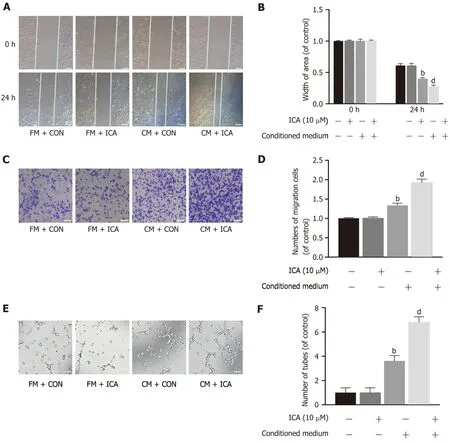
Figure 4 lcariin induced osteogenesis-angiogenesis coupling of bone marrow mesenchymal stem cells. A: Scratch wound assay to show migration of human umbilical vein endothelial cells (HUVECs) cultured with fresh medium (FM) or conditioned medium (CM) and with or without icariin (ICA) (scale bar: 100 μm);B: Quantification of the scratch wound assays;C: Transwell migration assay of HUVECs cultured with FM or CM and with or without ICA (scale bar: 100 μm);D: Quantification of the transwell migration assays;E: Tube formation assay of HUVECs cultured with FM or CM and with or without ICA (scale bar: 100 μm);F: Quantification of the tube formation assays.Data are mean ± SEM (n=5).bP < 0.01 vs control group;dP < 0.01 vs CM+control group.ICA: Icariin;FM: Fresh medium;CM: Conditioned medium;Con: Control.
ICA improved bone repair capacity by promoting osteogenesis in T1DM rats
After the tibial defect operation,the rats received ICA for 4 wk.The effect of ICA on bone defect repair was evaluated after 2 wk and 4 wk of the ICA treatment.The three-dimensional reconstruction revealed that ICA enlarged the area of bone regeneration at both time points (Figure 5A and B).Hematoxylin and eosin staining and Masson’s trichrome staining confirmed these results (Figure 5C-F).Accordingly,further quantitative analysis of the defect area revealed that the values of bone mineral density,bone volume/tissue volume,and trabecular number were lowest in the T1DM group,and the value of trabecular separation was highest in the T1DM group (Figure 5G).Thus,while T1DM led to a decrease in bone formation in the tibial defects in this rat model,ICA could improve the bone repair capacity by promoting osteogenesis in the bone defect area.

Figure 5 lcariin improved bone repair capacity by promoting osteogenesis in type 1 diabetes mellitus rats. A and B: Three-dimensional reconstruction images of the defect area at week 2 (A) and week 4 (B) (scale bars: 1 mm);C and D: Hematoxylin and eosin staining of the defect area at week 2 (C) and week 4 (B) (scale bars: 200 μm);E and F: Masson’s trichrome staining of the defect area at week 2 (E) and week 4 (F) (scale bars: 200 μm);G: Micron-scale computed tomography analysis of bone mineral density (BMD),bone volume/tissue volume (BV/TV),trabecular number (Tb.N),and trabecular separation (Tb.Sp) at weeks 2 and 4.Data are mean ± SEM of the mean (n=8).bP < 0.01 vs the control group;dP < 0.01 vs the type 1 diabetes mellitus group.ICA: Icariin;Con: Control;T1DM: Type 1 diabetes mellitus.
ICA accelerated bone regeneration by inducing osteogenesis-angiogenesis coupling of BMSCs in T1DM rats
At week 2,immunohistochemical staining of the osteogenesis-related marker OCN and the angiogenesis-related markers VEGFA and CD31 revealed an increase in these markers in the ICA group compared to those in the T1DM group (Figure 6A-C).At week 4,the same trend was observed for these markers (Figure 6D-F).The CD31 and EMCN double immunofluorescence staining revealed that ICA facilitated CD31hiEMCNhitype H-positive capillary formation in the bone defect area at both time points (Figure 6G and H).Furthermore,sequential fluorescent labeling revealed that the distance between double labels was wider in the ICA group than in the T1DM group (Figure 6I),which indicated that ICA accelerated the rate of bone formation in the defect area.Quantitative analysis of the bone mineral apposition rate confirmed the above finding (Figure 6J),indicating that ICA accelerated bone regeneration by inducing the osteogenesisangiogenesis coupling in the T1DM rat model.

Figure 6 lcariin accelerated bone regeneration by inducing osteogenesis-angiogenesis coupling of bone marrow mesenchymal stem cells in type 1 diabetes mellitus rats. A-C: Immunohistochemical results of osteocalcin (OCN) (A),vascular endothelial growth factor A (VEGFA) (B),and platelet endothelial cell adhesion molecule 1 (CD31) (C) in the defect area at week 2 (scale bars: 100 μm);D-F: Immunohistochemical results of OCN (D),VEGFA (E),and CD31 (F) in the defect area at week 4 (scale bars: 100 μm);G: CD31 and endomucin (EMCN) double immunofluorescence staining in the defect area at week 2 (scale bar: 100 μm);H: CD31 and EMCN double immunofluorescence staining in the defect area at week 4 (scale bar: 100 μm);I: Sequential fluorescent labeling (scale bar: 10 μm);J: Quantitative analysis of mineral apposition rate.Data are mean ± SEM (n=8).bP < 0.01 vs the control group;dP < 0.01 vs the type 1 diabetes mellitus group.ICA: Icariin;Con: Control;T1DM: Type 1 diabetes mellitus;OCN: Osteocalcin;VEGFA: Vascular endothelial growth factor A;CD31: Platelet endothelial cell adhesion molecule 1.
DISCUSSION
It was widely accepted that the relationship between osteogenesis and angiogenesis is unidirectional[41].However,later studies indicated that the relationship is actually closely coordinated[42-44].It is worth noting that DM normally impairs angiogenesis[45-47].Chinipardazetal[48] observed that angiogenesis was significantly reduced in a fractured region in a T1DM mouse model compared to that in normal mice.The decrease in angiogenesis could suppress trabecular bone regeneration and delay bone healing[49].Therefore,it is essential to focus on vascular regeneration when studying bone regeneration.Through this study,we discovered that ICA enhanced the expression of osteogenesis-related and angiogenesis-related markers in a T1DM mouse model,and the optimal concentration for promoting osteogenesis and angiogenesis was the same.In addition,although ICA cannot directly promote angiogenesis our results indicated that it can work synergistically with BMSCs to promote angiogenesis.
The connection between BMSCs and endothelial cells is attributed to osteoblastic and angiogenic factor production (e.g.,VEGFA)[50],which was consistent with ourinvitrofindings.Our subsequentinvivostudies revealed that ICA promoted osteogenesis and H-positive capillary formation in the defect area.Previous studies have observed that H-type blood vessels could couple osteogenesis and angiogenesis[51-53].This indicated that the roles of ICA in promoting osteogenesis and angiogenesis were not independentinvivo.They are coupled by H-type blood vessels to play synergistic roles.This study is the first to demonstrate that ICA possesses the ability to induce osteogenesis-angiogenesis coupling in BMSCs.
Natural products with structural diversity and biological activity are important sources of innovative drugs[54].ICA is a natural flavonol glycoside used in traditional Chinese medicine[55] and research has shown that it can promote osteogenesis in various ways[56-58].Xiaetal[59] found that ICA could promote osteogenic differentiation of BMSCs by upregulating GLI-1,and Luoetal[60] observed that ICA restored osteogenic differentiation of BMSCs in ovariectomized (commonly known as OVX) rats.A growing body of research has shown that ICA is beneficial in models of diabetes[61-63].Significantly,the safety of ICA has been demonstrated by multiple studies[64-66].Thus,ICA is expected to exert more vital roles in improving diabetes and promoting bone regeneration due to its natural origin and safety.
The osteogenic differentiation of BMSCs is important for bone regeneration,and angiogenesis plays an indispensable role during the bone regeneration process[67-69].Wuetal[70] showed that ICA could promote repair of a normal bone defectviaenhancement of osteogenesis and angiogenesis.In this study,we demonstrated that ICA induced osteogenesisangiogenesis coupling in BMSCsinvitro,and that ICA facilitated bone formation and CD31hiEMCNhitype H-positive capillary formation in the defect area of a T1DM rat model.We used single and double fluorochrome labeling as a direct histologic marker of bone formation.This test showed that in the T1DM model there was a reduced rate of bone formation in the defect area compared to the CON group.When the T1DM rats were treated with ICA,the double fluorochrome labeling demonstrated that ICA accelerated the rate of bone formation in the defect area.Although this studyrevealed that the pro-osteogenesis effect of ICA is strictly connected to its pro-angiogenesis effect and together contribute to the bone regeneration in the context of TIDM,the effects of ICA on bone resorption and bone homeostasis were not investigated;indeed,these latter topics are the focus of our ongoing research.
CONCLUSlON
Taken together,we conclude that ICA could accelerate bone regeneration by inducing osteogenesis-angiogenesis coupling of BMSCs in the T1DM rat model.This finding also implies that ICA may be a potential drug for treating bone defect in the context of TIDM,and more importantly that further investigations into the effective coupling of osteogenesis and angiogenesis should be undertaken in the field of bone regeneration in T1DM patients.
ARTlCLE HlGHLlGHTS
Research background
Icariin (ICA) has multiple pharmacological activities.However,its effect on bone defect repair in the context of type 1 diabetes mellitus (T1DM) remains unclear.
Research motivation
ICA possesses the ability to promote osteogenesis and exert protective effects in T1DM.Therefore,ICA may have therapeutic potential for repairing bone defects in patients with T1DM.
Research objectives
To explore the role of ICA on bone defect repair in T1DM models.
Research methods
The effects of ICA on osteogenesis and angiogenesis were evaluated by molecular biology techniquesinvitro.After a T1DM rat model was established,we evaluated the effect of ICA on bone formation in a defect area.
Research results
ICA promoted bone marrow mesenchymal stem cell (BMSC) osteogenic differentiation and induced osteogenesisangiogenesis coupling of BMSCsinvitro.Subsequently,we observed that ICA facilitated bone formation and type H vessel formation in the defect area of the T1DM rat model.Sequential fluorescent labeling confirmed that ICA could effectively accelerate the rate of bone formation in the defect area.
Research conclusions
ICA accelerated bone regeneration by inducing osteogenesis-angiogenesis coupling of BMSCs in the T1DM rat model.
Research perspectives
Our study highlighted the importance of effective coupling of osteogenesis and angiogenesis in bone regeneration in the context of T1DM.
FOOTNOTES
Co-corresponding authors:Jia Zheng and Yi-Kai Li.
Author contributions:Zheng J and Li YK contributed equally to this work and are co-corresponding authors on this paper according to the critical roles they played throughout the research in experimental design,data analysis,and provision of intellectual ideas and methods;Zheng S,Zheng J,and Li YK designed the research study;Zheng S,Hu GY,and Li JH performed the research;Li JH and Zheng J contributed new reagents and analytic tools;Zheng S,Zheng J,and Li YK analyzed the data and wrote the manuscript;and all authors read and approved the final manuscript.
Supported bythe Postdoctoral Fellowship Program of China Postdoctoral Science Foundation,No.GZC20 231088;and President Foundation of The Third Affiliated Hospital of Southern Medical University,China,No.YP202210.
lnstitutional animal care and use committee statement:The study was reviewed and approved by the Animal Ethics Committee of Southern Medical University (Approval No.SMUL2022023).
Conflict-of-interest statement:The authors declare having no competing interests for this article.
Data sharing statement:Data will be made available upon reasonable request to the corresponding author.
ARRlVE guidelines statement:The authors have read the ARRIVE guidelines,and the manuscript was prepared and revised according to the ARRIVE guidelines.
Open-Access:This article is an open-access article that was selected by an in-house editor and fully peer-reviewed by external reviewers.It is distributed in accordance with the Creative Commons Attribution NonCommercial (CC BY-NC 4.0) license,which permits others to distribute,remix,adapt,build upon this work non-commercially,and license their derivative works on different terms,provided the original work is properly cited and the use is non-commercial.See: https://creativecommons.org/Licenses/by-nc/4.0/
Country/Territory of origin:China
ORClD number:Sheng Zheng 0000-0001-6525-8721;Guan-Yu Hu 0000-0002-1148-0631;Jun-Hua Li 0000-0001-6860-3877;Jia Zheng 0000-0002-6480-3422;Yi-Kai Li 0000-0003-0766-6051.
S-Editor:Chen YL
L-Editor:A
P-Editor:Zhang YL
杂志排行
World Journal of Diabetes的其它文章
- Metabolic syndrome’s new therapy: Supplement the gut microbiome
- Pancreatic surgery and tertiary pancreatitis services warrant provision for support from a specialist diabetes team
- Effect of bariatric surgery on metabolism in diabetes and obesity comorbidity: lnsight from recent research
- Application of three-dimensional speckle tracking technique in measuring left ventricular myocardial function in patients with diabetes
- Long-term effects of gestational diabetes mellitus on the pancreas of female mouse offspring
- Novel insights into immune-related genes associated with type 2 diabetes mellitus-related cognitive impairment
