Urinary exosomal microRNA-145-5p and microRNA-27a-3p act as noninvasive diagnostic biomarkers for diabetic kidney disease
2024-01-26LuLuHanShengHaiWangMingYanYaoHongZhou
Lu-Lu Han,Sheng-Hai Wang,Ming-Yan Yao,Hong Zhou
Abstract BACKGROUND Diabetic kidney disease (DKD),characterized by increased urinary microalbumin levels and decreased renal function,is the primary cause of end-stage renal disease.Its pathological mechanisms are complicated and multifactorial;Therefore,sensitive and specific biomarkers are needed.Urinary exosome originate from diverse renal cells in nephron segments and partially mirror the pathological changes in the kidney.The microRNAs (miRNAs) in urinary exosome are remarkably stable and highly tissue-specific for the kidney.AIM To determine if urinary exosomal miRNAs from diabetic patients can serve as noninvasive biomarkers for early DKD diagnosis.METHODS Type 2 diabetic mellitus (T2DM) patients were recruited from the Second Hospital of Hebei Medical University and were divided into two groups: DM,diabetic patients without albuminuria [urinary albumin to creatinine ratio (UACR) < 30 mg/g] and DKD,diabetic patients with albuminuria (UACR ≥ 30 mg/g).Healthy subjects were the normal control (NC) group.Urinary exosomal miR-145-5p,miR-27a-3p,and miR-29c-3p,were detected using real-time quantitative polymerase chain reaction.The correlation between exosomal miRNAs and the clinical indexes was evaluated.The diagnostic values of exosomal miR-145-5p and miR-27a-3p in DKD were determined using receiver operating characteristic (ROC) analysis.Biological functions of miR-145-5p were investigated by performing Gene Ontology analysis and Kyoto Encyclopedia of Genes and Genomes pathway enrichment.RESULTS Urinary exosomal expression of miR-145-5p and miR-27a-3p was more upregulated in the DKD group than in the DM group (miR-145-5p: 4.54 ± 1.45 vs 1.95 ± 0.93,P < 0.001;miR-27a-3p: 2.33 ± 0.79 vs 1.71 ± 0.76,P < 0.05) and the NC group (miR-145-5p: 4.54 ± 1.45 vs 1.55 ± 0.83,P < 0.001;miR-27a-3p: 2.33 ± 0.79 vs 1.10 ± 0.51,P < 0.001).The exosomal miR-145-5p and miR-27a-3p positively correlated with albuminuria and serum creatinine and negatively correlated with the estimated glomerular filtration rate.miR-27a-3p was also closely related to blood glucose,glycosylated hemoglobin A1c,and low-density lipoprotein cholesterol.ROC analysis revealed that miR-145-5p had a better area under the curve of 0.88 [95% confidence interval (CI): 0.784-0.985,P < 0.0001] in diagnosing DKD than miR-27a-3p with 0.71 (95%CI: 0.547-0.871,P=0.0239).Bioinformatics analysis revealed that the target genes of miR-145-5p were located in the actin filament,cytoskeleton,and extracellular exosome and were involved in the pathological processes of DKD,including apoptosis,inflammation,and fibrosis.CONCLUSION Urinary exosomal miR-145-5p and miR-27a-3p may serve as novel noninvasive diagnostic biomarkers or promising therapeutic targets for DKD.
Key Words: Urinary exosome;MicroRNA-145-5p;MicroRNA-27a-3p;Diabetic kidney disease;Diagnostic biomarkers
INTRODUCTION
Diabetic kidney disease (DKD) is a serious complication of diabetes mellitus (DM).It is characterized by increased urinary microalbumin levels and decreased estimated glomerular filtration rate (eGFR),resulting in rapid progression to end-stage renal disease[1].According to a recent report,the number of adults with diabetes worldwide is approximately 0.537 billion,and the total number may rise to 0.643 billion by 2030.Notably,approximately 20%-40% of DM patients progress to DKD[2].The pathological mechanisms of DKD,including abnormal glucose metabolism,advanced glycation end product generation,inflammation,oxidative stress,and renal hemodynamic changes that eventually lead to renal injury,are complicated and multifactorial[3,4].Podocytes are the key cell type damaged early in DKD due to their highly differentiated postmitotic phenotype with restricted abilities for self-repair and renewal.Hyperglycemia-induced podocyte injury can lead to glomerular filtration dysfunction and proteinuria[5].Early diagnosis and specific treatment can prevent DKD progression;however,renal biopsy,the golden standard for DKD diagnosis,cannot be widely used because it is invasive,and microalbuminuria is often unable to reflect early renal injury or kidney dysfunction progression in DKD patients[6,7].Sensitive biomarkers and effective therapeutic targets are needed for DKD in current clinical settings.
Exosome are lipid bilayer cup-shaped extracellular vesicles with 40-160 nm in diameter.Many cell types can excrete exosome;Hence,they can be detected in various body or tissue fluids,such as blood,urine,and saliva[8,9].The bioactive cargo derived from parental cells,which includes proteins,metabolites,and genetic information,is delivered by the exosome to adjacent or distant cells that regulate the phenotypes or functions of recipient cells[10].Circulating exosome cannot cross the glomerular filtration barrier.Urinary exosome are generally derived from diverse renal cells in nephron segments and are not easily confounded by circulating exosome.Urinary exosome have specific responses to the renal pathological changes[11].Exosome can carry genetic information.MicroRNAs (miRNAs) are the most abundant exosomal RNAs,comprising roughly 22 nucleotides.miRNAs participate in post-transcriptional gene silencing or target mRNA degrading[12,13].Several miRNAs play vital roles in the pathological processes of DKD[12].MiR-145-5p,miRNA-27a,and miR-29c are associated with podocyte injury[14].miRNAs contained in urinary exosome can avoid degradation by nuclease.They are remarkably stable,independent of urine,highly tissue-specific for the kidney,and can be collected in large quantities noninvasively[15-17].Therefore,urinary exosomal miRNAs are better candidates for diagnostic markers than free miRNAs[18].Ghaiet al[17] showed that miR-31-5p and miR-200c-3p were upregulated in urinary exosome from DKD patients compared with patients with normoalbuminuria.Parket al[19] identified 22 differentially expressed miRNAs in urinary exosome from DKD patients;of these,14 differentially expressed miRNAs were associated with renal inflammation and glomerular injury.Zhaoet al[20] found that the urinary exosomal miR-4534 was significantly increased and positively correlated with microalbuminuria in DKD patients with type 2 DM (T2DM).The exosomal miR-4534 can serve as a novel biomarker for early DKD diagnosis.Zanget al[21] also found that urinary exosomal miR-21-5p increased and miR-30b-5p decreased in T2DM patients with DKD compared with patients without DKD.These exosomal miRNAs were closely related to poor renal function and may serve as promising biomarkers for DKD prognosis.
In the present study,we extracted urinary exosome from T2DM patients to determine the expression of miR-145-5p,miR-27a-3p,and miR-29c-3p and evaluate their sensitivity and specificity in diagnosing DKD.Moreover,the molecular biological function of exosomal miR-145-5p was predicted by conducting a bioinformatics analysis to seek novel noninvasive diagnostic biomarkers and potential therapy targets for DKD.
MATERIALS AND METHODS
Characteristics of the study participants
The Ethics Committee of the Second Hospital of Hebei Medical University approved the trial protocols (approval number: 2022-R059).Written informed consent was obtained from each participant before the study.The diagnosis of T2DM and DKD was based on the criteria of the American Diabetes Association[22,23].All T2DM patients were recruited from the Endocrinology Department of the Second Hospital of Hebei Medical University from February 2022 to May 2022.The age of the enrolled patients was 18-75 year,and their eGFR was ≥ 60 mL/min/1.73 m2.T2DM patients with normoalbuminuria [urinary albumin to creatinine ratio (UACR) < 30 mg/g] were included in the DM group (n=20),and T2DM patients with albuminuria (UACR ≥ 30 mg/g) were included in the DKD group (n=20).In the DKD group,five patients were confirmedviakidney biopsy.Twenty healthy volunteers from the Medical Examination Department were included in the normal control (NC) group.
The exclusion criteria of the patients were as follows: (1) Those with T1DM and other specific types of diabetes;(2) Those with severe metabolic disorder or infectious disease within the last 1 mo;(3) Those with severe cardiovascular and cerebrovascular diseases within the last 3 mo;(4) Those with proliferative retinopathy or diabetic foot;(5) Those with nondiabetic renal diseases,kidney stones,and urinary tract infection;and (6) Those with a malignant tumor or autoimmune and chronic liver diseases.
The general and clinical data,including age,gender,body mass index (BMI),fasting blood glucose (FBG),glycosylated hemoglobin A1c (HbA1c),fasting insulin (FINS),serum C-peptide (C-P),total cholesterol (TC),triglyceride (TG),lowdensity lipoprotein cholesterol (LDL-C),blood urea nitrogen (BUN),serum creatinine (Scr) and UACR of all participants were obtained from the medical record system.Fasting urine samples were collected for urinary exosome separation and exosomal miRNAs detection.The eGFR was calculated using the Chronic Kidney Disease Epidemiology Collaboration 2021 formula[24].
Isolation of urinary exosome
Each participant provided 200 mL of fasting urine,which was centrifuged at 4 °C and 3000 ×gfor 20 min.The supernatant was filtered with a 0.22-μm filter to remove cell debris,bacteria,and impurities.The urinary exosome were separated in a sterile environment according to our previous experiments[25] and stored at -80 °C for subsequent testing.
Nanoparticle tracking analysis
The urinary exosome particle sizes were measuredviananoparticle tracking analysis (NTA) using ZetaView PMX 110 (Particle Metrix,Germany).An exosome suspension of 50 μL was appropriately diluted using 1× phosphate buffer saline.NTA was recorded and analyzed at pH 7.0.The entered conductivity was 15000 μS/cm sensed.
Western blotting analysis
Total proteins were separated from exosome using radioimmunoprecipitation assay lysis buffer (Solarbio,China).Protein concentrations were estimated using a bicinchoninic acid protein analysis kit (Solarbio,China) according to our previous study[25].Denatured proteins,30 μg from each sample,were separatedvia10% SDS-polyacrylamide gel electrophoresis (Epizyme,China) and transferred to polyvinylidene fluoride membranes (Millipore,United States).The membranes were blocked with 5% skim milk (BioFroxx,Germany) for 2 h at 24 °C and then incubated with the primary antibodies CD63 (1:1000,Abcam),CD9 (1:1000,Abcam),and TSG101 (1:1000,Abcam) at 4 °C overnight.The next day,the membranes were incubated with the secondary antibody (1:5000,Affinity) for 1 h at 24 °C.The protein bands were detected using chemiluminescence reagents (Sharebio,China) and quantified using ImageJ software (Bio-Rad,United States).
Real-time quantitative polymerase chain reaction
Real-time quantitative polymerase chain reaction (RT-qPCR) was performed according to a previous study[26].Total RNA in the exosome was isolated using an RNA-easy isolation reagent (Vazyme,China).Complementary DNA (cDNA) was reverse transcribed from 1 μg total RNA using miRNA 1stStrand cDNA Synthesis Kit (Vazyme,China).RT-qPCR was performed on a CFX96 PCR system (Bio-Rad,United States) using GoTaq®qPCR Master Mix (Promega,United States).The relative expression of miRNAs were normalized to that of the internal reference U6 and then calculated using the 2-ΔΔCtmethod.
The following primers were used for RT-qPCR: hsa-miR-145-5p: F-5’-AAGCGACCGTCCAGTTTTCCC-3’,R-5’-ATCCAGTGCAGGGTCCGAGG-3’;hsa-miR-27a-3p: F-5’-AATCGGCGTTCACAGTGGCTAA-3’,R-5’-ATCCAGTGCAGGGTCCGAGG-3’;hsa-miR-29c-3p: F-5’-CGCGGCATAGCACCATTTGAAA-3’,R-5’-ATCCAGTGCAGGGTCCGAGG-3’;U6: F-5’-CTCGCTTCGGCAGCACA-3’,R-5’-AACGCTTCACGAATTTGCGT-3’.
Bioinformatics analysis
The potential target genes of miR-145-5p were predicted using three different gene databases: TargetScan7.2 (http://wwtargetscan.org/vert_72/),miRDB (https://mirdb.org/mirdb/index.html),and miRTarBase (https://mirtarbase.cuhk.edu.cn).Gene Ontology (GO) analysis and Kyoto Encyclopedia of Genes and Genomes (KEGG) pathway enrichment were implemented on the Database for Annotation,Visualization,and Integrated Discovery (DAVID) 6.8 (https://david.ncifcrf.gov/).GO analysis elucidated detailed biological functions of potential target genes of miR-145-5p from the molecular function (MF),biological process (BP),and cellular component (CC) aspects.AP-value and a false discovery rate (FDR) of < 0.05 were considered statistically different.-Log10 (P-value) and FDR also represented the enrichment degree of gene function and signaling pathway.
Statistical analyses
All data were processed by GraphPad Prism 8.0 software (La Jolla,United States).A normality test was first performed in each group.Normally distributed data are presented as mean ± SD and analyzed using one-way analysis of variance (ANOVA),followed by the Tukey test.The heterogeneity of the variance data was detected using Welch’s ANOVA test.Pearson correlation analysis was used to analyze the correlation between two normally distributed parameters.Receiver operating characteristic (ROC) curves were used to assess the diagnostic efficiency of urinary exosomal miRNAs in DKD.APvalue of < 0.05 was considered statistically significant.
RESULTS
General and clinical data
The general and clinical data of participants are shown in Table 1.There were no differences in age,gender,BMI,and C-P levels among the three groups (P> 0.05).FBG,HbA1c,FINS,TC,and LDL-C in the DM and DKD groups were higher than those in the NC group (P< 0.05),but there were no differences in these indexes between the DM and DKD groups (P> 0.05).Scr and UACR were higher and eGFR was lower in the DKD group than in the DM group (P< 0.001).Patients with DKD exhibited mesangial cell proliferation,extracellular matrix accumulation,and basement membrane thickening (Figure 1A).

Figure 1 The characterization of urinary exosome. A: The glomeruli histological features of diabetic kidney disease patients (Periodic acid-Schiff staining),Bar=100 μm;B: The exosomal surface markers CD9,CD63 and TSG101 were detected by western blotting;C: The particle screenshots of nanoparticle tracking analysis (NTA) in the urine samples from participants;D: The diameter sizes and diameter concentration distributions of the particles were measured by NTA.NC: Normal control;DM: Diabetes mellitus;DKD: Diabetic kidney disease.

Table 1 General and clinical data in participants
Urinary exosome characteristics
Western blot analysis verified the expression of exosome marker proteins,including CD9,CD63,and TSG101,in the particles from the NC,DM,and DKD groups (Figure 1B).NTA screen capture showed that abundant nanoparticles existed in the urine specimens of the three groups (Figure 1C).The particle diameters at peak concentrations were 120.8 nm (91.9% of the total),126.6 nm (95.5% of the total),and 122.6 nm (85.4% of the total) for the NC,DM,and DKD groups,respectively.The average diameter sizes of exosome were 135.3 ± 1.59 nm,149.2 ± 1.81 nm,and 147.7 ± 10.55 nm,respectively (Figure 1D).These results indicated that urinary exosome were successfully extracted from the urine samples.
Expression of urinary exosomal miR-145-5p, miR-27a-3p and miR-29c-3p
The relative expression of the urinary exosomal miR-145-5p,miR-27a-3p,and miR-29c-3p in the three groups was measured using RT-qPCR.The expression of exosomal miR-145-5p and miR-27a-3p was remarkably increased in the DKD group compared with the DM group (miR-145-5p: 4.54 ± 1.45vs1.95 ± 0.93,P< 0.001;miR-27a-3p: 2.33 ± 0.79vs1.71 ± 0.76,P< 0.05) and the NC group (miR-145-5p: 4.54 ± 1.45vs1.55 ± 0.83,P< 0.001;miR-27a-3p: 2.33 ± 0.79vs1.10 ± 0.51,P< 0.001).The expression of exosomal miR-27a-3p in the DM group was higher than that in the NC group (1.71 ± 0.76vs1.10 ± 0.51,P< 0.05),but no significant difference was found in the expression of exosomal miR-145-5p between the DM and NC groups (1.95 ± 0.93vs1.55 ± 0.83,P> 0.05).Similarly,the expression of exosomal miR-29c-3p did not differ among the three groups (P> 0.05) (Figure 2A).
Correlations between urinary exosomal miR-145-5p and miR-27a-3p and renal function in T2DM patients
Exosomal miR-145-5p was found to positively correlate with Scr (r=0.781,P< 0.0001) and UACR (r=0.801,P< 0.0001) and negatively correlate with eGFR (r=-0.784,P< 0.0001) (Figure 2B).Similarly,miR-27a-3p positively correlated with Scr (r=0.380,P=0.016) and UACR (r=0.439,P=0.005) and negatively correlated with eGFR (r=-0.477,P=0.002) (Figure 2C).Moreover,miR-27a-3p positively correlated with glycolipid metabolism indexes,including FBG,HbA1c,and LDL-C (Figure 2D).
Diagnostic efficiency of exosomal miR-145-5p and miR-27a-3p in DKD
ROC analyses defined the diagnostic potential of urinary exosomal miR-145-5p and miR-27a-3p in DKD.MiR-145-5p hada better area under the curve (AUC) of 0.88 [95% confidence interval (CI): 0.784-0.985,P< 0.0001] than miR-27a-3p with an AUC of 0.71 (95%CI: 0.547-0.871,P=0.0239) in DKD patients (Figure 3).For DKD diagnosis,exosomal miR-145-5p exhibited a higher sensitivity of 90% and a specificity of 75% at the best cutoff value of 2.67 than miR-27a-3p with a sensitivity of 65% and a specificity of 70% at the optimal cutoff value of 2.12.The combination of miR-145-5p and miR-27a-3p contributed to an increased AUC of 0.97 (95%CI: 0.927-1.000,P< 0.0001) with a sensitivity of 95% and a specificity of 90% for DKD diagnosis.Urinary exosomal miR-145-5p and miR-27a-3p may serve as potential biomarkers of early DKD diagnosis,especially miR-145-5p.
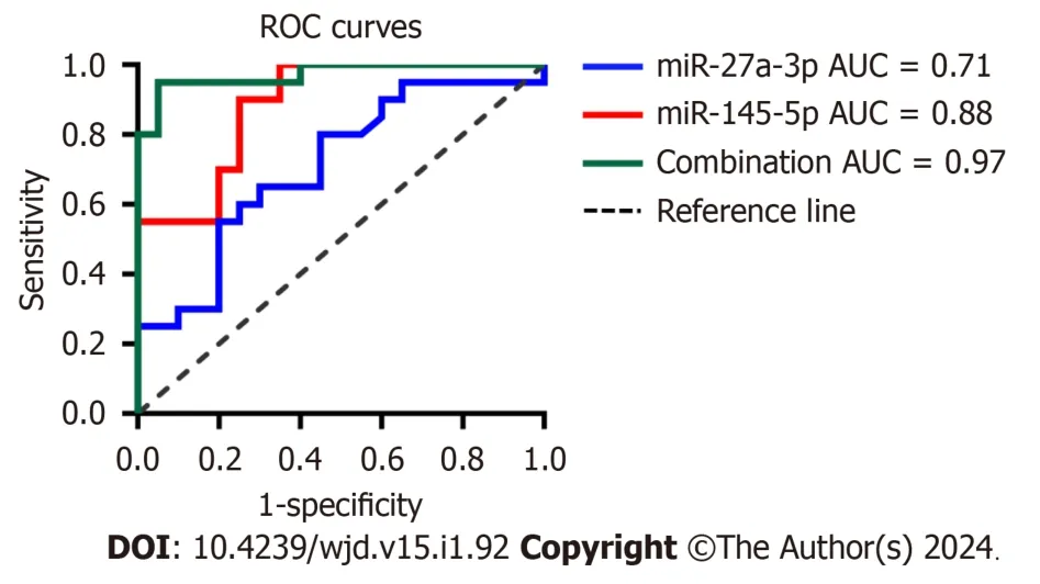
Figure 3 Receiver operating characteristic curves of urinary exosomal miR-145-5p,miR-27a-3p and their combination to discriminate diabetic kidney disease from type 2 diabetes mellitus patients. The area under the curve for miR-27a-3p was 0.71 [95% confidence interval (CI): 0.547-0.871,P =0.0239],0.88 (95%CI: 0.784-0.985,P < 0.0001) for miR-145-5p,and 0.97 (95%CI: 0.927-1.000,P < 0.0001) for their combination.ROC: Receiver operating characteristic;AUC: Area under the curve.
Bioinformatics analysis of miR-145-5p
Since urinary exosomal miR-145-5p has the potential to serve as a promising noninvasive diagnostic biomarker of DKD,its biological function was further explored using bioinformatics analysis.In total,907,909,and 248 potential target mRNAs of miR-145-5p were predicted using TargetScan,miRDB,and miRTarBase,respectively.A total of 77 mRNAs were detected simultaneously using the three gene databases (Figure 4A).We listed some gene names according to the target score,such as SMAD3,SOX9,and SRGAP2,which may be involved in the pathophysiological processes of DM and DKD[27-29] (Figure 4B).

Figure 4 The potential target genes of exosomal miR-145-5p were predicted by three different gene databases. A: The Venn diagram showed that 907 genes were detected by TargetScan,909 genes were tested by miRDB and 248 genes were predicted by miRTarBase.A total of 77 items gene were simultaneously predicted by the three gene databases;B: Among the 77 target genes,we listed some gene names according to the target score.
GO analysis classified and described these target genes on CC,MF,and BP aspects.The CC catalog contained various cell locations,including the cytosol (count: 42,FDR=1.97E-05),nucleoplasm (count: 28,FDR=0.009056116),SMAD protein complex (count: 3,FDR=0.009056116),extracellular exosome (count: 19,FDR=0.012172645),actin cytoskeleton (count: 6,FDR=0.029013556),and actin filament (count: 5,FDR=0.009056116) (Figure 5A).The terms including actin binding,protein binding,sequence-specific DNA binding,SMAD binding,mitogen-activated protein kinase (MAPK) binding,and small GTPase binding were enriched in the MF catalog (Figure 5B).Regarding the biological regulatory processes,terms such as transforming growth factor β (TGF-β) receptor signaling pathway,cellular response to TGF-β stimulus,response to hypoxia,positive regulation of cell proliferation,cell differentiation,cell motility,and actin filament organization were listed in the BP catalog (Figure 5C).

Figure 5 Bioinformatics analysis of the genes of miR-145-5p potential target. A-C: Biological functions of the 77 intersectant target genes of miR-145-5p were described by Gene Ontology on cellular component,molecular function,and biological process aspect respectively;D: Kyoto Encyclopedia of Genes and Genomes pathways analysis predicted a total of 11 enriched signal pathways of the potential target genes of miR-145-5p.GO: Gene Ontology;KEGG: Kyoto Encyclopedia of Genes and Genomes;FDR: False discovery rate.
KEGG pathway enrichment analysis manifested the target genes of miR-145-5p.These genes were mainly enriched in 11 signaling pathways (FDR < 0.05),such as the MAPK signaling pathway (FDR=0.002477523),TGF-β signaling pathway (FDR=0.00784279),forkhead box O (FOXO) signaling pathway (FDR=0.018431373),Ras signaling pathway (FDR=0.036227759),and advanced glycosylation end products-the accumulation of their receptors (AGE-RAGE) signaling pathway in diabetic complications (FDR=0.036960173),which may be involved in the pathological processes of DKD[3,30].Besides,the pathways related to adherens junction,cellular senescence,and cancer were included (Figure 5D).
DISCUSSION
DKD is the primary cause of end-stage renal disease and seriously threatens the lives of patients with DM due to its irreversible and progressive evolution[1].Early identification of high-risk patients may prevent DKD progression;However,sensitive and noninvasive diagnostic biomarkers for DKD are scarce.
miRNA dysregulation participates in various pathological processes of diabetic kidney injury and possesses great potential in the early diagnosis of DKD.A previous study showed that serum miR-145-5p was significantly lower in T1DM patients with nephropathy than T1DM controls[31].MiR-145-5p overexpression suppressed podocyte apoptosis under high glucose (HG) conditions by inhibiting the Notch signaling pathway[32].MiR-27a-3p was reported to be higher in the serum of T2DM patients than in the serum of non-diabetic individuals[33].Upregulation of miR-27a accelerated renal tubular epithelial-mesenchymal transition and apoptosis of podocytes exposed to HG by activating the βcatenin signaling pathway[34].MiRNA-29c was increased in the serum of DKD patients and HG-stimulated podocytes and induced a renal inflammatory response by downregulating tristetraprolin[35].Exosome are found in almost all body fluids.Abundant miRNAs are cornered in exosome stably and specifically[35].Urinary exosome are primarily generated from the kidney cells in almost all nephron segments;urinary exosomal miRNAs may reflect pathophysiological events of the kidney and are proposed as noninvasive biomarkers for DKD progression[36].Choet al[18] identified 21 differentially expressed urinary exosomal miRNAs between T2DM patients taking dipeptidyl peptidase-4 inhibitor+metformin and patients taking sulfonylurea+metformin using miRNA sequencing.Delićet al[37] also showed that the urinary exosomal miR-29b and miR-29c expression decreased in 5/6 nephrectomized rats compared with the sham group.Telmisartan or linagliptin treatment could significantly restore the levels of urinary exosomal miR-29c,which negatively correlated with an increase in UACR in rats.Thus,urinary exosomal miRNAs may serve as future indicators for DKD clinical therapeutic evaluation.
In the present study,urinary exosomal miR-145-5p was remarkably upregulated in T2DM patients with DKD.Baruttaet al[38] demonstrated that urinary exosomal miR-145 was higher in T1DM patients with microalbuminuria than in normoalbuminuric and nondiabetic subjects.Moreover,miR-145 was enriched in both urinary exosome and their glomeruli tissues in DKD mice.Similarly,miR-145 was rapidly increased in HG-stimulated mesangial cells and their exosomein vitro,indicating that miR-145 might serve as a diagnostic biomarker for DKD.Zhanget al[39] reported that miR-145-5p enriched in exosome could lead to albuminuria and podocyte injury in healthy mice.These results suggest exosomal miR-145-5p is involved in kidney damage in DKD.The correlation analysis performed in the present study showed that exosomal miR-145-5p was positively correlated with albuminuria and Scr and negatively correlated with eGFR,suggesting that miR-145-5p can reflect the occurrence and development of DKD.Therefore,miR-145-5p can be regarded as a noninvasive biomarker for DKD.Castañoet al[40] confirmed that the circulating exosomal miR-27a-3p was overexpressed in high-fat diet-induced obese and prediabetic mouse models.Compared with control,the expression of miR-27a-3p was markedly decreased in the epididymal white adipose tissue of obese mice.The author highlighted that miR-27a-3p was one of the obesity-associated miRNAs and that the exosomal miR-27a-3p may play vital roles in the pathological processes of dyslipidemia and insulin resistance[40].Our data revealed an increased level of exosomal miR-27a-3p in DKD patients.Upregulated miR-27a-3p was related to impaired renal function and was positively associated with glycolipid metabolism factors,including FBG,HbA1c,and LDL-C.Thus,exosomal miR-27a-3p is a valuable candidate to respond to the dysregulated glycolipid metabolism and kidney damage in T2DM.Urinary exosomal miR-29c has been reported to decrease in DKD and nondiabetic nephropathy patients,positively correlate with eGFR,and negatively correlate with the fibrosis score[41].Our results exhibited a downregulated trend of exosomal miR-29c-3p in DKD;However,this trend was not statistically significant.
ROC analysis revealed that exosomal miR-145-5p had a better AUC with higher sensitivity and specificity than miR-27a-3p in determining diabetic kidney damage in T2DM patients.Combining the two exosomal miRNAs led to the improvement of diagnostic efficiency and were expected to serve as novel markers for early identification and diagnosis of DKD.The prediction and biological functional analysis of the target genes of miR-145-5p can guide future research on their pathological effects on DKD.GO analysis revealed the cell locations where the genes acted.The terms “actin filament”,“actin cytoskeleton”,and “cytoskeleton” were often related to the structure injury of podocytes[42].The term “extracellular exosome” supported the biological regulatory roles of miR-145-5p in exosome.Consistent with this,our previous study confirmed that urinary exosomal miR-145-5p from patients with DKD induced podocyte apoptosis by inhibiting Srgap2 and activating the RhoA/Rho kinase (ROCK) pathway[25].The term “insulin receptor complex” suggested that miR-145-5p may be involved in the pathophysiological process of glucose metabolism.MF suggested that miR-145-5p participates in actin binding,protein binding,GI-SMAD binding,MAPK binding and small GTPase binding,which were involved in the BP,including TGF-β receptor signaling pathway;cellular response to TGF-β stimulus;response to hypoxia;and cell proliferation,motility,or migration.Exposure of renal cells to HG can activate various intracellular signaling pathways,such as TGF-β-Smad-MAPK and small GTPase-related pathways.Complex perturbation and reciprocal modulation between these signalings pathways directly accelerate the pathological process of DKD[43].The target genes of miR-145-5p were mainly involved in 11 signal pathways,including the MAPK pathway,TGF-β pathway,FOXO pathway,Ras pathway,and AGE-RAGE pathway in diabetic complications.Our previous research indicated that RhoA is a member of the Ras superfamily of GTP-binding proteins;ROCK is the downstream molecule of RhoA;and the RhoA/ROCK pathway is closely implicated in pathological processes such as inflammation,apoptosis,and fibrosis of DKD[44].TGF-β and MAPK signaling pathways have been considered central to extracellular matrix accumulation and renal fibrosis in DKD[43,45].The FOXO pathway is known to focus on oxidative stress and inflammatory responses in the pathological process of DKD[20].The activation of the AGE-RAGE pathway can increase the release of reactive oxygen species and trigger various intracellular signaling cascades,including TGF-β,PKC,MAPK,nuclear factor-kappaB,and GTP-binding protein pathways.Thus,the AGE-RAGE pathway plays a central role in the multiple pathogenesis of DKD[43,46].
This study had some limitations.First,it was a cross-sectional,observational study with a small sample size without clinical follow-up and evaluation of these biomarkers in the middle or long term.Second,data on global miRNA sequencing of urinary exosome of participants in the three groups were lacking.Thus,a further prospective data analysis with a larger sample is needed to confirm the clinical applicability of these urinary exosomal miRNAs.
CONCLUSION
Our results show that urinary exosomal miR-145-5p and miR-27a-3p were markedly increased in DKD patients and were associated with the progression of kidney injury in T2DM patients.These findings imply that urinary exosomal miR-145-5p and miR-27a-3p are promising noninvasive biomarkers for DKD diagnosis.In particular,miR-145-5p was highly specific and sensitive to DKD.MiR-145-5p may be involved in signaling pathways,including MAPK,TGF-β,FOXO,Ras,and AGE-RAGE pathways,which are related to the pathological processes,including inflammation,apoptosis,and fibrosis of DKD.These hint that miR-145-5p is a potential therapeutic target for DKD.
ARTICLE HIGHLIGHTS
Research background
Diabetic kidney disease (DKD) is the primary cause of end-stage renal disease due to its irreversible and rapidly progressive evolution.DKD remains a serious threat to the lives of diabetic patients.
Research motivation
Early diagnosis and specific treatment can prevent DKD progression.Urinary exosomal microRNAs (miRNAs) are generally derived from renal cells and directly mirror the pathological changes in the kidney.Urinary exosomal miRNAs are remarkably stable and highly tissue-specific for the kidney and may act as promising biomarkers for DKD.
Research objectives
To explore whether urinary exosomal miRNAs from diabetic patients can serve as noninvasive biomarkers for the early diagnosis of DKD.
Research methods
Patients with type 2 diabetes mellitus (T2DM) were enrolled and divided into a DM group,diabetic patients without albuminuria,and a DKD group,diabetic patients with a urinary albumin to creatinine ratio of ≥ 30 mg/g.Healthy subjects were included in the normal control group.The relative expressions of urinary exosomal miR-145-5p,miR-27a-3p,and miR-29c-3p were detected using real-time quantitative polymerase chain reaction.Correlation analysis,receiver operating characteristic analysis,and bioinformatics analysis were used to explore the potential of urinary exosomal miR-145-5p and miR-27a-3p as DKD biomarkers.
Research results
The expression of urinary exosomal miR-145-5p and miR-27a-3p was significantly upregulated in the DKD group.They were closely related to kidney damage and abnormal glycolipid metabolism in T2DM patients.Exosomal miR-145-5p had higher sensitivity and specificity for diagnosing DKD;combining miR-145-5p and miR-27a-3p increased their diagnostic efficiency.Bioinformatics analysis suggested that miR-145-5p regulated various molecular biological functions and signaling pathways involved in the pathological processes of DKD,including apoptosis,inflammation,and fibrosis.
Research conclusions
Urinary exosomal miR-145-5p and miR-27a-3p may serve as novel noninvasive diagnostic biomarkers for DKD.
Research perspectives
Urinary exosomal miR-145-5p and miR-27a-3p may complement traditional DKD diagnostic methods.They may also be effective therapeutic targets for DKD cell-free therapy in the future.
FOOTNOTES
Co-first authors:Lu-Lu Han and Sheng-Hai Wang.
Author contributions:Zhou H contributed to the conceptualization,methodology,resources,writing-reviewing and editing of this manuscript;Han LL participated in the visualization,writing-original draft preparation of this study;Wang SH took part in the investigation and data curation of this article;Yao MY was major in the interpretation of data,manuscript revision,language and format editing;and all authors were responsible for the content and proposed critical comments on the manuscript,as well as approving final version submission.
Supported bythe Nature Science Foundation of Hebei Province,No.H2023104011.
Institutional review board statement:The study was conducted in accordance with the Declaration of Helsinki,all research protocols were approved by Ethical Committee of the Second Hospital of Hebei Medical University (approval number: 2022-R059,28 February 2022) and the Chinese Clinical Trial Registry (approval number: ChiCTR2200066055).A written informed consent was demanded from each participant before they joined this study.
Conflict-of-interest statement:All the authors report no relevant conflicts of interest for this article.
Data sharing statement:The raw data supporting the conclusions of this article will be made available by the authors,without undue reservation.
Open-Access:This article is an open-access article that was selected by an in-house editor and fully peer-reviewed by external reviewers.It is distributed in accordance with the Creative Commons Attribution NonCommercial (CC BY-NC 4.0) license,which permits others to distribute,remix,adapt,build upon this work non-commercially,and license their derivative works on different terms,provided the original work is properly cited and the use is non-commercial.See: https://creativecommons.org/Licenses/by-nc/4.0/
Country/Territory of origin:China
ORCID number:Hong Zhou 0000-0003-3356-9175.
S-Editor:Wang JJ
L-Editor:A
P-Editor:Xu ZH
杂志排行

World Journal of Diabetes的其它文章
- Effects of Tai Chi in diabetes patients: Insights from recent research
- Management of monogenic diabetes in pregnancy: A narrative review
- Prediabetes: An overlooked risk factor for major adverse cardiac and cerebrovascular events in atrial fibrillation patients
- Predictive value of bilirubin and serum γ-glutamyltranspeptidase levels in type-2 diabetes mellitus patients with acute coronary syndrome
- Clinical study of different prediction models in predicting diabetic nephropathy in patients with type 2 diabetes mellitus
- Heterogeneously elevated branched-chain/aromatic amino acids among new-onset type-2 diabetes mellitus patients are potentially skewed diabetes predictors