Optical coherence tomography angiography for macular microvessels in ischemic branch retinal vein occlusion treated with conbercept: predictive factors for the prognosis
2023-12-14LiTangGuangLiSunYueZhaoTingTingYangJinYao
Li Tang, Guang-Li Sun, Yue Zhao, Ting-Ting Yang, Jin Yao
Department of Ophthalmology, the Affiliated Eye Hospital of Nanjing Medical University, Nanjing 210029, Jiangsu Province, China
Abstract
● KEYWORDS: optical coherence tomography angiography; branch retinal vein occlusion; macular edema;foveal avascular zone; conbercept
INTRODUCTION
It has reported that the intraocular level of vascular endothelial growth factor (VEGF) elevates in patients with BRVO[3].Intravitreal anti-VEGF injection is the preferred treatment for ME secondary to BRVO[4].However, the mechanisms underlying the development of refractory ME remain unknown.ME is often prone to relapse, and some patients require repeated multiple injections.BRVO was previously classified as ischemic or non-ischemic based on fundus fluorescein angiography (FFA) findings, and the presence of ME and ischemia was determined.But, in the late phase of FFA, dyes often leak into retina[5], thereafter disrupting the observation of retinal microvasculature that may show pathologic changes in the recurrence of ME.
Optical coherence tomography angiography (OCTA) provides depth-resolved visualization of the retinal microvasculature without using intravenous dye[6-8].The non-invasive OCTA can image the superficial and deep retinal capillary network,providing visualization details of the foveal avascular zone(FAZ) and vessel flow density[8].Samaraet al[9]reported that low vessel flow density both in the superficial and deep layers of FAZ was correlated with reduced visual function.The newly created software, such as A-circularity index (AI) and vessel densities (VDs) 300 μm area around FAZ (FD-300), can show the development of macular ischemia by using parameters such as area and FAZ perimeter (PERIM).
Previous studies have described the changes in the superficial and deep capillary networks observed by OCTA in BRVO eyes[9-10].In this study, we investigated and evaluated the predictive factors for final visual acuity recovery in eyes with ME secondary to BRVO by investigating multiple quantitative parameters of the macular area.
SUBJECTS AND METHODS
Ethical ApprovalThis retrospective study, which took place between March 2021 and January 2022, included 60 ischemic BRVO patients who were followed-up at the Affiliated Eye Hospital of Nanjing Medical University.This study followed the principles of the Declaration of Helsinki and was approved by the Ethics Committee of the Affiliated Eye Hospital of Nanjing Medical University (2020015).Written informed consent was obtained from all patients and their families.
Inclusion and Exclusion CriteriaInclusion criteria included:1) All patients showed the involvement of major BRVO in the superior or inferior temporal sector; 2) ME with flameshaped hemorrhages was found with ophthalmoscope examination; 3) The diagnosis was confirmed using FFA and/or optical coherence tomography (OCT).FFA showed dilated and tortuous veins in the BRVO area and late fluorescence staining leakage.OCT showed ME with central macular thickness (CMT)≥250 μm; 4) A broken foveal capillary ring was regarded as the evidence of existent macular ischemia by OCTA (Figure 1); 5) All patients had not received any treatment before enrollment; 6) All patients received intravitreal conbercept (IVC) and 3+pro re nata(PRN)regimen after enrollment.The follow-up lasted at least 6mo.
Exclusion criteria included macular vein occlusion, central retinal vein occlusion, multiple occlusions of the retinal veins, concomitant ocular diseases (such as uveitis, diabetic retinopathy, age-related macular degeneration, retinal macroaneurysm, glaucoma), keratoconus, myopia (more severe than 3 diopters), and media opacity that was dense enough to hamper the interpretation of fundus photography results (such as dense cataract, and corneal problems other than dry eye).
Ophthalmological ExaminationAll patients were enrolled for ophthalmic examination using a +90 diopter non-contact lens slit lamp biomicroscopy.Topcon TRC 50DX fundus camera (Topcon Corporation, Tokyo, Japan) was employed to scan capillary non-perfusion areas, diffuse fluorescein leakage and fluorocover-up from bleeding.FFA images were obtained to confirm the diagnose of ischemic BRVO.The thickness of the central macula in the affected eyes before and after treatment was recorded by spectral domain OCT (SD-OCT)(Heidelberg Spectralis; Heidelberg Engineering Inc, Franklin,Massachusetts, USA).The morphologic changes of macular microvessels were obtained by OCTA (Optovue RTVue XR Avanti; Optovue Inc, Fremont, California, USA) using the split-spectrum amplitude decorrelation angiography algorithm.The software automatically generated superficial capillary plexus (SCP) and deep capillary plexus (DCP).The SCP was measured over a range extending from the inner limiting membrane (ILM) to 10 μm above the inner plexiform layer(IPL).The DCP was measured from 10 μm above the IPL to 10 μm below the outer plump layer (OPL).Sections (3×3 mm2)were captured from the scanned foveal area to obtain the FAZ area, PERIM, AI and FD-300.The FAZ area was measured from the ILM to OPL.The FAZ parameters were based on a set of data automatically obtained by stacking the whole retinal layer, that was, the superficial and DCP.The macular nonperfusion area (NPA) was defined as the capillary dropout area within a 3×3 mm2section, including the FAZ.The FAZ and the parafoveal capillary dropouts (parafoveal NPA) were independently reviewed by two fully trained retina specialists blind to the study information (Figure 2).The AI was defined as the ratio of PERIM to the same area in the standard circle.The FD-300 referred to the retinal vessel flow density within 300 μm of the FAZ.
Surgical TechniqueAll patients received IVC and 3+PRN regimen by the same experienced doctor.During the operation,the principle of asepsis was strictly followed.After disinfection,topical anesthesia was performed with oxybuproacaine eye drops, povidone iodine was used for eye washing, and a 30-gauge needle was used at 3 or 4 mm posterior to the corneoscleral margin (3 mm posterior to the corneoscleral margin in aphakic patients ); 0.5 mg/0.05 mL conbercept was injected into the vitreous cavity with a needle perpendicular to the eyeball (Chengdu Kanghong Biotechnology Co., LTD.,Chengdu, China; National Drug approval S20130012), and sterile cotton swabs were applied at the injection site for 30s.The patient’s eye was wrapped with gauze after the presence of manual vision in front of the eye and the finger intraocular pressure was normal.Patients were treated with levofloxacin eye drops 3 times a day, 1 drop a time for 7 consecutive days.When OCT showed evident ME and/or serous retinal detachment at the fovea, the patients received monthly IVC(0.5 mg/0.05 mL), until a dry macula (absence of intraretinal or subretinal fluid) appeared on SD-OCT.
特殊食品产业发展潜力与庞大的市场需求息息相关。中国企业联合会中国企业家协会理事长朱宏任举例说,2017年,我国60岁及以上人口达2.41亿人,占总人口比例为17.3%,其中65岁及以上人口为1.58亿人,占比达11.4%,人口结构变动,带动了特殊食品消费;同时,随着“两孩”政策全面实施,2015年至2017年新出生人口达5114万人,新生儿、低龄儿都有特殊食品消费需求。此外,随着人们生活水平的不断提高,中等收入群体不断扩大,对保健食品等特殊食品具有需求的人群显著增加,无疑都将带动特殊食品产业高速发展。
Additionally, all patients received scattered laser photocoagulation of according to FFA 2wk after the first injection.
Data AnalysisStatistical analysis was carried out using a statistical package (SPSS Inc., version 23.0, Chicago, IL,USA).The best-corrected visual acuity (BCVA) was converted to the logarithm of the minimal angle of resolution (logMAR)for statistical evaluation.The final visual gain (FVG) was the differential BCVA value between the baseline and month 6.All data were collected monthly during the follow-up.Quantitative data were expressed as mean±standard deviation (SD), and qualitative variables were described in percentages.The parameters of FAZ and BCVA at the baseline and 6mo were compared with pairedt-test.The Pearson correlation coefficient was used to study the correlation between the variables.The level of statistical significance was set atP<0.05.
RESULTS
Study Participants and Baseline CharacteristicsWe measured 60 eyes of 60 patients (21 men and 39 women, mean age 48.62±4.32y, range 41-69y).The baseline characteristics are shown in Table 1.BRVO occurred in superotemporal (37 patients) or inferotemporal (23 patients) quadrants.Forty-five patients (75%) were diagnosed with hypertension.The mean disease duration was 1.56±2.21mo (range 0.5-3mo).
Best-corrected Visual Acuity and Central Macular ThicknessBefore the treatment, the mean BCVA in the diseased eyes with ME was 0.82±0.32 (range 0.5-1.2) logMAR.At 6mo after treatment, the mean BCVA in the diseased eyes improved significantly (0.39±0.11,P<0.001).The mean CMT significantly decreased from 476.22±163.54 μm at baseline to 298.66±109.23 μm at 6mo (P<0.001; Table 2).

Figure 2 Macula ischemia varies considerably in size of NPA in BRVO on OCTA A: Mild macular ischemia had scattered tiny NPA around macular NPA with a capillary plexus around the entire circumference.B and C: The enlargement of NPA (red arrow).NPA: Nonperfusion area; BRVO: Branch retinal vein occlusion; OCTA: Optical coherence tomography angiography.

Table 1 Baseline characteristics

Table 2 The mean BCVA (logMAR) and CMT results
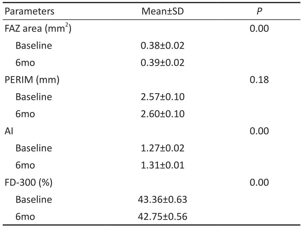
Foveal Avascular Zone ParametersCompared to those at the baseline, the parameters at post-treatment 6mo were as follows:the FAZ area increased (0.38±0.02vs0.39±0.02 mm2,P<0.05); the AI increased (1.27±0.02vs1.31±0.01,P=0.000); the FD-300 (%) decreased (43.36±0.63vs42.75±0.56,P<0.05); the PERIM showed no significant differences (P>0.05; Table 3).Accordingly, the macular ischemia continued during the follow-up even though the effective treatment was given (Figure 3).

Figure 3 Ischemic BRVO with ME in a 55-year-old woman was first seen in 2021 (Case 1) The patient had a total of 3 times of IVC injections,the first injection at 2021-10-18, the second one at 2021-11-18, and the last one at 2022-01-03.During the follow-up, the BCVA increased from 0.6 to 0.2.FAZ area increased from 0.452 to 0.626 mm2.PERIM increased from 2.752 to 3.566 mm.AI increased from 1.16 to 1.27.FD-300 (%)increased from 51.40 to 49.42.B-scan showed the ME from onset to regression.BRVO: Branch retinal vein occlusion; ME: Macular edema;FAZ: Foveal avascular zone; IVC: Intravitreal conbercept; BCVA: Best-corrected visual acuity; FD-300: Vessel densities 300 μm area around FAZ;PERIM: Foveal avascular zone perimeter; AI: A-circularity index.

Table 3 FAZ parameters results
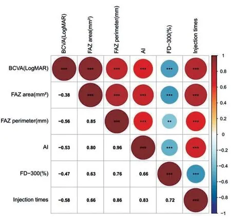
Correlation Analysis Among BCVA and FAZ ParametersThe baseline BCVA showed positive correlation with the FAZ area, PERIM and AI (r=0.471, 0.798, and 0.658, respectively;P<0.05), but negative correlation with the FD-300 (r=-0.533,P<0.05).Moreover, a positive correlation was found between the baseline BCVA and the times of IVC injections (r=0.833,P<0.05; Figure 4).
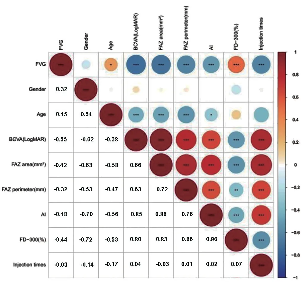
Correlation Analysis Among Final Visual Gain and Other VariablesNo correlation was found between the FVG and gender (r=0.137,P>0.05).The FVG was positively correlated with age and the FD-300 (r=0.323 and 0.537,P<0.05;respectively).Instead, the FVG was strongly and inversely correlated with the baseline BCVA, FAZ area, PERIM, AI, and injection times (r=-0.722, -0.701, -0.621, -0.527 and -0.628,P<0.05, respectively; Figure 5).
Injection times was positively correlated with the baseline FAZ area, PERIM, and AI (r=0.856, 0.665 and 0.716,P<0.05), but negatively correlated with the FD-300 (r=-0.579,P<0.05).
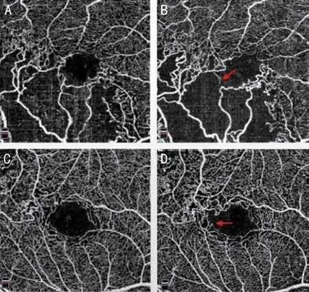
Macular IschemiaOCTA images showed macular ischemia in 41 eyes (68.3%) before treatment, and 46 eyes (76.7%)at 6mo after treatment (P<0.05).During the follow-up, the vascular continuity of the arch ring was disrupted, and macular ischemia developed in 5 new cases (Figure 6).

Figure 4 Heat map of correlation between the baseline BCVA and FAZ parameters The baseline BCVA had significantly positive correlation with FAZ area, PERIM and AI, but negative correlation with FD-300.BCVA: Best-corrected visual acuity; FAZ: Foveal avascular zone; FD-300: Vessel densities 300 μm area around FAZ;PERIM: Foveal avascular zone perimeter; AI: A-circularity index.

Figure 5 Heat map of correlations among FVG and other variables FVG was positively related to age and FD-300, but strongly and inversely related with baseline BCVA, FAZ area, PERIM, AI, and the number of injections.FVG: Final visual gain; FD-300: Vessel densities 300 μm area around FAZ; BCVA: Best-corrected visual acuity; FAZ:Foveal avascular zone; PERIM: Foveal avascular zone perimeter; AI:A-circularity index.
DISCUSSION

Figure 6 The OCTA images of the macular area of the two BRVO eyes A and C: At baseline, basically complete continuity of the arch ring was showed; B and D: At 6mo, the vascular continuity of the arch ring was disrupted, and macular ischemia developed (red arrow).OCTA: Optical coherence tomography angiography; BRVO: Branch retinal vein occlusion.
The pathogenesis of ME secondary to BRVO is multifactorial.First, ME may be caused by over-expression of VEGF, a process that disrupts the blood-retina barrier (BRB) with vessel caliber modification.Then, cause of venous pressure, retinal capillary non-perfusion and tissue ischemia, BRVO may cause the damage of blood capillary endothelial cells and the loss of structural integrity[11-12].Due to the destruction of the inner BRB (iBRB), the permeability of capillaries increases,and the fluid leaks out of the blood vessels and accumulates in the macular area, which may also be the cause of ME.In addition, due to retinal circulation disorder, retinal ischemia and hypoxia, the structure and function of retinal pigment epithelium (RPE) cells are damaged, as well as the changes in the ellipsoid zone-RPE complex, which can cause outer BRB(oBRB) damage, resulting in the accumulation of intraretinal or subretinal fluid, thus forming ME[13-15].Tomiyasuet al[16]believed that microaneurysms with local leakage could lead to BRVO-induced refractory ME.
OCTA was introduced as a non-invasive, promising imaging technique that enables the detection of retinal and choroidal diseases and allows more detailed imaging of vascular microstructures without the use of exogenous dyes compared to FFA.The presence of ME implied segmentation errors.ME may still cause small segmentation errors in the foveal region,which could explain the differences in the foveal vessel density before and after treatment.However, FAZ parameters were based on retinal slab instead of the separated SCP and DCP,and thus were not affected by segmentation errors.
Several previous clinical studies used OCTA to evaluate the retinal vascular changes that occur in BRVO[9-10,13].However,there were few studies on OCTA follow-up analysis of BRVO patients with ME after conbercept treatment.In our study,OCTA built-in software was used to analyze the OCTA parameters of BRVO eyes, including FAZ parameters.By studying multiple quantitative parameters of the macular area,the predictive factors of final visual acuity recovery in ME eyes secondary to BRVO were explored and evaluated.
Previous randomized multicenter studies have shown that ME healed spontaneously only in 30%-34% cases.Anti-VEGF drugs can effectively inhibit the formation of microaneurysms and reduce leakage, which is the top priority for the effective treatment of BRVO-induced ME[5].However, some patients still have refractory ME despite long-term and multiple intraocular injections[15,17-18].Our findings confirmed that with the development of the disease at post-treatment 6mo,the FAZ area, PERIM and AI increased, while the FD-300 decreased.We think the cause may be related to the continued progression of the disease.Since blood flow density is defined as the percentage of area marked by vessel flow signal,increased vessel diameter may result in a higher baseline FD-300.However, after treatment with intraocular injection,the reduction of vessel diameter and the development of macular ischemia reduced the level of FD-300.Therefore,the quantitative FAZ parameters may be closely related to the recurrence of ME and more times of IVC injection.
Our findings recorded the development of macular ischemia during 6mo.It suggested that severe macular ischemia at baseline is the primary risk factor for recurrence of ME.Meanwhile, although IVC treatment was given, more new cases of macular ischemia still appeared.In the present, there have been a few case reports about macular ischemia after anti-VEGF injection treatment[19-20], but in the observation of 5 cases with macular ischemia, we found that the ischemia was relatively slow, so we hold that it originated from BRVO, not anti-VEGF.
Multiple prospective studies have demonstrated that visual acuity improved and ME subsided significantly after intravitreal anti-VEGF drugs[21-22].In our study, BRVO patients also gained favorable BCVA at post-treatment 6mo.That means, the worse baseline BCVA indicated severer injury in foveal capillary ring and more times of IVC injection.The FAZ parameters had a significant correlation with FVG.So, a high baseline FD-300 might point to a better visual prognosis.Contrastively, the large baseline FAZ area, high AI and baseline BCVA might imply a bad prognosis.These findings were consistent with previous studies[23]of poor visual outcomes in patients with broken foveal capillary ring.
The application of OCTA technology has played an important role in promoting the clinical diagnosis and treatment of ophthalmology and the improvement of scientific level, and it is also one of the most important new advances in the field of ophthalmology in recent years.At present, the foreign studies mainly focus on the stratification and quantitative observation of OCTA in optic nerve, retinal and choroidal neovascularization diseases, glaucoma and other diseases,which make up for the limitations of previous examination methods and improves the limited understanding of diseases.Although foreign studies have certain advanced advantages,there indicators are not Asian populations, so the diagnosis of some diseases is still different from the actual clinical characteristics of China.In our study, OCTA parameters were used to evaluate the efficacy of intraocular injection for BRVO,which may have a certain predictive role in future clinical treatment.
In conclusion, through this study, we found that quantitative analysis of macular ischemia using FAZ parameters can provide evidence for how to better use anti-VEGF drugs to improve the visual prognosis of BRVO patients.Therefore,we can use OCTA to observe macular ischemia and quantify parameters in order to better predict the patient’s final visual recovery before treatment.At the same time, these indicators can be used to further understand the development of the disease in detail during follow-up.Prospective, multicenter studies with large samples are required, as are long-term follow-up periods on changes in the FAZ parameters in BRVO patients, as well as the factors influencing them.
ACKNOWLEDGEMENTS
Conflicts of Interest:Tang L,None;Sun GL,None;Zhao Y,None;Yang TT,None;Yao J,None.
猜你喜欢
杂志排行
International Journal of Ophthalmology的其它文章
- Dynamic tear meniscus parameters in complete blinking:insights into dry eye assessment
- Effects of diquafosol sodium in povidone iodine-induced dry eye model
- Morroniside ameliorates lipopolysaccharide-induced inflammatory damage in iris pigment epithelial cells through inhibition of TLR4/JAK2/STAT3 pathway
- Role of reactive oxygen species in epithelial-mesenchymal transition and apoptosis of human lens epithelial cells
- Electroacupuncture alleviates ciliary muscle cell apoptosis in lens-induced myopic guinea pigs through inhibiting the mitochondrial signaling pathway
- De novel heterozygous copy number deletion on 7q31.31-7q31.32 involving TSPAN12 gene with familial exudative vitreoretinopathy in a Chinese family
