Systemic right ventricle complications in levo-transposition of the great arteries: A case report and review of literature
2023-12-02MohamedRamziAlmajedAbdullaAlmajedNaoshinKhanMarkObriKarthikeyanAnanthasubramaniam
Mohamed Ramzi Almajed,Abdulla Almajed,Naoshin Khan,Mark S Obri,Karthikeyan Ananthasubramaniam
Abstract BACKGROUND Congenitally corrected levo-transposition of the great arteries (L-TGA) is a congenital heart disease in which the ventricles and great arteries are transposed from their typical anatomy.In L-TGA,the double discordance,atrioventricular and ventriculoarterial,create an acyanotic milieu which allows patients to survive their early decades,however,progressive systemic right ventricle (sRV) dysfunction creates complications later in life.sRV dysfunction and remodeling predisposes patients to intracardiac thrombus (ICT) formation.CASE SUMMARY A 40-year-old male with L-TGA presented with symptoms of acute decompensated heart failure.In childhood,he had surgical repair of a ventricular septal defect.In adulthood,he developed sRV dysfunction,systemic tricuspid valve(sTV) regurgitation,and left-bundle branch block for which he underwent cardiac resynchronization therapy.Transthoracic echocardiogram showed a sRV ejection fraction of 40%,severe sTV regurgitation,and a newly identified sRV ICT.ICT was confirmed by ultrasound-enhancing agents and transesophageal echocardiography.Our patient was optimized with guideline-directed medical therapy and diuresis.Anticoagulation was achieved with a vitamin K antagonist (VKA) and he was later referred for evaluation by advanced heart failure and heart transplant services.CONCLUSION Anticoagulation with VKA is the mainstay of treatment in the absence of conclusive data supporting direct oral anticoagulant use in ICT in patients with congenital heart disease.This case illustrates the natural history of L-TGA and highlights the importance of surveillance and monitoring with dedicated cardiac imaging to identify complications.
Key Words: Levo-transposition of the great arteries;Systemic right ventricle;Congenital heart disease;Intracardiac thrombus;Anticoagulation;Direct oral anticoagulant;Case report
INTRODUCTION
Congenitally corrected transposition,or levo-transposition of the great arteries (L-TGA) is a rare congenital heart disease with an estimated prevalence of 0.4%-1.0% among patients with congenital heart disease[1,2].The double discordance,atrioventricular and ventriculoarterial,creates an acyanotic milieu in which both pulmonary and systemic circulations exist and freely communicate[3].This phenomenon,which is alluded to as “congenitally corrected”,allows patients to survive their early decades with minimal cardiac complications.A large prospective survival study found that 95.5% of patients survive the first month of life;72.7% survive to the age of 3 and this percentage of patients continues to live to the age of 15[1].Survival data among adults is variable due to the presence of different associated cardiac lesions and the heterogenous surgical interventions these patients undergo[4].Among all patients with L-TGA,a minority survive beyond the fifth decade of life due to cardiac complications[5].In those who undergo surgical correction as children,the 10-year survival rate ranges from 60%-70%[6,7].
The natural history of L-TGA involves a wide spectrum of cardiac complications that manifest during early adulthood.The atypical anatomy and pathophysiologic circulation with the systemic right ventricle (sRV) in L-TGA predisposes patients to progressive sRV dysfunction,systemic tricuspid valve (sTV) regurgitation,and conduction defects including heart block[8].Patients commonly present with clinical manifestations of heart failure as the lifetime prevalence is 34% in L-TGA as opposed to 1%-2% across the general population[9,10].
A large multicenter study found that by the age of 45,heart failure develops in 67% of patients with L-TGA and associated cardiac lesions whereas it develops in 25% of patients with L-TGA and no associated cardiac lesions[9].Anatomical surgical repair,which aims to correct the double discordance by making the morphologic left ventricle the systemic ventricle and the morphologic right ventricle the pulmonary ventricle,is associated with higher survival rates and lower morbidity[11-13].Physiologic surgical repair,which targets associated cardiac lesions without correcting the double discordance,is associated with higher rates of sRV dysfunction and mortality in adulthood[14].
We report a case of sRV thrombus in a patient with L-TGA who presented with acute decompensated heart failure(ADHF) and discuss the state of current literature regarding anticoagulation management.
CASE PRESENTATION
Chief complaints
A 40-year-old male with L-TGA presented to the hospital with ADHF.Upon review of systems,he had no chest pain,palpitations,lightheadedness,or syncope;he also denied cough,sputum production,or fever.He was not recently ill and had no sick contacts.
History of present illness
Symptoms started three-weeks prior to presentation with progressive shortness of breath on exertion,orthopnea,paroxysmal nocturnal dyspnea,and bilateral lower limb swelling.His symptoms were severe enough to make conversation difficult and limited his activities of daily living.
History of past illness
His medical history was remarkable for L-TGA with an associated ventricular septal defect,he underwent physiologic surgical closure of the septal defect at three years of age.He was monitored by a pediatric cardiologist in childhood and early adulthood during;he remained free of cardiac symptoms during this time and was eventually lost to follow-up.At the age of thirty-five,he was hospitalized for ADHF and was found to have sRV dysfunction with severely reduced global systolic function and severe sTV regurgitation.He was also noted to have a progressive conduction disease as his previously known first degree atrioventricular block was replaced by a newly identified left-bundle branch block.He was medically managed for heart failure and underwent cardiac resynchronization therapy with biventricular pacemaker implantation.He followed with the advanced heart failure and transplant service but was then lost to follow-up for several years until this latest presentation to the hospital.
Personal and family history
The patient did not have a family history of congenital heart disease,heart failure,or respiratory illness.
Physical examination
During this presentation,the patient was tachycardic (108 beats per minute) and tachypneic (25-40 breaths per minute);blood pressure was 104/68 mmHg and he was not hypoxic on room air.Height was 168 cm and weight was 92 kg.Physical exam was positive for decreased bilateral breath sounds with mild crepitation.Jugular venous distension was present and pitting edema was noted in the bilateral lower limbs.
Laboratory examinations
Laboratory tests were notable for a brain natriuretic peptide of 472 pg/mL,high-sensitivity troponin of 16 ng/L,venous lactate of 1.2 mmol/L,and creatinine of 0.80 mg/dL (Table 1).Electrocardiogram showed an atrial-sensed ventricularlypaced rhythm without acute abnormalities (Figure 1).

Table 1 Pertinent laboratory investigations
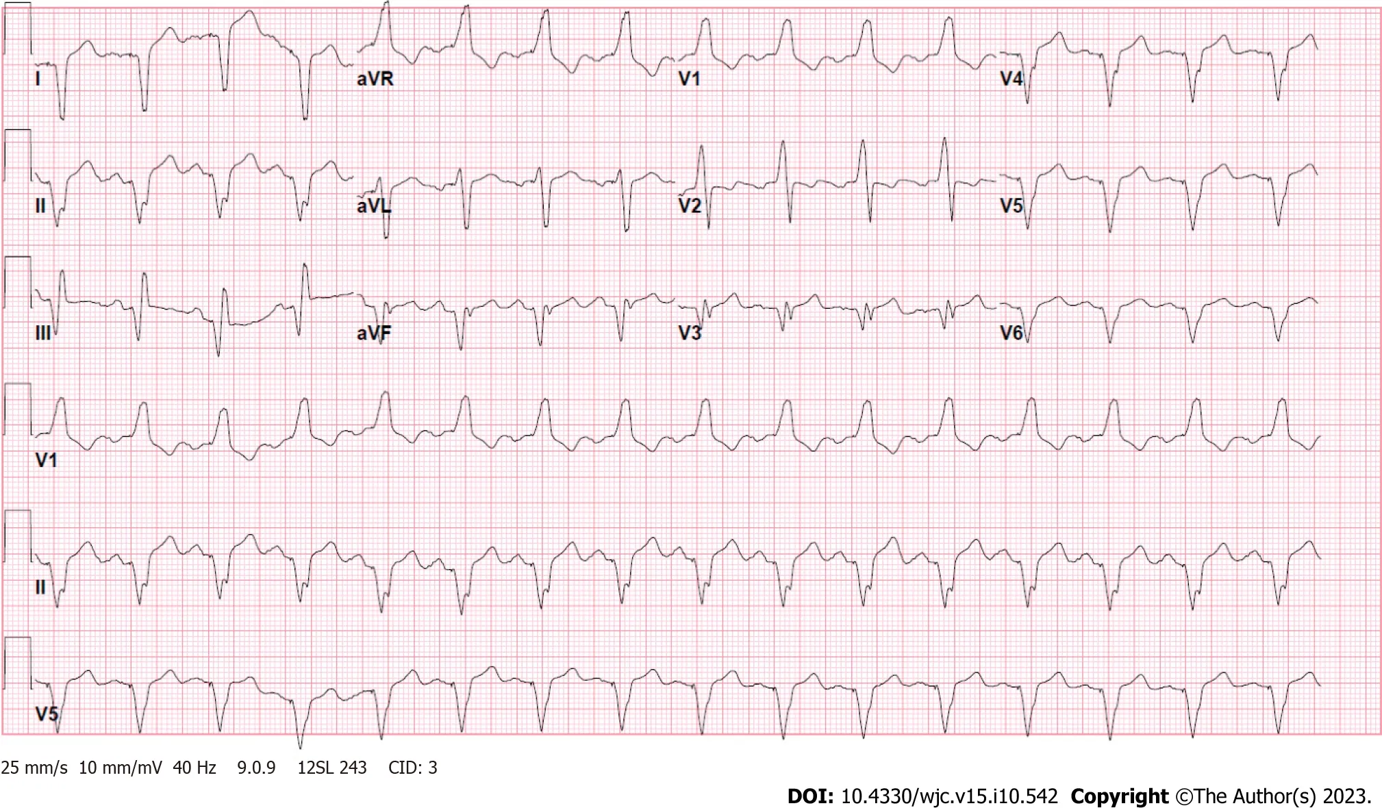
Figure 1 Electrocardiogram showing an atrial-sensed ventricularly-paced rhythm without acute abnormalities.
Imaging examinations
Chest X-ray was remarkable for cardiomegaly with small bilateral pleural effusions (Figure 2).Chest pulmonary angiography was negative for pulmonary embolism,pericardial effusion,or pneumothorax (Figure 3).His cardiac anatomy of L-TGA is visualized on chest computed tomography (Figure 4).
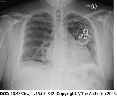
Figure 2 Chest X-ray showing cardiomegaly with small bilateral pleural effusions with unremarkable pulmonary vasculature.Biventricular pacemaker is present at the right chest.
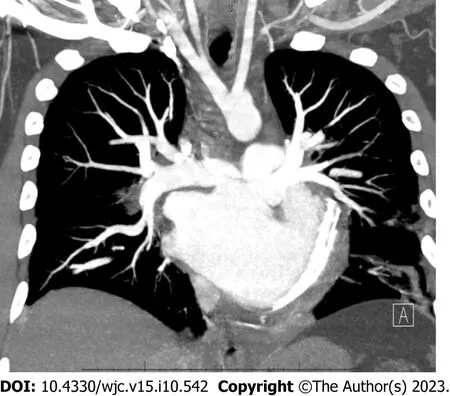
Figure 3 Chest pulmonary angiography demonstrating a levo-transposition of the great arteries cardiac anatomy.Examination was negative for pulmonary embolism,pericardial effusion,or pneumothorax.
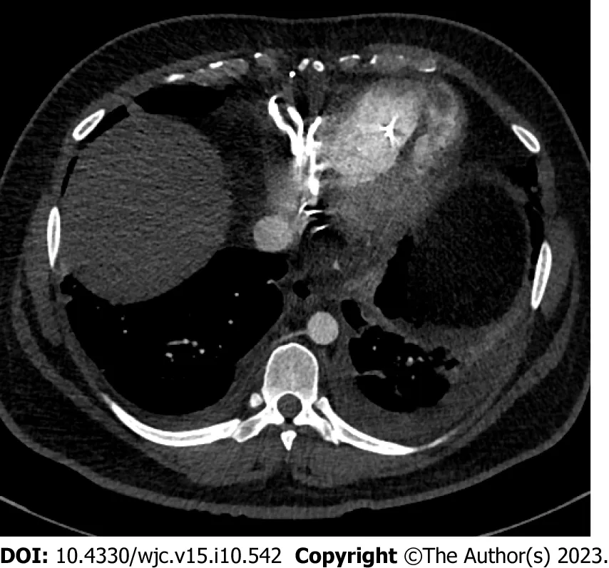
Figure 4 Computed tomography of the chest detailing the patient’s cardiac anatomy of levo-transposition of the great arteries.Venous circulation consists of the right atrium,left ventricle,and pulmonary artery.Systemic circulation consists of the pulmonary vein,left atrium,right ventricle,and aorta.Patient has a left-sided aortic arch with typical configuration.
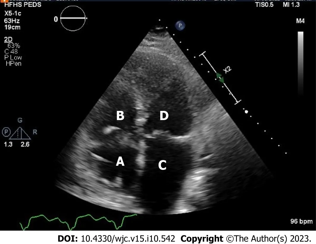
Figure 5 Transthoracic echocardiography demonstrating the patient’s classic levo-transposition of the great arteries anatomy. A: Right atrium;B: Venous left ventricle;C: Left atrium;D: Systemic right ventricle.Visualized is an apically displaced systemic tricuspid valve opening into systemic right ventricle.
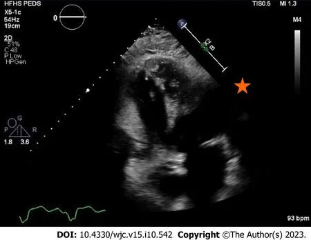
Figure 6 Transthoracic echocardiography in apical four chamber view showing an systemic right ventricle apical thrombus. This view highlights the importance of visualizing the true apex of the systemic right ventricle as the thrombus is not seen in Figure 5 where the apex is foreshortened.
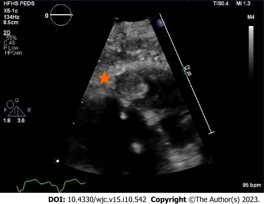
Figure 7 Transthoracic echocardiography highlighting the systemic right ventricle apical thrombus. A 1.3 cm × 1.3 cm well circumscribed mass with an echolucent center.

Figure 8 Transthoracic echocardiography with intravenous contrast demarcating the apical thrombus in the systemic right ventricle.
FURTHER DIAGNOSTIC WORK-UP
Our patient was managed for ADHF with guideline-directed medical therapy including diuresis with intravenous furosemide 40 mg once daily,oral metoprolol succinate 50 mg once daily,oral losartan 50 mg once daily,and oral dapagliflozin 10 mg daily.He had rapid clinical improvement with resolution of his symptoms and was transitioned to oral diuretic as needed.
Transthoracic echocardiogram obtained prior to discharge showed a mildly reduced global sRV ejection fraction of 40%,severe sAV regurgitation,and a newly identified apical thrombus in the sRV (Figures 5-8).Transesophageal echocardiogram confirmed this finding with visualization of a 2.07 cm by 1.43 cm well-circumscribed mass.
FINAL DIAGNOSIS
The final diagnosis was ADHF complicated by a systemic right ventricular thrombus in the setting of L-TGA.
TREATMENT
The presence of a sRV thrombus posed a dilemma given the limited literature on this topic.Our patient was anticoagulated with a vitamin K antagonist (VKA) and later referred for evaluation by advanced heart failure and heart transplant services.
OUTCOME AND FOLLOW-UP
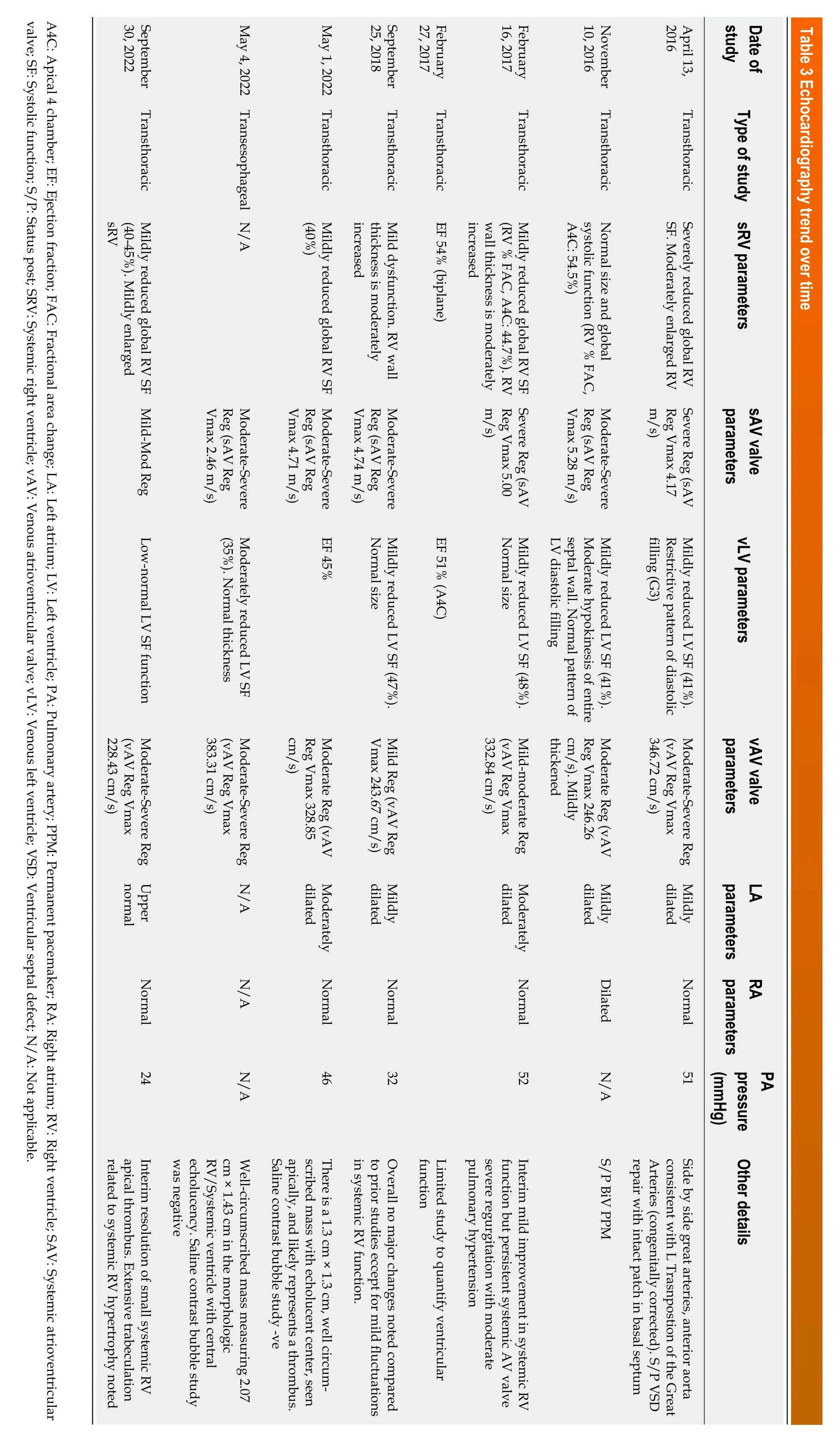
On subsequent follow-up visits,our patient’s symptoms of heart failure continued to improve with medical optimization and cardiac rehabilitation.Transthoracic echocardiogram was performed 5 mo after the index echocardiogram that identified the sRV thrombus;it demonstrated interim resolution of the sRV thrombus with improvement in sAV regurgitation and estimated pulmonary artery systolic pressure (Table 2).Cardiopulmonary exercise testing (CPX)provides objective measures of cardiovascular fitness and allows them to be tracked over time;our patient’s peak oxygen uptake (peak VO2),a strong prognostic indicator in heart failure,showed improvement (Table 3).Tables 2 and 3 allow readers to understand the natural history and progression of L-TGA in adults by demonstrating findings from CPX and echocardiography from the patient’s initial visit in 2016 to the last known follow-up in 2022.

Table 2 Cardiopulmonary exercise testing trend over time
DISCUSSION
Intracardiac thrombus (ICT) involves the formation of a blood clot within the heart chambers.It typically occurs in the setting of acute myocardial infarction,left ventricular aneurysm,and cardiomyopathy with dilated chambers[15].Pathophysiology of thrombus formation involves an interplay of prothrombotic state,tissue endothelial injury,and bloodstasis as described by Virchow’s triad[16-18].Systemic embolization of left-sided ICT results in clinical manifestations of stroke and transient ischemic attack,mesenteric ischemia and infarction,renal infarction,and acute limb ischemia.Pulmonary embolization of right-sided ICT results in pulmonary embolism.Diagnostic gold standard is identification of a thrombus on cardiac magnetic resonance imaging,although echocardiography with the use of echocardiographic contrast agents is a widely used initial modality[19].
ICT of the sRV is sparsely covered in the literature with limited data and guidelines available regarding the approach to management.Clinicians resort to extrapolating from practice standards for systemic left ventricular (sLV) thrombus.A review of literature identified no published case reports of sRV thrombus.
Current American and European guidelines for ICT in patients with structurally typical hearts are covered by class IIa,LOE C recommendations.Standard of care consists of anticoagulation with a VKA for 3-6 mo with an international normalized ratio (INR) target range of 2.0-3.0 followed by repeat imaging to assess for thrombus resolution[20,21].Anticoagulation in this population has been shown to decrease major cardiovascular events including cerebral and systemic sequala of thrombus embolization[22].Patients who have interval resolution of ICT on repeat imaging typically have anticoagulation discontinued,although some experts continue anticoagulation as a preventative measure in select patients with persistent and significant sLV wall hypokinesis due to the higher risk of recurrence[23].In the absence of data on this population,patients with sRV dysfunction who develop right ventricular thrombus,such as our patient,can be similarly managed with anticoagulation using a VKA followed by repeat imaging.
Oral anticoagulant (OAC) agent of choice for the treatment of ICT has classically been a VKA as opposed to a direct OAC (DOAC),as early available literature demonstrated clinically significant difference in outcomes between the two agents.The largest study to date,a multicenter cohort study compared 514 patients with sLV thrombus and demonstrated a higher risk for stroke and systemic embolism with DOAC therapy compared to VKA which suggests against efficacy equivalence[24].However,more recent data derived from small-scale randomized controlled trials,cohort studies,and case series report similar outcomes and support the use of DOAC for sLV thrombus which has led experts to adopt it as an off-label alternative[23,25-31].A recent scientific statement by the American Heart Association carried out a comprehensive meta-analysis of all published studies that compared VKA and DOAC use in sLV thrombus,it demonstrated no differences in therapeutic efficacy and safety;the statement concluded that the use of DOAC for sLV thrombus is a reasonable alternative to VKA[32].This practice-changing statement is a yet to be reflected in society guidelines and adopted by other organizations.
Clinical consensus for the management of sRV thrombus in patients with congenital heart disease has been derived from the management principles of sLV thrombus in patients without congenital heart disease;it consists of OAC,interval repeat imaging,and case-by-case evaluation of anticoagulation duration.The advent of DOAC therapy has seen it become the OAC agent of choice for most anticoagulation indications which has raised questions regarding its applicability in the treatment of sRV thrombus in the setting of the limited data.The international NOTE registry evaluated 530 adults with congenital heart disease treated with OAC for various indications and concluded non-inferior efficacy and safety of DOAC use compared to VKA;subgroup analysis of 76 patients with sRV found high efficacy and safety rates[33,34].Another similar study of 215 patients with different congenital heart diseases quoted high efficacy rates but nonnegligible bleeding risks[35].However,a retrospective cohort review of a German nationwide registry of 6504 adults with congenital heart disease on OAC determined that DOAC use was associated with higher rates of thromboembolism,bleeding,major adverse cardiac events,and all-cause mortality compared to VKA[36].The discrepancy of results between the former and latter studies raises concerns can be explained by the significant heterogeneity including differences in age and complexity of lesions in the latter’s study population.In the absence of conclusive data supporting DOAC use in sRV thrombus in patients with congenital heart disease,VKA remains the OAC of choice.
Patients with sRV thrombus are typically younger than those with sLV thrombus given the earlier development of heart failure in the setting of congenital heart disease.The structural cardiac anomalies and abnormal hemodynamics are likely contributors to abnormal flow and blood stasis,this predisposes patients to thrombus formation.Younger patients are more likely to have educational and workplace commitments that cause time constraints.VKA therapy is particularly challenging in this population as dietary restrictions and frequent laboratory testing results in difficulty achieving and maintaining therapeutic INR levels;lower time in therapeutic range is associated with higher risk of stroke in patients with sLV[37].Further studies evaluating the safety and efficacy of DOAC agents in sRV thrombus are necessary.
CONCLUSION
Systemic right ventricular thrombus is a rare complication of congenital heart disease.We describe the first reported case of sRV thrombus in a patient with L-TGA who presented with ADHF.Management of this condition is driven by expert opinion and extrapolation of treatment principles from ICT in patients with structurally normal hearts.
In the absence of conclusive data supporting DOAC use in sRV thrombus in patients with congenital heart disease,VKA bridged with intravenous heparin or subcutaneous low-molecular weight heparin and remains the time-honored approach particularly in patients with recent large thrombi and in the setting of dysfunctional ventricles or slow flow states.Transitioning to a DOAC in these cases should be individualized to a patient characteristics and imaging features;it should involve shared decision making regarding limited and conflicting literature.This case illustrates the natural history of L-TGA and highlights the importance of surveillance and monitoring in this patient population to prevent and treat complications.
FOOTNOTES
Author contributions:Almajed MR,Almajed A,Khan N,Obri M,Ananthasubramaniam K contributed equally to this work;All authors evaluated the case,reviewed the literature,and wrote the manuscript;All authors have read and approve of the final manuscript.
Informed consent statement:Informed written consent was obtained from the patient for publication of this report and any accompanying images.
Conflict-of-interest statement:Dr.Ananthasubramaniam has nothing to disclose.
CARE Checklist (2016) statement:The authors have read the CARE Checklist (2016),and the manuscript was prepared and revised according to the CARE Checklist (2016).
Open-Access:This article is an open-access article that was selected by an in-house editor and fully peer-reviewed by external reviewers.It is distributed in accordance with the Creative Commons Attribution NonCommercial (CC BY-NC 4.0) license,which permits others to distribute,remix,adapt,build upon this work non-commercially,and license their derivative works on different terms,provided the original work is properly cited and the use is non-commercial.See: https://creativecommons.org/Licenses/by-nc/4.0/
Country/Territory of origin:United States
ORCID number:Mohamed Ramzi Almajed 0000-0001-6161-8494;Karthikeyan Ananthasubramaniam 0000-0001-5837-496X.
S-Editor:Lin C
L-Editor:A
P-Editor:Yuan YY
杂志排行
World Journal of Cardiology的其它文章
- Candida endocarditis: Update on management considerations
- Do cardiopulmonary resuscitation real-time audiovisual feedback devices improve patient outcomes? A systematic review and metaanalysis
- Cardiovascular complications following medical termination of pregnancy: An updated review
- Establishment of a prediction model for prehospital return of spontaneous circulation in out-of-hospital patients with cardiac arrest
- Integrated analysis of comorbidity,pregnant outcomes,and amniotic fluid cytogenetics of fetuses with persistent left superior vena cava
- Value of cardiac magnetic resonance on the risk stratification of cardiomyopathies