Pulmonary cytomegalovirus infection: A case report and systematic review
2023-11-18AwotarKanikaJonathanSoldera
Awotar Kanika,Jonathan Soldera
Abstract
Key Words: Cytomegalovirus; Immunocompromised; Immunocompetent; Severe acute respiratory syndrome coronavirus 2; Coronavirus disease 2019; Ganciclovir
INTRODUCTION
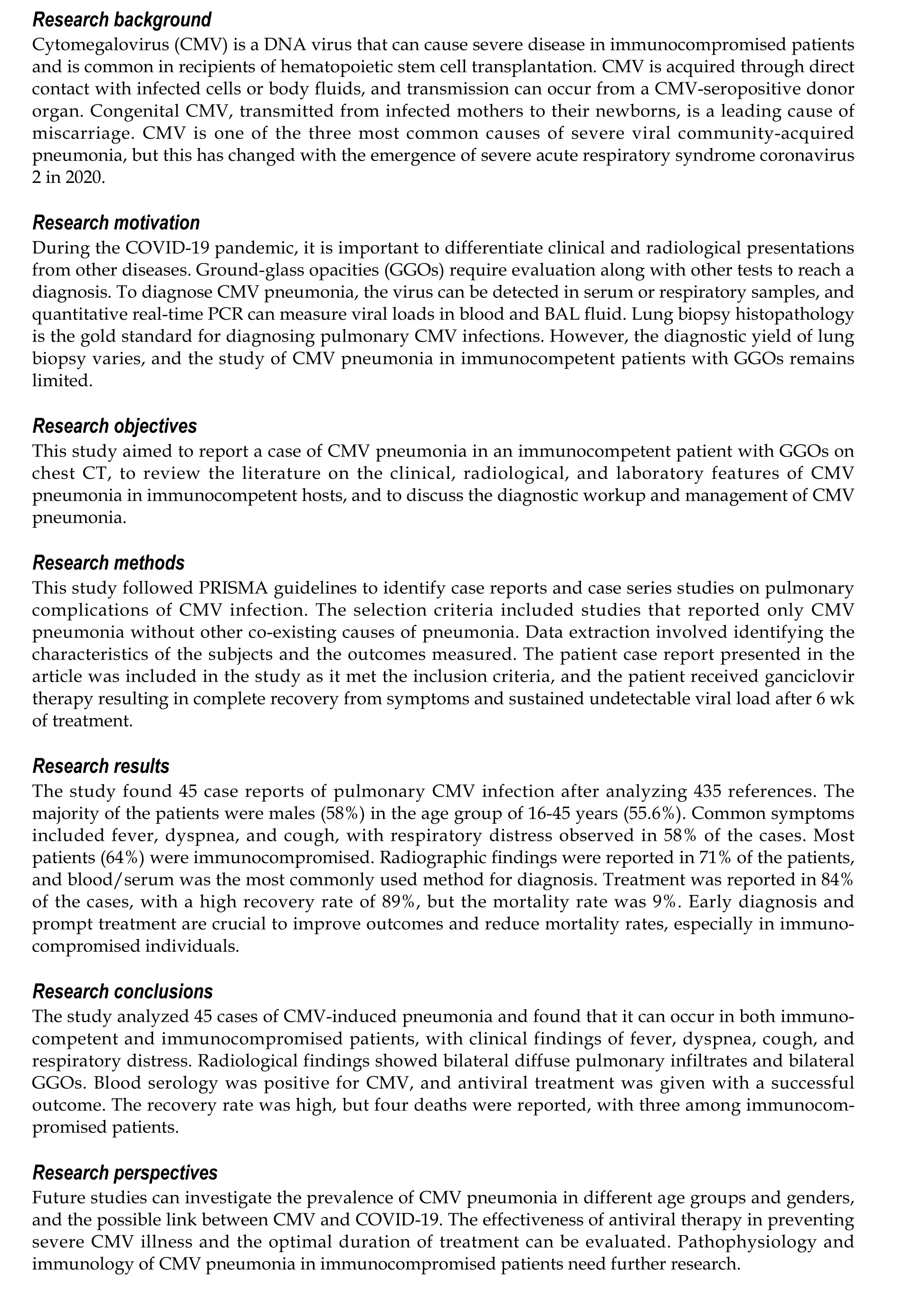
Cytomegalovirus (CMV) is a DNA virus that belongs to the herpesviridae family and shares similarities with other herpes viruses.In immunocompetent adults, CMV infection is usually asymptomatic and causes mild mononucleosis-like syndrome, typically in childhood or adolescence.However, CMV can cause severe disease and pneumonia in immunocompetent individuals, albeit rarely[1,2].CMV infection may lead to severe viral pneumonitis in immunocompromised patients, such as those with autoimmune deficiency syndrome (AIDS), allogeneic bone marrow transplantation recipients, or those on immunosuppressive drugs or high-dose steroids.The incidence of CMV infection is approximately 25%-30% in recipients of hematopoietic stem cell transplantation[3].The gastrointestinal tract and central nervous system are the most frequent sites of severe CMV infection.CMV was one of the three most common causes of severe viral community-acquired pneumonia (CAP), along with influenza and adenovirus.However, this has changed with the emergence of severe acute respiratory syndrome coronavirus 2(SARS-CoV-2) in 2020[4].The pulmonary manifestations of CMV infection may vary from a dry cough to severe interstitial pneumonia, with patients presenting with diffuse pulmonary infiltrates resembling a ground glass appearance.The diagnosis of CMV pneumonia is based on radiological patterns and serology (CMV IgM antibody) or polymerase chain reaction (PCR)[4].In 1968, the first case of CMV CAP was reported by Carlstorm and colleagues in their case series of CMV infection in immunocompetent hosts[5].CMV CAP in immunocompetent hosts presents as prolonged fever and interstitial infiltrates on chest X-ray that resolved slowly over 6 wk.Patients with CMV CAP present with relative lymphopenia, atypical lymphocytes, and mildly elevated serum transaminases.Primary CMV infection persists for life and is generally acquired through close physical contact involving direct inoculation with infected cells or body fluids.The spread of viral infection is through coughing, direct contact with body fluids such as blood, urine, feces, semen, vaginal fluid, and breast milk, orviamucous membranes,including the mouth or genitals.CMV infection following transplantation can be acquired if the transmission is from the organ from a CMV-seropositive donor.Mothers infected with CMV during pregnancy may transmit this infection to their newborn baby, leading to congenital CMV.CMV infection is one of the leading causes of miscarriage[1,6].Babies with congenital CMV sometimes may be healthy for months or years after birth but may have late occurring signs such as hearing loss, and develop vision problems and developmental delay.Latent CMV can reactivate and replicate rapidly when the immune system is suppressed.It can lead to high levels of CMV viremia, and infection of multiple organ systems can cause severe illness such as retinitis, colitis, hepatitis, pneumonia, or encephalitis.Fatal CMV pneumonia is more common in patients who have received marrow transplants than those who received transplant of solid organs like the lung, heart, liver, or kidney[7,8].CMV accentuates the sepsis-induced immunologic effects, leading to an increase in the risk for secondary infections.CMV infection in critically ill patients is associated with prolonged ventilator support,nosocomial infections, prolonged hospital/intensive care unit (ICU) stay, and increased mortality rates[9].
As the coronavirus disease 2019 (COVID-19) pandemic continues and becomes an endemic, it is crucial to recognize that not all clinical and radiological presentations are solely attributable to COVID-19[10].Therefore, diagnostic differentiation is essential, and ground-glass opacities (GGOs) must be evaluated in conjunction with other imaging findings, laboratory tests, and clinical features to reach a definitive diagnosis.CMV pneumonia can be diagnosed by detecting the virus in serum and/or respiratory samples such as bronchoalveolar lavage (BAL) or tracheal aspiration[10].Quantitative realtime PCR (qRT-PCR) can be utilized to measure viral loads in blood and BAL fluid[11].Lung biopsy histopathology is considered the gold standard for diagnosing pulmonary CMV infections, with the presence of CMV inclusion bodies (owl’s eye) in biopsy specimens being confirmatory of lung infection[12].However, the diagnostic yield of lung biopsy for diagnosing lung CMV infections can vary as inclusions may not always be visualized.Immunohistochemical (IHC) staining for CMV in cytological specimens of bronchial washing fluid can also detect CMV[13,14].
The first-line treatment for CMV disease is intravenous ganciclovir and its prodrug, oral valganciclovir, which inhibits viral deoxyribonucleic acid (DNA) polymerase, thereby interfering with DNA elongation.Mild disease in immunosuppressed patients may be treated with oral valganciclovir,whereas severe illness requires initial treatment with intravenous ganciclovir or foscarnet at full doses(adjusted for renal function)[15].Treatment at full doses should be continued until symptom resolution and blood antigenemia (or DNAemia) clears.Adjuvant treatment with intravenous immunoglobulin or CMV hyper-immunoglobulin is recommended in immunocompromised patients and may be used in cases of severe CMV disease and hypogammaglobinemia[12].
This study aimed to report a case of disseminated CMV in an immunocompetent patient, and systematically review published cases of pulmonary CMV infection in both immunocompromised and immunocompetent patients.
Case report
Chief complaints:A 32-year-old man presented with a cough, dyspnea, high-grade fever, and jaundice.
History of present illness:The patient had no significant medical history and was not taking any medication.Physical examination revealed a temperature of 39.5°C, tachypnea, icteric sclera, and hepatosplenomegaly.He had no skin rash or lymphadenopathy.The initial blood tests showed pancytopenia, elevated liver enzymes, elevated bilirubin, and hypoalbuminemia.CT of the thorax showed GGOs, while CT of the face showed sinusitis, raising suspicion of an infectious etiology.
History of past illness:The patient had no significant past medical history.
Personal and family history:No significant personal or family history was reported.
Physical examination:The patient presented with a temperature of 39.5°C, tachypnea, icteric sclera, and hepatosplenomegaly.He had no skin rash or lymphadenopathy.
Laboratory examinations:Complete blood count revealed a platelet count of 87000/mm³, hemoglobin level of 8.2 g/dL, and leukocyte count of 4830/mm³.Liver function tests showed alkaline phosphatase of 1174 U/L, gamma-glutamyl transferase of 804 U/L, aspartate aminotransferase of 403 U/L, total bilirubin of 17.2 mg/dL, albumin of 1.7 g/dL, and international normalized ratio of 1.11.Autoimmune antibody testing for fluorescence antinuclear antibody was negative.COVID-19 antigen swab test was negative.
Imaging examinations:After a liver biopsy, the patient's results were suggestive of drug-induced liver injury, and subsequent immunochemistry testing returned negative results for CMV.Magnetic resonance imaging (MRI) of the abdomen showed a liver with enlarged dimensions, regular contours,and heterogeneous signal intensity, with predominance of hyper signal in the T2-weighted sequences,suggestive of an inflammatory process (hepatitis), and splenomegaly and pancreatic edema suggestive of pancreatitis.CT of the thorax showed GGOs (Figure 1), while CT of the face showed sinusitis.
Final diagnosis:The patient's clinical condition worsened, and he developed hypotension and sepsis,requiring admission to the ICU.Broad-spectrum antibiotics were started, and he was investigated for possible Wegener's granulomatosis.However, auto-antibodies were negative and his final diagnosis was disseminated CMV infection, confirmed by the high viral load of 325192.5 copies/mL.
Treatment:The patient was started on ganciclovir therapy.
Outcome and follow-up: After 6 wk of treatment, the patient recovered completely from his symptoms,achieving a sustained undetectable viral load.
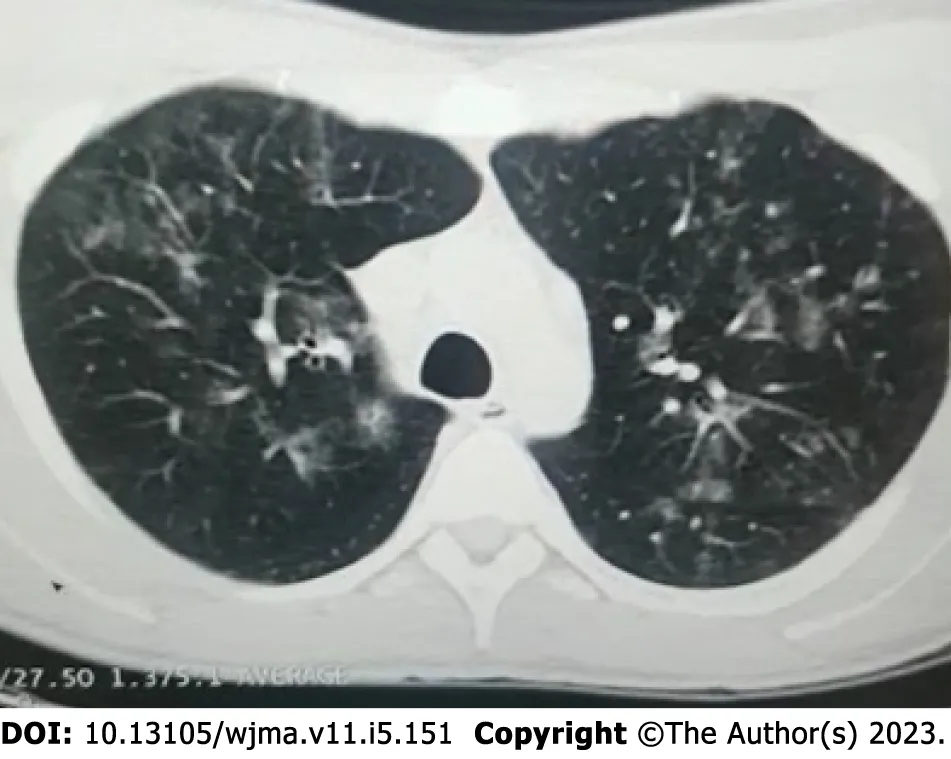
Figure 1 Computed tomography of the thorax showing ground glass opacities.
MATERIALS AND METHODS
This study followed the recommendations outlined in the Preferred Reporting Items for Systematic Reviews and Meta-Analysis (PRISMA) guidelines[16].
Data sources
The electronic database MEDLINE (PubMed) was searched using the terms described in the Supplementary material.The searches were conducted in September and October 2022, with no date of publication restrictions and language restricted to English.References of included studies were screened for relevant records, and the reference lists of the retrieved studies were submitted to a manual search.
Inclusion and exclusion criteria
Case report or case series studies were eligible for selection.If there was more than one study published using the same case, the most recent study was selected for analysis.Studies published only as abstracts were also included, as long as the data available made data collection possible.Studies written in languages other than English were excluded.Studies having other co-existing causes of pneumonia were excluded from our study, for example, superimposed bacterial, parasitic, or fungal infections in existing CMV pneumonia, and other lung pathologies.
Study selection and data extraction
Titles were screened initially to select the cases of pulmonary complications of CMV infection and filter out non-relevant studies.Then, abstracts of chosen studies were read to select potentially relevant papers.The third step was the analysis of the full-length papers, and those which were not case reports of pulmonary CMV were filtered out.Data was extracted on the characteristics of the subjects and the outcomes measured from each eligible study.A table of extracted data on eligible studies was made in order to measure and identify patterns.
RESULTS
Using the search strategy, a total of 435 references were retrieved.After reviewing titles, 232 studies were found to be relevant for our topic and 203 studies were excluded.By analyzing abstracts, 172 studies were found to be potential relevant papers for our topic and therefore 60 studies were excluded.After reading and analyzing full length papers, 43 studies with 45 case reports of pulmonary CMV infection were included.The data of 45 case reports was extracted and prepared in Table 1 to measure and identify the patterns to get the results to reach a conclusion.Figure 2 shows the PRISMA search strategy.Every study included was a case report.
The baseline features are described in Table 2 and Table 3 for the 45 patients who were included for data extraction.All patients were diagnosed with CMV pneumonia.The majority of patients were males(58%) and in the age group of 16-45 years (55.6%).The most common symptoms reported were fever(82%), dyspnea (76%), and cough (53%).Respiratory distress was observed in 58% of the patients.Almost two-thirds of the patients (64%) were immunocompromised.Radiographic findings were reported in 71% of the patients by chest X-ray and 69% by CT.Blood/serum was the most commonly used method for serology testing (89%), and bronchoalveolar fluid was used in 45% of the cases.

Table 1 Summary of systematically reviewed clinical cases of cytomegalovirus pneumonia
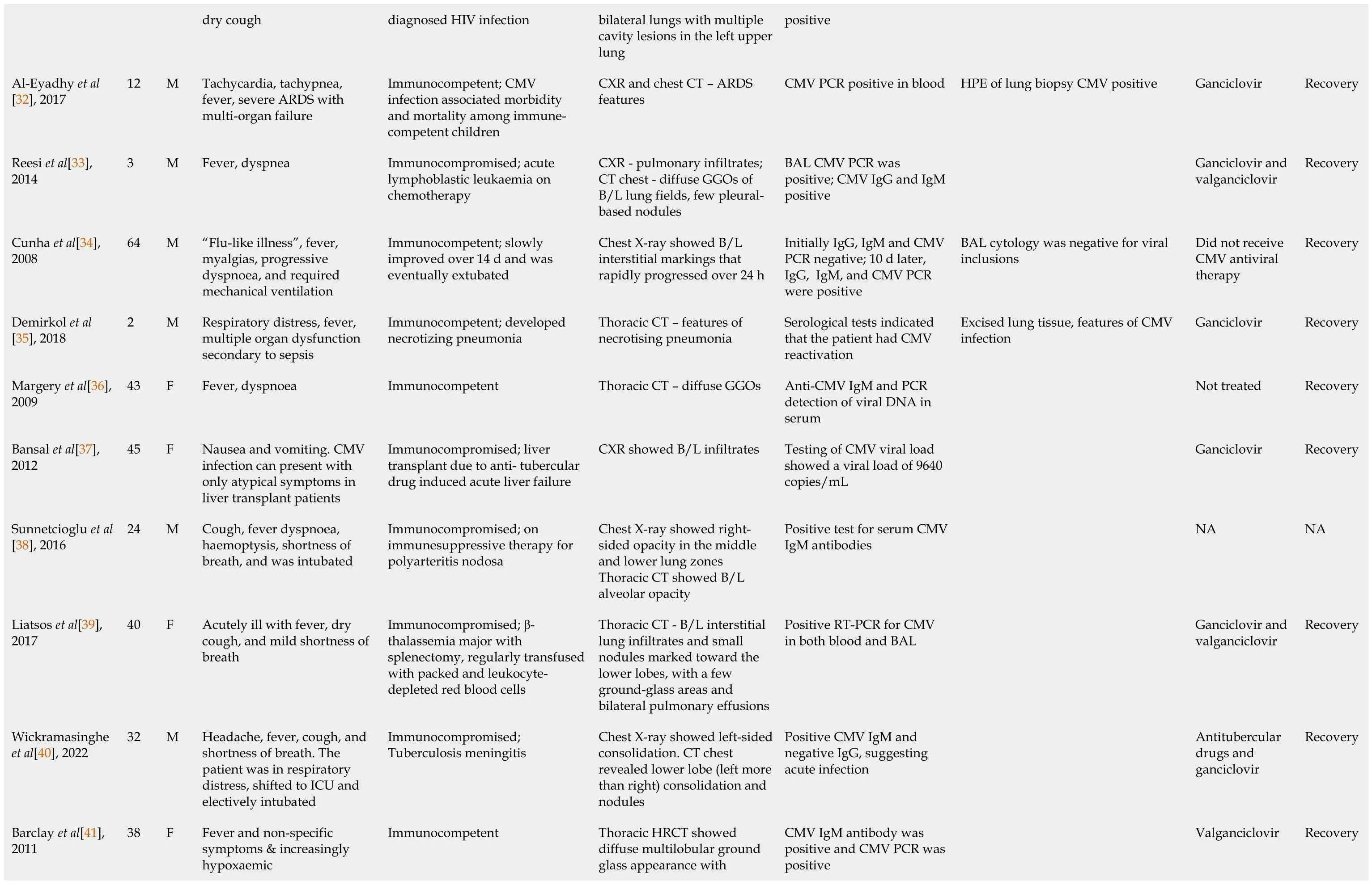
dry cough diagnosed HIV infection bilateral lungs with multiple cavity lesions in the left upper lung positive Al-Eyadhy et al[32], 2017 12 M Tachycardia, tachypnea,fever, severe ARDS with multi-organ failure Immunocompetent; CMV infection associated morbidity and mortality among immunecompetent children CXR and chest CT – ARDS features CMV PCR positive in blood HPE of lung biopsy CMV positive Ganciclovir Recovery Reesi et al[33],2014 3 M Fever, dyspnea Immunocompromised; acute lymphoblastic leukaemia on chemotherapy CXR - pulmonary infiltrates;CT chest - diffuse GGOs of B/L lung fields, few pleuralbased nodules BAL CMV PCR was positive; CMV IgG and IgM positive Ganciclovir and valganciclovir Recovery Cunha et al[34],2008 64 M “Flu-like illness”, fever,myalgias, progressive dyspnoea, and required mechanical ventilation Immunocompetent; slowly improved over 14 d and was eventually extubated Chest X-ray showed B/L interstitial markings that rapidly progressed over 24 h Initially IgG, IgM and CMV PCR negative; 10 d later,IgG, IgM, and CMV PCR were positive BAL cytology was negative for viral inclusions Did not receive CMV antiviral therapy Recovery Demirkol et al[35], 2018 2 M Respiratory distress, fever,multiple organ dysfunction secondary to sepsis Immunocompetent; developed necrotizing pneumonia Thoracic CT – features of necrotising pneumonia Serological tests indicated that the patient had CMV reactivation Excised lung tissue, features of CMV infection Ganciclovir Recovery Margery et al[36],2009 43 F Fever, dyspnoea Immunocompetent Thoracic CT – diffuse GGOs Anti-CMV IgM and PCR detection of viral DNA in serum Not treated Recovery Bansal et al[37],2012 45 F Nausea and vomiting.CMV infection can present with only atypical symptoms in liver transplant patients Immunocompromised; liver transplant due to anti- tubercular drug induced acute liver failure CXR showed B/L infiltrates Testing of CMV viral load showed a viral load of 9640 copies/mL Ganciclovir Recovery Sunnetcioglu et al[38], 2016 24 M Cough, fever dyspnoea,haemoptysis, shortness of breath, and was intubated Immunocompromised; on immunesuppressive therapy for polyarteritis nodosa Chest X-ray showed rightsided opacity in the middle and lower lung zones Thoracic CT showed B/L alveolar opacity Positive test for serum CMV IgM antibodies NA NA Liatsos et al[39],2017 40 F Acutely ill with fever, dry cough, and mild shortness of breath Immunocompromised; βthalassemia major with splenectomy, regularly transfused with packed and leukocytedepleted red blood cells Thoracic CT - B/L interstitial lung infiltrates and small nodules marked toward the lower lobes, with a few ground-glass areas and bilateral pulmonary effusions Positive RT-PCR for CMV in both blood and BAL Ganciclovir and valganciclovir Recovery Wickramasinghe et al[40], 2022 32 M Headache, fever, cough, and shortness of breath.The patient was in respiratory distress, shifted to ICU and electively intubated Immunocompromised;Tuberculosis meningitis Chest X-ray showed left-sided consolidation.CT chest revealed lower lobe (left more than right) consolidation and nodules Positive CMV IgM and negative IgG, suggesting acute infection Antitubercular drugs and ganciclovir Recovery Thoracic HRCT showed diffuse multilobular ground glass appearance with Barclay et al[41],2011 38 F Fever and non-specific symptoms & increasingly hypoxaemic Immunocompetent CMV IgM antibody was positive and CMV PCR was positive Valganciclovir Recovery

Shimada et al[50],2004 27 F Fever Immunocompromised; on immunosuppressive treatment for viral-associated hemophagocytic syndrome CXR and chest HRCT –diffuse small pulmonary nodules CMV DNA PCR was positive on bronchoalveolar lavage cells; immunoassay pp65 CMV antigen positive Lung biopsy inclusion-bearing cells for CMV Gancyclovir Recovery Simsir et al[51],2001 43 M Malaise, fever, pleuritic chest pain, epigastric pain,diarrhea, nausea, vomiting Immunocompromised;underwent renal transplant secondary to diabetic nephropathy CXR showed a nodule in the upper lobe of the right lung;chest CT revealed bilateral smaller pulmonary nodules CMV antigen test was positive, with negative CMV IgG CMV was established by fine-needle aspiration biopsy of the lung nodule Gancyclovir Recovery Abbey et al[52],2014 51 M Fever, dry, cough, dyspnoea,general malaise Immunocompromised; Crohn’s disease on azathioprine; also had mild pancreatic insufficiency and bile salt malabsorption CXR showed bilateral infiltrates in middle and lower zones; chest CT showed B/L small pleural effusions and B/L basal lung consolidation CMV IgM positive, acute CMV infection Ganciclovir and valganciclovir Recovery Belin et al[53],2003 47 F Shortness of breath, fever,stomatitis, genital ulcerations,burning sensations Immunocompromised; severe rheumatoid arthritis, on prednisolone, methotrexate, and cyclosporine CXR showed interstitial infiltrates in both lung bases BAL showed CMV mRNA Ganciclovir Recovery Kaşifoğlu et al[54], 2006 21 F Polyarthralgias, fatigue, fever,muscle weakness, nonproductive cough, dyspnea Immunocompromised;dermatomyo-sitis, treated with azathioprine, prednisolone, and cyclosporine Chest XR showed bilateral interstitial infiltration; chest HRCT - bilaterally ill-defined multifocal GGOs Positivity for anti-CMV,IgM, and anti-CMV IgG antibodies and presence of CMV DNA by PCR Ganciclovir Recovery Chen et al[55],2010 5 M Fever, cough, dyspnea,hypoxemia, ARDS Immunocompetent; the patient developed ventilator-associated pneumonia, and died of burkhoderia sepsis Chest XR – multiple parenchymal consolidations;chest XR disclosed “white lung” during the second week Positive PCR; bronchoalveolar and seroconversion of CMV IgM and IgG NR Died Tambe et al[56],2019 32 F Fever, dyspnea, generalized rash, weakness Immunocompromised; stage IV,classical Hodgkin’s lymphoma,treated with chemotherapy Chest CT revealed bilateral pulmonary infiltrates and bilateral pleural effusion CMV was detected on BAL culture; serum quantitative CMV PCR was positive Ganciclovir and valganciclovir Recovery Boussouar et al[57], 2018 47 F Dry cough, chest pain and fever Immunocompromised; orthotopic heart transplant and immunosuppressive treatment was initiated with corticosteroids, cyclosporine,and mycophenolate Chest XR - alveolar opacities with upper lobe predominance; chest CT revealed consolidation in the right upper lobe associated with septal thickening and multiple nodules Blood CMV PCR, which has been undetectable Lung biopsy showed nuclear inclusions suggestive of CMV infection; IHC showed nuclear positivity for CMV Ganciclovir and valganciclovir Recovery Haddad et al[58],1984 18 M Fever, chills, non-productive cough, severe hypoxia requiring intubation Immunocompromised; sickle cell thalassemia Chest XR suggested early pulmonary edema and cardiomegaly On postmortem culture of lung parenchyma, CMV grew in 5 d NR Died Katagiri et al[59],2008 35 F Deterioration of lupus nephritis and received treatment with a high dose of steroid and cyclosporine Immunocompromised; SLE with increased risk of opportunistic infection Chest X-ray showed bilateral pleural effusion; chest CT revealed a cavitary lesion in the right middle lobe of the lung Positive for CMV;antigenemia Ganciclovir Recovery

Ayyappan et al[60], 2006 72 M Fever, productive cough,worsening breathlessness and tenderness in epigastrium Immunocompromised;rheumatoid arthritis-related interstitial lung disease, on corticosteroids and cyclophosphamide Chest XR showed bilateral consolidation; chest CT revealed cavitating masses in the right upper lobe & lingula and diffuse interstitial fibrosis PCR assay of BAL fluid was positive for CMV Gastric biopsy - intracytoplasmic viral inclusions consistent with CMV gastritis; transbronchial lung biopsy showed intracytoplasmic viral inclusion Gancyclovir Recovery Manian et al[61],1993 32 F Fever, non-productive cough,worsening oxygenation Immunocompetent Chest X ray - bilateral interstitial infiltrates Enzyme immune-assay showed that CMV IgG and CMV IgM were positive Ganciclovir Recovery McCormack et al[62], 1998 31 M Fever, abdominal pain,jaundice, cough, palpitations,shortness of breath with atrial fibrillation Immunocompetent Chest radiograph showed bilateral interstitial pulmonary infiltrates EIA for antibodies to CMV showed a strong reaction to IgM and a weak reaction to IgG A urine culture yielded CMV; a cytopathic effect was observed and con-firmed by immunofluorescence Ganciclovir Recovery Najjar et al[63],2004, Case 1 34 F Fever Immunocompromised; SLE with renal failure on haemodialysis Chest XR - bilateral infiltrates;chest CT - bilateral peripheral parenchymal infiltrates and a cavitating mass in right lower lobe A CMV antigenaemia assay was positive and CMV isolation in blood Histological findings included numerous intranuclear and intracytoplasmic CMV inclusions confirmed by IHC IV ganciclovir and IV IgG Recovery Najjar et al[63],2004, Case 2 33 M Fever, dyspnoea, worsening renal function Immunocompromised; SLE, class IV lupus, nephritis treated with chronic steroid therapy,azathioprine, and cyclophosphamide Chest CT revealed a right upper lobe thick-walled cavitary lesion Serology revealed raised CMV IgM & IgG HPE - evidence of focal interstitial fibrosis, accumulation of intraalveolar macrophages, and CMV with intracytoplasmic and nuclear inclusions in the lining alveolar cells Gancyclovir Recovery Kanika et al 32 M Fever, dyspneia, hypotension,jaundice Immunocompetent MRI showed hepatitis and pancreatitis; CT showed GGO Serum PCR with a high viral load Liver biopsy suggestive of drug induced liver injury and immunochemistry negative for CMV Ganciclovir Recorvery
Immunohistochemistry (IHC) was reported in 24% of the cases, and biopsy-histopathology was performed in 27% of the patients.The treatment was reported in 84% of the cases, with a high recovery rate of 89%.Unfortunately, the mortality rate was 9%, with four patients reported to have died.
DISCUSSION
This paper analyzed 45 cases of CMV-induced pneumonia.Patients were divided into two main categories: Immunocompetent and immunocompromised.Twenty-nine (64%) patients were immunocompromised, and 16 (36%) were immunocompetent and developed CMV pneumonia.This suggests that CMV infection prevalence is higher in immunocompromised patients[2].The reported case highlights the importance of considering CMV infection in patients who present with fever, respiratory symptoms, and abnormal liver function tests.Although CMV infection is more common in immunocompromised patients, this case demonstrates that it can also occur in immunocompetent individuals.Itis important to note that CMV is a common cause of pneumonia, particularly in immunocompromised patients, and should be considered in the differential diagnosis of patients with respiratory symptoms who do not respond to standard treatment.Early diagnosis and treatment are essential in improving patient outcomes, especially in severe cases.Therefore, clinicians should be aware of the clinical features and radiological findings of CMV pneumonia to enable early diagnosis and appropriate management[17-20].

Table 2 Baseline features of 45 patients with cytomegalovirus pneumonia

Table 3 Summary of data collected
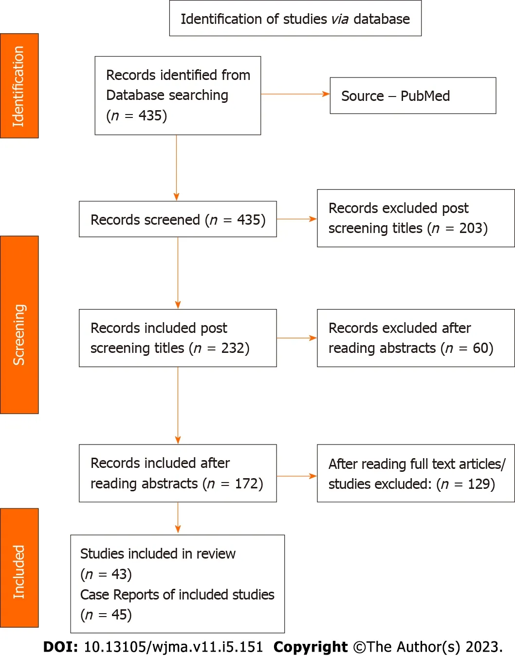
Figure 2 PRISMA search strategy for systematic review.
The differential diagnosis of this case includes severe COVID-19 infection, which shares some clinical features with CMV pneumonia, such as cough, dyspnea, and fever.However, some features of the case,such as jaundice, hepatosplenomegaly, and pancytopenia, are not typically seen in severe COVID-19 cases.Additionally, GGOs on CT imaging can be seen in both CMV pneumonia and COVID-19.Therefore, it is important to consider other infectious and non-infectious etiologies in patients with respiratory symptoms and abnormal liver function tests.
A systematic review was performed a total of 45 patients, of which 26 (58%) were male and 19 (42%)were female.Infection was more prevalent in males, with 11 immunocompetent and 15 immunocompromised male patients and 5 immunocompetent and 14 immunocompromised female patients.This suggests that CMV infection is more prevalent in immunosuppressed patients in both males and females.Immunocompromised states are an important host-associated risk factor to get CMV infection[2].
Regarding age, 25 patients were adults (13 males and 12 females), indicating that the adult population is more prone to developing pulmonary CMV infection.As it is estimated that more than half of the adult population are infected with CMV in the United States, and 80% of the adult population have this infection by the age of 40 years, the prevalence of CMV-induced pneumonia may increase with age[1].The clinical findings of most patients were fever (82%), dyspnea (75%), cough (53%), and respiratory distress (53%) in both immunocompetent and immunocompromised patients.These findings are consistent with previous studies on CMV pneumonia[4].
Regarding radiological findings, 32 patients were submitted to a chest X-ray mostly showing bilateral diffuse pulmonary infiltrates.CT of the thorax was done in 31 patients, and the main finding was bilateral GGOs.In some patients, there were small bilateral pulmonary nodules, confluent consolidations, and bronchiectasis.In case of atypical radiological findings other than bilateral infiltrates and GGOs, further investigation, such as blood and BAL serology, lung biopsy histopathological examination (HPE), and IHC, should be considered to rule out CMV pneumonia[7].
Blood serology was done in 40 (89%) patients, and IgM and IgG were positive for CMV.Other tests,such as BAL fluid serology, lung biopsy histopathology, and IHC, were done to confirm the diagnosis in some patients.IgM CMV positive in blood represents acute CMV infection, and antiviral treatment was given to the patients with a successful outcome[2,5].
A study by Basingeret al[24] demonstrated that immunocompromised states, particularly those with a history of allogenic hematopoietic stem cell transplant, can result in rapidly deteriorating conditions and respiratory status post-CMV infection.Radiologically, patients may present with rapidly progressive bilateral pulmonary nodules approximately 2 mo after receiving a bone marrow transplant.This patient died shortly after admission, and the diagnosis was made on post-mortem microscopic examination of the pulmonary nodules that demonstrated viral cytopathologic changes consistent with CMV infection, confirmed by IHC.It is essential to note that the radiographic presentation is not always GGOs, and rapidly enlarging pulmonary nodules in an immunosuppressed patient are highly suggestive of an infectious process.Therefore, careful histologic examination for viral cytopathologic changes is essential[3].
Regarding treatment, 38 (85%) patients received antiviral therapy, and 2 patients recovered without receiving antiviral treatment.In total, 89% of patients recovered, indicating that the prognosis of CMV pneumonia is good if diagnosed early and treated in time, in both immunocompetent and immunocompromised patients[2].A study by Al-Eyadhyet al[32] in 2017 presented the case of a 12-year-old immunocompetent patient who was admitted with severe ARDS and developed multi-organ failure,which is an important differential diagnosis from severe acute respiratory syndrome coronavirus 2 infection.Due to the correct diagnosis and treatment of CMV infection in time, the patient recovered.Another study by Coussementet al[42] in 2016 showed that a 63-year-old immunocompromised patient who did a bilateral lung transplant for chronic obstructive pulmonary disease admitted with severe CMV infection and due to timely diagnosis and antiviral treatment, the patient recovered well.
In immunocompetent patients, the recovery rate was 94%, while in immunocompromised patients, it was 86%.The study showed that there were four deaths, three of which were among immunocompromised patients.This suggests that immunocompromised patients may develop more severe CMV illness that deteriorates quickly, sometimes making it challenging to make a timely diagnosis.Therefore,it is crucial to consider CMV infection as one of the important differentials in immunocompromised patients[1,4].
The final result of this analysis showed that 89% of total patients recovered, indicating that the prognosis of CMV pneumonia is good if patients are diagnosed early and treated promptly, even for immunocompromised patients[1,4].
To reach a definitive diagnosis, clinical findings must be correlated with imaging tests and laboratory tests.Polymerase chain reaction (PCR) is the most sensitive method of detecting CMV, and qRT-PCR can be used to quantify viral loads in blood and BAL fluid.BAL CMV-PCR is considered the most accepted approach for viral isolation in the lungs due to its high sensitivity.Lung biopsy histopathology is considered the gold standard for the diagnosis of pulmonary CMV infections, and the presence of CMV inclusions in the HPE report is confirmatory of lung infection.Additionally, CMV can be detected by IHC staining for CMV in cytologic specimens of bronchial lavage fluid[1,2].
In critically ill patients, CMV infection is associated with prolonged mechanical ventilation,nosocomial infections, prolonged hospital and ICU stay, and increased mortality.The first-line treatment for CMV disease is intravenous ganciclovir and its prodrug, oral valganciclovir.Mild disease in immunosuppressed patients may be treated with oral valganciclovir, while severe illness is treated with IV ganciclovir or foscarnet at full doses (adjusted for renal function), followed by valganciclovir.Treatment at full doses should be continued until the resolution of symptoms and blood antigenemia (or DNAemia) is cleared.The prognosis of CMV pneumonia is good if patients are diagnosed and treated at an early stage[1,2,4].This systematic review aimed to understand the pattern, presentations, clinical course, and outcome of patients with COVID-19 and CMV coinfection and analyzed data from 34 reports with 59 patients.The results showed that middle-aged and elderly patients with comorbidities were more susceptible to coinfection, and CMV colitis was the most common manifestation of endorgan involvement.The findings of this study may assist in detecting and treating patients with unusual clinical courses or severe, prolonged, or unexplained deterioration of end-organ function[64].
CONCLUSION
In conclusion, CMV pneumonia is a serious complication in both immunocompromised and immunocompetent patients, with a higher morbidity and mortality rate in the former group.The diagnosis of CMV pneumonia can be challenging as it may present with nonspecific clinical and radiological features similar to COVID-19 pneumonia.Therefore, it is crucial to consider CMV infection as a differential diagnosis in immunocompromised patients with respiratory symptoms.Early diagnosis and treatment with antiviral therapy can lead to a good prognosis, while delayed diagnosis and treatment can lead to a more severe illness and potentially fatal outcomes.Clinicians should have a high index of suspicion for CMV pneumonia in immunocompromised patients and perform appropriate diagnostic tests, such as PCR and histopathological examination.Further research is needed to better understand the pathogenesis, risk factors, and optimal management of CMV pneumonia.
ARTICLE HIGHLIGHTS
ACKNOWLEDGEMENTS
We would like to extend our sincere appreciation to the Acute Medicine MSc program at the University of South Wales for their invaluable assistance in our work.We acknowledge and commend the University of South Wales for their commitment to providing advanced problem-solving skills and lifelong learning opportunities for healthcare professionals.
FOOTNOTES
Author contributions:Both authors contributed to writing and reviewing the final draft of the manuscript.
Conflict-of-interest statement:All the authors declare no conflict of interest for this article.
PRISMA 2009 Checklist statement:The authors have read the PRISMA 2009 Checklist, and the manuscript was prepared and revised according to the PRISMA 2009 Checklist.
Open-Access:This article is an open-access article that was selected by an in-house editor and fully peer-reviewed by external reviewers.It is distributed in accordance with the Creative Commons Attribution Non-Commercial (CC BYNC 4.0) license, which permits others to distribute, remix, adapt, build upon this work non-commercially, and license their derivative works on different terms, provided the original work is properly cited and the use is noncommercial.See: https://creativecommons.org/Licenses/by-nc/4.0/
Country/Territory of origin:United Kingdom
ORCID number:Jonathan Soldera 0000-0001-6055-4783.
S-Editor:Liu JH
L-Editor:Wang TQ
P-Editor:Liu JH
杂志排行

World Journal of Meta-Analysis的其它文章
- Exploratory systematic review and meta-analysis on period poverty
- Vitamin D deficiency among outpatients and hospitalized patients with diabetic foot ulcers: A systematic review and meta-analysis
- Advances in the mechanism of action of metformin in pituitary tumors
- Real-world effectiveness of mRNA COVID-19 vaccines in the elderly during the Delta and Omicron variants: Systematic review
- Haploidentical hematopoietic stem cell transplantation as promising therapy in the improved survival of pediatric patients with leukemias and myelodysplasias
- Diabetes mellitus: An overview of the types, prevalence, comorbidity, complication, genetics, economic implication, and treatment