再生美Wnt-5ARor2信号通路的软骨细胞增殖方法
2023-04-13杜剑波
杜剑波
【摘 要】目的 探讨再生美Wnt-5ARor2信号通路的软骨细胞增殖方法。方法 配置软骨细胞的培养基,制备与培养软骨细胞,分析不同浓度细胞增殖制剂对软骨细胞生长的影响,同时添加两种增殖制剂对软骨细胞生长的影响,采用WESTERN检测芦荟大黄素和咖啡酸对于细胞增殖的信号途径。结果 将相应浓度的芦荟大黄素及咖啡酸添加培养基中能够促进软骨细胞的培养,其增殖效果優于其他培养基。结论 再生美能够通过对Wnt-5ARor2信号通路进行抑制的途径调控IL-1β诱导软骨细胞增殖及再生。
【关键词】再生美;Wnt-5ARor2信号通路;软骨细胞增殖
中图分类号:R68 文献标识码:A 文章编号:1004-4949(2023)02-0011-03
Chondrocyte Proliferation Method of REBORN Wnt-5ARor2 Signaling Pathway
DU Jian-bo
(Regenerative Beauty Medical Technology Development Co., Ltd., Shanghai 200000, China)
【Abstract】Objective To investigate the method of chondrocyte proliferation in REBORN Wnt-5ARor2 signaling pathway. Methods The culture medium of chondrocytes was prepared to prepare and culture chondrocytes. The effects of different concentrations of cell proliferation preparations on the growth of chondrocytes were analyzed. At the same time, the effects of two proliferation preparations on the growth of chondrocytes were added. WESTERN was used to detect the signal pathways of aloe-emodin and caffeic acid on cell proliferation. Results The addition of aloe-emodin and caffeic acid at the corresponding concentration could promote the culture of chondrocytes, and its proliferation effect was better than other media. Conclusion REBORN can regulate IL-1β-induced chondrocyte proliferation and regeneration by inhibiting Wnt-5ARor2 signaling pathway.
【Key words】REBORN; Wnt-5ARor2 signaling pathway; Chondrocyte proliferation
软骨细胞(chondrocyte)是在软骨中发现的唯一一种细胞。虽然成软骨细胞仍被经常用来描述不成熟的软骨细胞,但在技术上是不准确的叫法,原因在于软骨细胞的前体(间充质干细胞)还能分化为成骨细胞[1,2]。软骨细胞在软骨里的组织排列因软骨的种类和位置不同而异,一是为了对软骨细胞表型进行改善或维持,二是为了对体内培养模式进行模拟[3-5]。本研究选用DMEM培养基为基础培养基,配制软骨细胞的培养基,制备软骨细胞,旨在探讨再生美Wnt-5ARor2信号通路的软骨细胞增殖方法。
1 材料与方法
1.1 材料 基础培养基:DMEM培养基(GIBCO,货号:SH30022.01,规格:500 ml)。
1.2 方法
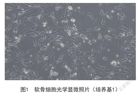
1.2.1 软骨细胞的培养基的配制 培养基1:基础培养基+10%胎牛血清(对照)。培养基2:培养基1+芦荟大黄素。培养基3:培养基1+咖啡酸。培养基4:培养基2+咖啡酸。培养基2-1、2-2、2-3分别为基础培养基+6%胎牛血清+40 μmol/L芦荟大黄素、60 μmol/L芦荟大黄素、80 μmol/L芦荟大黄素。
1.2.2 软骨细胞的制备方法 使用95%乙醇消毒处理人关节软骨组织10 min,先在37 ℃的温度下充分利用0.5%胰蛋白酶进行处理2 h,使用磷酸缓冲液洗涤,再使用0.08% Ⅱ型胶原酶进行消化;在培养基1中离心洗涤,将原代人软骨细胞获取。
1.2.3 Wnt-5A蛋白表达检测 采用Western-blot法:定量蛋白,使用BCA蛋白浓度测定试剂盒进行操作。TBST洗膜后将兔抗人Wnt-5A、tublin一抗(1∶1000稀释)加入,在4 ℃的温度下过夜杂交,洗膜后再将二抗(1∶3000稀释)加入,室温杂交1 h,洗膜后以ECL化学发光,暗室拍照,以tublin为内标蛋白,从而计算蛋白表达水平。
2 结果
2.1 不同浓度细胞增殖制剂对软骨细胞生长的影响 将相应浓度的芦荟大黄素及咖啡酸添加培养基中能够将有利条件提供给培养软骨细胞,见图1、图2、图3。
2.2 同时添加两种增殖制剂对软骨细胞生长的影响 将相应浓度的芦荟大黄素及咖啡酸同时添加在培养基中的增殖效果更优,见图4、图5。

[8] 樊薰勤,张明勇,李雯婷,等.sox-9通过Wnt3a/β-catenin通路促进大鼠骨髓间充质干细胞分化[J].中国骨质疏松杂志,2021,27(8):1169-1173.
[9] Chawla S,Mainardi A,Majumder N,et al.Chondrocyte Hypertrophy in Osteoarthritis:Mechanistic Studies and Models for the Identification of New Therapeutic Strategies[J].Cells,2022,11(24):4034.
[10] Shen H,He Y,Wang N,et al.Enhancing the potential of aged human articular chondrocytes for high-quality cartilage regeneration[J].FASEB J,2021,35(3):e21410.
[11] Thielen NGM,Neefjes M,Vitters EL,et al.Identification of Transcription Factors Responsible for a Transforming Growth Factor-β-Driven Hypertrophy-like Phenotype in Human Osteoarthritic Chondrocytes[J]. Cells,2022,11(7):1232.
[12] Riegger J,Brenner RE.Pathomechanisms of Posttraumatic Osteoarthritis:Chondrocyte Behavior and Fate in a Precarious Environment[J].Int J Mol Sci,2020,21(5):1560.
[13] Lauer JC,Selig M,Hart ML,et al.Articular Chondrocyte Phenotype Regulation through the Cytoskeleton and the Signaling Processes That Originate from or Converge on the Cytoskeleton:Towards a Novel Understanding of the Intersection between Actin Dynamics and Chondrogenic Function[J].Int J Mol Sci,2021,22(6):3279.
[14] Collins JA,Arbeeva L,Chubinskaya S,et al.Articular chondrocytes isolated from the knee and ankle joints of human tissue donors demonstrate similar redoxregulated MAP kinase and Akt signaling[J].Osteoarthritis Cartilage,2019,27(4):703-711.
編辑 扶田
