Huoxue Jiedu Huayu recipe (活血解毒化瘀方) alleviates contralateral renal fibrosis in unilateral ureteral obstruction rats by inhibiting the transformation of macrophages to myofibroblast
2023-02-15XIONGYunzhaoLIULingjinLIUZiqianCHENGegeHAOJuanGAOXiaomengQIANGPanpanWANGZhengXUQingyou
XIONG Yunzhao,LIU Lingjin,LIU Ziqian,CHEN Gege,HAO Juan,GAO Xiaomeng,QIANG Panpan,WANG Zheng,XU Qingyou
XIONG Yunzhao,Graduate School,Hebei University of Chinese Medicine;Hebei Key Laboratory of Integrative Medicine on Liver-Kidney Patterns,Hebei University of Chinese Medicine;Department of Internal Medicine,Hebei University of Chinese Medicine,Shijiazhuang 050091,China
LIU Lingjin,LIU Ziqian,CHEN Gege,HAO Juan,GAO Xiaomeng,QIANG Panpan,Graduate School,Hebei University of Chinese Medicine;Hebei Key Laboratory of Integrative Medicine on Liver-Kidney Patterns,Hebei University of Chinese Medicine,Shijiazhuang 050091,China
WANG Zheng,XU Qingyou,Graduate School,Hebei University of Chinese Medicine;Hebei Key Laboratory of Integrative Medicine on Liver-Kidney Patterns,Hebei University of Chinese Medicine;Department of Internal Medicine,Hebei University of Chinese Medicine,Shijiazhuang 050091,China
Abstract OBJECTIVE: To investigate the action and underlying mechanisms of Huoxue Jiedu Huayu recipe (活血解毒化瘀方,HJHR) against unilateral ureteral obstruction(UUO)-induced injury in the contralateral kidney.METHODS: Forty-eight male Sprague-Dawley rats weighing (200 ± 10) g were used in this study and randomly assigned to 4 groups: a sham group,a UUO group,a UUO+eplerenone (EPL) group,and a UUO +HJHR group.The contralateral kidneys were harvested for further study 180 d after surgery.Histological analysis,immunohistochemistry and immunofluorescence were used to study the fibrosis of the contralateral kidneys obtained from UUO rats.Contralateral kidney damagerelated pathway proteins were detected by real-time polymerase chain reaction and Western blot analysis.RESULTS: HJHR significantly inhibited fibrosis of the contralateral kidney in UUO rats by attenuating the UUOinduced macrophage-to-myofibroblast transition (MMT)in the contralateral kidney.Moreover,HJHR attenuated fibrosis in the contralateral kidney of UUO rats by preventing MMT through the aldosterone/mineralocorticoid receptor/serum/glucocorticoid regulated kinase 1 pathway.CONCLUSIONS: Our findings suggest that HJHR may be a potential treatment for renal interstitial fibrosis of obstructive nephropathy.
Keywords: ureteral obstruction;renal fibrosis;aldosterone;receptors,mineralocorticoid;macrophages;myofibroblast;Huoxue Jiedu Huayu recipe
1.INTRODUCTION
Chronic kidney disease (CKD) is a public health challenge,as it has a prevalence of 11% and is the 14thleading cause of death worldwide.1Persistent chronic inflammation and fibrosis product accumulation aggravate tubulointerstitial fibrosis (TIF),leading to the progression of chronic kidney disease.2One of the major causes of CKD is obstructive nephropathy.Obstructive nephropathy is a relatively common entity that is treatable and often reversible.In recent decades,researchers have shown that aldosterone (ALD) plays a major role in the obstructed kidney.In addition to its epithelial effect on sodium retention and potassium excretion in the distal tubule,aldosterone promotes inflammation and fibrosis in the heart,kidneys,and blood vessels.3,4
Mineralocorticoid receptor (MR) is widely expressed in the cardiovascular system and is a major determinant of endothelial function,smooth muscle tone,vascular remodeling,fibrosis,and blood pressure.An important new dimension is the appreciation of the role MR plays in immune cells and target organ damage in the heart,kidney and vasculature and in the development of insulin resistance.5Accumulating evidence indicates protective actions of mineralocorticoid antagonists (MR antagonists) on cardiovascular pathology,which includes blunting vascular inflammation and myocardial fibrosis.6,7Aldosterone also has effects on kidney function.An aldosterone infusion can counteract the beneficial effects of treatment with angiotensin-converting-enzyme inhibitors,causing more severe proteinuria and an increased number of vascular and glomerular lesions;treatment with aldosterone antagonists can reverse these alterations.8
It is well accepted that myofibroblasts are the major collagen-producing cell type during active fibrosis,but the origin of myofibroblasts continues to be a subject of intense debate.9,10Recent studies in the mouse model of unilateral ureteral obstruction (UUO) and in patients with progressive CKD have shown that bone marrow-derived monocytes/macrophages are capable of transition into myofibroblasts as identified by coexpression ofmacrophage markers (F4/80 and CD68) and α-smooth muscle actin (α-SMA) in conjunction with production of collagen I.These observations suggest that macrophageto-myofibroblast transition (MMT) may be another pathway leading to myofibroblast accumulation during renal fibrosis.11,12
UUO rapidly leads to acute kidney injury in the obstructed kidney,but in the chronic phase,both the obstructed kidney and contralateral kidney exhibit fibrosis leading to progressive CKD.Moreover,our group has reported that in UUO,the activation of MR by elevated aldosterone in plasma not only participates in renal injury and fibrosis in the obstructed kidney but also contributes to fibrosis in the contralateral kidney,exacerbating CKD and renal failure.13MR signaling is a pivotal pathway in renal fibrosis.This is supported by the finding that UUO induces fibrosis and proliferation in the contralateral kidney and MR plays an important role through cell proliferationviathe MR/serum/glucocorticoid regulated kinase 1 (SGK-1) and extracellular regulated protein kinase (ERK)/nuclear factor kappa-B(NF-κB) pathways.13In particular,we recently reported that MR/SGK-1 pathway is involved in the mechanism of MMT in chronic contralateral kidney injury in UUO,which mediates interstitial fibrosis and eventually leads to CKD and renal failure.This process was prevented by treatment with eplerenone.14Huoxue Jiedu Huayu recipe(活血解毒化瘀方,HJHR) consists of Huangqi (Radix Astragali Mongolici) 20 g,Dilong (Pheretima Aspergillum) 10 g,Biejia (Carapax Trionycis) 10 g,Chishao (Radix Paeoniae Rubra) 10 g,Huangqin (Radix Scutellariae) 10 g.This recipe comes from Zhao Yuyong's theory of pathogenesis of “blood stagnation in kidney meridian”.On basis of years of clinical experience,combining TCM theory with the pathology and clinical signs and symptoms of chronic renal disease in modern medicine,Professor Zhao Yuyong raised the pathogenesis of chronic kidney disease as ‘blood stagnation in kidney meridian’,which had important clinical significance.15Our previous studies have shown that HJHR ameliorates mesangial cell pyroptosis in contralateral kidney of UUO rats.16However,whether HJHR also attenuates fibrosis in the contralateral kidney in obstructive nephropathy and by what mechanism are important questions that remain to be answered.
In this study,we hypothesized that HJHR attenuates chronic fibrosis in the contralateral kidneys of UUO rats by suppressing the macrophage-to-myofibroblast transition through inhibition of the ALD/MR/SGK-1 signaling pathways.
2.METHODS
2.1.HJHR preparation
HJHR consists of Huangqi (Radix Astragali Mongolici)20 g,Dilong (Pheretima Aspergillum) 10 g,Biejia(Carapax Trionycis) 10 g,Chishao (Radix Paeoniae Rubra) 10 g,Huangqin (Radix Scutellariae) 10 g,and the crude drug at 1 g/mL liquid.These herbs were purchased from Shijiazhuang Lerentang Pharmaceutical Co.,Ltd.,and the herbs were identified and decocted in water.16
2.2.Animals and in vivo experimental models
Male Sprague-Dawley rats [approximately 7 weeks old,body weight (200 ± 10) g] were purchased from Hebei Medical University Animal Center for this study.The rats were maintained with standard rat chow and tap water at room temperature under a 12-h light/12-h dark cycle.Animal care followed the criteria of the Ethics Committee on Animal Experimentation of Hebei Medical University.Forty-eight SD rats were randomly assigned to the sham group,UUO group,UUO with eplerenone treatment group (UUO+EPL,n=12 each),and UUO with Huoxue Jiedu Huayu recipe treatment group (UUO+HJHR,n=12 each).UUO and sham surgeries were performed as described previously.17After surgery,eplerenone (Pfizer,USA) was given to the UUO+EPL groupviathe diet at a dose of 1.25 g/kg diet(equal to 100 mg·kg-1·d-1) for 6 months.The UUO+HJHR group was intragastrically administered 11.7 g·kg-1·d-1HJHR for 6 months,and the other groups of rats were fed regular chow.Then,180 d after UUO,all of the animals were sacrificed,and the kidneys on the contralateral side of UUO and from the sham surgeries were collected for histological and protein analysis.
2.3.Histological analysis,immunohistochemistry and immunofluorescence
The contralateral kidneys were dehydrated with alcohol and embedded in paraffin blocks after fixation overnight in 4% paraformaldehyde (PFA).The paraffin blocks were cut into 5 μm sections for HE,Masson,Sirius red staining and immunohistochemistry for α-SMA (Abcam,Shanghai,China),vimentin (Abcam,Shanghai,China),SGK-1 (Abcam,Shanghai,China) and neutropil gelatinase-associated lipocalin (NGAL) (AB clonal,Wuhan,China).Light images were observed and imaged using a Leica BX53 optical microscope.For fluorescent staining,the kidneys were irrigated with 4% PFA,dehydrated in 30% sucrose solution and frozen in OCT compound.Kidney sections (7 μm) were cut using a freezing microtome and prepared for staining with the following fluorescent-conjugated antibodies: Alexa Fluor 555-conjugated α-smooth muscle actin (Abcam,Shanghai,China),FITC-conjugated F4/80 (EterLife,Tianjin,China) or unconjugated antibodies anticollagen-III (Col-I),anti-CD68,anti-inducible nitric oxide synthase (iNOS),anti-CD206,anti-macrophage colony-stimulating factor (M-CSF) and anti-NR3C2 (all from Abcam,Shanghai,China),followed by fluorescent secondary staining.After staining,the sections were incubated with 4,6-diamino-2-phenyl indole (DAPI) for nuclear staining and sealed for photography using a Leica SP8 confocal microscope.
HE staining sections were examined by two nephronlogists and scored from 0 to 3 (normal to severe).Other image quantifications were performed using Image J or Image-Pro Plus 6.0 software.18
2.4.Protein extraction and Western blot analysis
The contralateral kidneys were homogenized in radio immunoprecipitation assay (RIPA) lysis buffer for protein extraction.The protein samples were subjected to Western blotting with sodium dodecyl sulfate polyacrylamide gel electrophoresis (SDS-PAGE) and polyvinylidene fluoride film (PVDF) membranes.After blocking nonspecific binding with 5% nonfat milk,the membranes were incubated with primary antibodies against NF-κB,M-CSF,α-SMA,vimentin,SGK-1 and NGAL (all from Abcam,Shanghai,China) at a 1∶500-1∶1000 dilution overnight at 4 ℃.The next day,the blots were incubated with fluorescein-conjugated secondary antibodies for 1 h at room temperature and scanned with an Odyssey Infrared Imaging System (LICOR,Lincoln,NE,USA).β-actin,glyceraldehyde-3-phosphate dehydrogenase (GAPDH) and β-tubulin antibodies were used for normalization of the protein loading.
2.5.Reverse transcription and quantitative real-time polymerase chain reaction (PCR)
Total RNA was isolated using the EZNA Total RNA Kit II (Omega,Biotek,Norcross GA,USA) following the protocol recommended by the manufacturer.cDNA was used as a template for real-time PCR with MonAmp ChemoHS qPCR Mix (Monad Biotech Co.,Ltd.,Shanghai,China).The mRNA levels of SGK-1,NF-κB,monocyte chemoattractant protein-1 (MCP-1),transforming growth factor-β1 (TGF-β1) and GAPDH were detected by the ABI 7500 FAST System (Applied Biosystems,Lincoln Center Drive Foster City,CA,USA)using the previously described primers.All primers were supplied by Sangon Biotech (Shanghai,China).GAPDH was used as a housekeeping transcript for normalization,and relative real-time PCR results were calculated using the 2-ΔΔCTmethod.
2.6.Aldosterone measurement by radioimmunoassay
Samples and calibrators were incubated with125I-labeled aldosterone (Tianjin Jiuding Biological Engineering Co.,Tianjin,China),as a tracer in antibody-coated tubes.After incubation,the bound radioactivity was measured using an FJ-2021r RIA counter.
2.7.Statistical analysis
All data are expressed as the mean ± stand error of mean.Statistical analysis was performed using GraphPad Prism 5 (GraphPad Software Inc.,San Diego,CA,USA).The statistical comparisons between groups were determined using Student’st-test for two groups or one-way analysis of variance followed by Tukey’s post hoc tests for multiple groups,withPvalue < 0.05 considered statistically significant.
3.RESULTS
3.1.HJHR protects the contralateral kidney from UUOinduced chronic fibrosis
UUO has emerged as an important model for the study of the mechanisms of renal fibrosis and for the evaluation of the impact of potential therapeutic approaches to ameliorate renal disease.17To investigate the pathological changes,we performed UUO on Sprague-Dawley rats and harvested their contralateral kidneys at day 180.HE staining confirmed that long-term UUO induces injury in the contralateral kidney by revealing dilated distal tubules and cast formation.Moreover,Masson staining showed significantly obvious collagen deposition and fibrosis in the contralateral kidneys of long-term UUO rats compared to sham kidneys (Figure 1,Figure S1).
Similar results were also observed with Sirius red staining for collagens in these kidneys (Figure S2).Collagen type I (Col I) and collagen type III (Col III)accumulation are early events during renal fibrosis.The levels of collagen I and collagen III,major components of the extracellular matrix,lead to renal fibrosis and reduced renal function.19Hence,collagen is an important protein marker of renal fibrosis.To test the relationship between HJHR and the levels of collagen I and collagen III,we used three-color confocal microscopy to identify whether HJHR attenuates the development of interstitial fibrosis in the contralateral kidney of UUO rats.We found that the expression of collagen I/III in the HJHR and EPL groups was significantly reduced compared with that in the UUO group (Figure S2),indicating that HJHR alleviates fibrosis in the contralateral kidneys of UUO rats,which is closely related to MMT.
It was reported that plasma aldosterone is increased in the UUO model,13which was confirmed in our study.Notably,long-term treatment with HJHR and eplerenone of UUO rats protected their contralateral kidneys from chronic fibrosis.We have found that MR activation is involved in the development of chronic renal fibrosis in the contralateral kidneys of UUO rats (Figure S3).These findings suggest that HJHR attenuated massive fibrosis in the contralateral kidneys of UUO rats,indicating that the mechanism of HJHR may be related to blocking MR.
3.2.HJHR attenuated UUO-induced MMT in the contralateral kidney
MR activation promotes the development of renal fibrosis.20Renal fibrosis leads to kidney destruction and CKD.To examine the role of myofibroblasts in chronic injury of the contralateral kidney of long-term UUO rats,we stained these kidneys with antibodies against α-SMA and vimentin (Figure 1),specific markers used to identify myofibroblasts.Meanwhile,western blot was used to analyze the expression of SMA and vimentin.Consistent with the pathology observations,the expression of α-SMA and vimentin also increased in long-term UUO rats compared with sham rats.These effects were prevented by treatment with HJHR (Figure S4).
Monocytes/macrophages are involved in the process of renal injury,repair and fibrosis.Recent studies have demonstrated that aldosterone induces renal fibrosis and inflammatory macrophagesviaMR in rats.21Moreover,the macrophage-to-myofibroblast transition contributes to interstitial fibrosis in chronic renal injury.22To investigate whether macrophages are involved in HJHRalleviated UUO-induced renal fibrosis in the contralateral kidneys of UUO rats,we stained the UUOcontralateral kidneys with multiple antibodies against collagen III,F4/80 (macrophage-specific marker) and α-SMA.As expected,cells coexpressing F4/80 and α-SMA were surrounded by collagen-III,suggesting the production of collagen-III from MMT cells (Figure S5).Moreover,we explored subtypes of macrophages contributing to MMT by co-staining for α-SMA and the macrophage subfamily marker CD206 (M2 macrophages)or iNOS (M1 macrophages).The results further revealed that M2 macrophages are the major contributor to MMT in the contralateral kidneys of long-term UUO rats(Figures 2,3),which is consistent with a previous study using UUO kidneys.22

Figure 1 Effects of long-term UUO and treatment with Huoxue Jiedu Huayu recipe on renal histology and fibrosis of the contralateral kidneys
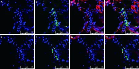
Figure 2 M2 macrophages are the major source of MMT in the contralateral kidneys of long-term UUO rats
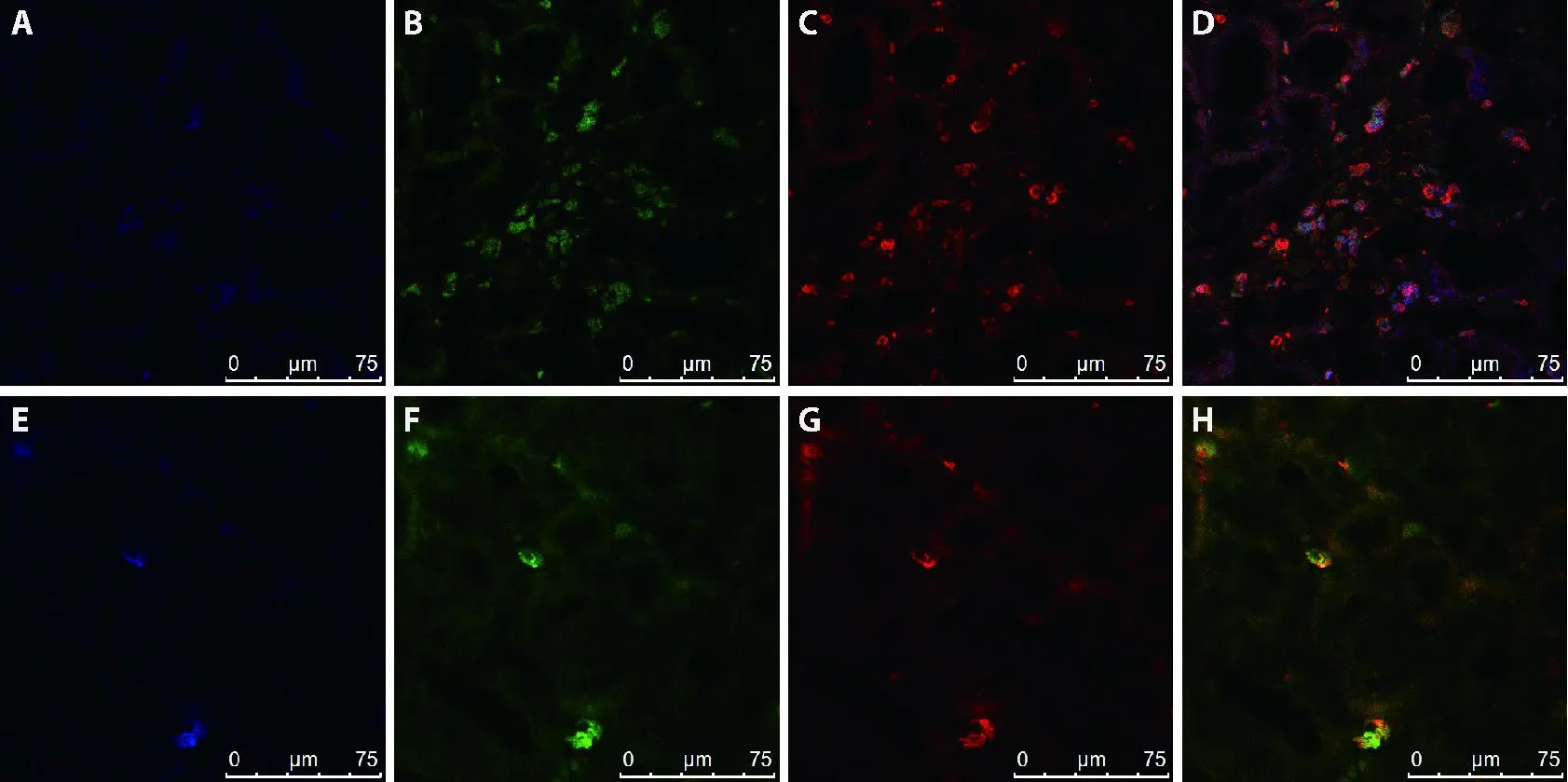
Figure 3 M2 macrophages are the major source of MMT in the contralateral kidneys of long-term UUO rats
As shown in Figures,the data suggest that a significant proportion of myofibroblasts is increased in response to MMT from renal infiltrated macrophages,contributing to collagen production and subsequently developing renal fibrosis.Moreover,these effects are inhibited by HJHR,leading us to speculate that HJHR alleviates fibrosis in the contralateral kidneys of UUO rats,which is closely related to MR activation and MMT.
3.3.HJHR attenuates fibrosis in the contralateral kidney of UUO rats by preventing MMT through the ALD/MR/SGK-1 pathway
The findings above revealed that HJHR blocking MR protects against MMT and interstitial fibrosis in the contralateral kidneys of UUO rats.However,the mechanism is unclear.Previous data have indicated that SGK-1 plays an important role during renal fibrosis.23To verify our hypothesis that HJHR attenuates fibrosis in the contralateral kidney of UUO rats by preventing MMT through the ALD/MR/SGK-1 pathway,immune-ohistochemical staining (Figure S6) and Western blot (Figure 4) was performed to identify the expression of SGK-1 and NGAL.The data showed higher expression of SGK-1 and NGAL in the contralateral kidneys of UUO rats than in the HJHR and EPL groups,suggesting that HJHR attenuates fibrosis in the contralateral kidneys of UUO rats,and the mechanism is related to the SGK-1 pathway.In addition,previous data have indicated that F4/80 plays an important role during contralateral renal fibrosis.13Immunofluorescence was performed to identify whether HJHR regulates F4/80 and M-CSF expression in the contralateral kidneys of UUO rats.There were many F4/80 and M-CSF cells in UUO.However,their expression was substantially inhibited in the HJHR group(Figure S7).

Figure 4 Effects of long-term UUO and treatment with Huoxue Jiedu Huayu recipe on the expression of MR downstream molecules
In the current study,we further confirmed the effect of HJHR in the contralateral kidneys of UUO rats by detecting macrophage colony stimulation (M-CSF) and molecules downstream of MR by performing western blot analysis.Consistent with our previous findings,14we found overexpression of M-CSF and NF-κB in the contralateral kidneys of UUO rats compared to the contralateral kidneys from sham rats,and these effects were inhibited by HJHR and the MR blocker eplerenone(Figure 4) .Consistent with the western blot results,realtime PCR analysis confirmed that the mRNA expression levels of SGK-1,NF-κB,MCP-1 and TGF-β1 in the contralateral kidney homogenates were significantly enhanced in the UUO group compared to the sham group(Table 1).However,their mRNA expression levels were significantly downregulated in the HJHR and EPL groups compared to the UUO group,indicating that HJHR attenuates fibrosis in the contralateral kidney of UUO rats,which is related to the ALD/MR/SGK-1 pathway.
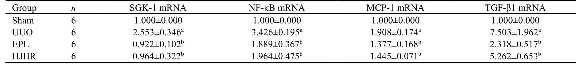
Table 1 Expression of SGK-1,NF-κB,MCP-1 and TGF-β1 mRNA in kidney tissue of UUO rats
Taken together,our results suggest that in the chronic phase of obstructive kidney disease,activated MR stimulates MMT,causing renal infiltrated macrophages to transdifferentiate into myofibroblasts,producing excessive collagen and exacerbating renal fibrosis in the contralateral kidney.Moreover,HJHR diminishes MMT and protects the contralateral kidney from fibrosis and further chronic renal injury.HJHR attenuates fibrosis in the contralateral kidney of UUO rats by preventing MMT through the ALD/MR/SGK-1 pathway.
4.DISCUSSION
Obstructive nephropathy is one of the most important causes of renal failure in infants and children.24UUO is a classic model in rodents to study obstructive kidney disease and renal fibrosis.In recent years,a growing body of evidence has shown that obstructive kidney injury-induced CKD or renal failure are related to both the obstructed kidney and the unobstructed contralateral kidney.25Our research confirmed that eplerenone attenuates fibrosis in the contralateral kidneys of UUO rats by preventing the macrophage-to-myofibroblast transition.Thus,in this experiment,our study aimed to investigate the effects of HJHR on inflammation and fibrosis in the contralateral kidneys of UUO rats with the ultimate goal of improving therapeutic strategies to help achieve better outcomes for patients suffering from obstructive kidney disease.
In this study,we found that HJHR efficiently prevented chronic development of fibrosis in the contralateral kidney of UUO rats and thereby protected the contralateral kidney from further damage.The mechanism of MR activation in tissue injury is still not clear.26By co-staining with macrophage and myofibroblast markers,our data further elucidated that attenuating MMT is an important mechanism by which HJHR prevents the accumulation of myofibroblasts in the contralateral kidneys of UUO rats.
We also found that HJHR attenuates fibrosis in the contralateral kidney of UUO rats by preventing MMT through the ALD/MR/SGK-1 pathway.Aldosterone acts on renal vessels and renal cells,inducing SGK-1 expression and activating nuclear factor kappa B (NFκB),to further promote cell proliferation and apoptosis.27SGK-1 has been shown to be upregulated in fibrotic tissue,including that associated with nephropathy.28SGK-1 is usually barely expressed in kidney tissue;however,its expression dramatically increases under certain pathophysiological conditions,such as glucocorticoid or mineralocorticoid excess.29Our studies suggest that NF-κB contributes to renal fibrosis and plays an important role in macrophage-to-myofibroblast transition-induced fibrosis of the contralateral kidney.Our data demonstrated that aldosterone stimulates macrophages to express higher levels of α-SMA,indicating enhanced MMT,and these effects are significantly blocked by HJHR.Similar results were observed by western blot and real-time PCR analyses.Holistic concepts are the central tenants of Traditional Chinese Medicine (TCM) to understand itself and the connections between humans and the environment.Therefore,kidney disease often affects other viscera and spreads.According to TCM,the pathogenesis of chronic kidney disease is the origin deficiency and symptom evil.11In addition,toxin and blood stasis damaging kidney collaterals run through the whole process of chronic kidney disease,and the HJHR method is the basic principle of TCM in treatment.In this prescription,Dilong (Pheretima Aspergillum) and Biejia (CarapaxTrionycis) dispel phlegm and dredge collaterals.Chishao(Radix Paeoniae Rubra) is used to remove blood stasis,Huangqin (Radix Scutellariae) is used to attack the toxicQi,and Huangqi (Radix Astragali Mongolici) is used to recover healthyQi.“Treatment before disease”highlights the preventive thinking of TCM to seize the opportunity to treat the disease early to prevent further development of the disease and cure the disease at the initial stage.
One limitation in this study is that we do not exactly know the molecular mechanism by which activated MR participates in macrophage-differentiation.One of the possible mechanisms for reducing MMT in the contralateral kidney of UUO rats is that the treatment with HJHR decreased renal infiltration or accumulation of macrophages.However,how HJHR reduces the accumulation of MMT in the contralateral kidney and which drug plays a more important role remains unclear.Therefore,future study will be needed to further address the precise molecular mechanisms of MR stimulating MTT.
In conclusion,protection against renal fibrosis is important for the management of obstructive nephropathy.We researched the roles and possible mechanism of HJHR in renal interstitial fibrosis,which may provide a potential endogenous target for renal interstitial fibrosis in obstructive nephropathy.Our data suggest that HJHR attenuates renal interstitial fibrosis in obstructive nephropathy by suppressing the macrophage-tomyofibroblast transition through inhibition of the ALD/MR/SGK-1 signaling pathways.
猜你喜欢
杂志排行

Journal of Traditional Chinese Medicine的其它文章
- Potential of natural medicines for treatment of osteoporosis: a narrative review
- Outlook on cultural inheritance and development
- Upholding fundamental principles and breaking new ground: ensuring positive efficacy and reaching new consensus
- General principle of high-quality academic development of traditional chinese medicine: “carrying on the essence,while pursuing innovations”
- Effects of moderate-intensity aerobic exercise combined with acupuncture on attention function of mentally-retarded adolescents: a randomised controlled trial
- Reveal the mechanisms of prescriptions for liver cancer' treatment based on two illustrious senior TCM physicians