A new species of Pararhizomys (Tachyoryctoidinae,Muroidea) from Linxia Basin of Gansu Province
2022-11-05WANGBanYue
WANG Ban-Yue
(Institute of Vertebrate Paleontology and Paleoanthropology, Chinese Academy of Sciences Beijing 100044 wangbanyue@ivpp.ac.cn)
Abstract An anterior part of skull was recently found near Xiayangwan in Guanghe County,Gansu Province, presumably from the Liushu Formation. The skull represents a new species of Pararhizomys, named as Pararhizomys parvulus. The new species is characterized by: small size,upper molars higher crowned and mesio-lingually hypsodont with sinus deeper than mesosinus;sinus and mesosinus in M3 being transverse and overlapping each other, but sinus longer than mesosinus on occlusal view. Based on shared apomorphies (lingually hypsodont upper molars and transverse sinus and mesosinus on M3 occulsal surface), P. parvulus and P. huaxiaensis are supposed to form a sister group. However, P. parvulus may be more derived than P. huaxiaensis as demonstrated by the more hypsodont molars and the deeper sinus in M3.
Key words Linxia Basin, Gansu; Late Miocene; Liushu Formation; Pararhizomys,Tachyoryctoidinae
Pararhizomysis one of the Late Miocene–Early Pliocene endemic rodent groups in the middle latitudes of East Asia. The genus was established by Teilhard de Chardin and Young (1931).The type species,Pararhizomyshipparionum, was based on a left mandible with m1–3 from the Late Miocene “Pontian Red Clay” (= Baodean ALMA) in Shaanxi Province. Later, more material including skulls and mandibles from Mongolia and Nei Mongol of China have been referred to it(Kowalski, 1968; Li, 2010). Recently, four species (P. hipparionum,P. qinensis,P. huaxiaensis,andP. longensis) andPararhizomysspp. were reported from Late Miocene of northern China(Zhang et al., 2005; Qiu and Li, 2008; Li, 2010; Wang and Qiu, 2018).
In 2018, while prospecting the Xiayangwan area in Guanghe County of the Linxia Basin,a field team of Northwest University found an anterior part of skull ofPararhizomyson surface of the deposits of the middle part of the Liushu Formation. Dr. Xie Kun, one of the Northwest University team members, kindly entrusted the skull ofPararhizomysto IVPP for identification and further study. Our study shows that the skull represents a new species ofPararhizomys,here named asPararhizomysparvulus. The discovery of this new species provides more information on the nature and phylogeny of the genusPararhizomys. For the terminology and measurements of the skull and cheek teeth, Wang and Qiu (2018) are followed.
Holotype IVPP V 26661, anterior part of a skull with two I2s, left M1–3 and right M1–2, found from the surface of the Liushu Formation near Xiayangwan, Guanghe County(Field Locality: E 103°31′55.54″, N 35°27′25.76″, H 2091 m above sea level).
Age Possible middle Late Miocene.
Diagnosis Small-sizedPararhizomys; molars being higher crowned than four known species, mesially and lingually hypsodont; M1–3 having sinus deeper than the mesosinus vertically; in M3 sinus and mesosinus transverse and overlapping each other but sinus is wider than mesosinus transversely.
EtymologyParvulus, Latin, little.
Description Skull: the skull is small in size among the muroid rodents with myomorphous zygomasseteric structure. The length of the anterior part of skull (length from anterior end of premaxilla to anterior margin of M1) is about 13.2 mm.
Dorsal view (Fig. 1E): The nasal (N) is mostly preserved, but its anterior end is lost.Fortunately, the print of the anterior part of the nasal is well preserved to show its anterior boundary. The nasal is rather wide and wedge-shaped, gradually narrowing posteriorly, with the posterior end pointed. The posterior end lies roughly in the same transverse line with the posterior ends of the premaxillo-maxillary sutures in position. The dorsal surface of the nasal is convex longitudinally, and slightly transversely. The transverse convexity is prominent on the anterior part, becoming flat on the posterior part. The premaxilla (Pm) is separated by the premaxillary laterodorsal crest (pmldc) into two parts: the long band-like dorsal part,which is slightly convex longitudinally and flat transversely, and a broad and slightly concave lateroventral part. The straight posterior part of the premaxillo-maxillary suture extends posteriorly to meet the frontal (F) to form obtuse angles with both the maxillo-frontal suture and the premaxillo-frontal suture. The maxillo-frontal suture extends posterolateraly to meet the small lacrimal bone (L), whereby forming a distinct lacrimal tubercle (lt).
Lateral view (Fig. 1C, D): The anterior end of the nasal is nearly in the same vertical line with that of the premaxilla (Pm). The infraorbital foramen (iof) lies within the maxilla. Both the left and right infraorbital foramina are poorly preserved, tending to show that they have an oval upper part and a ventral slit of infraorbital foramen (vsiof) below.
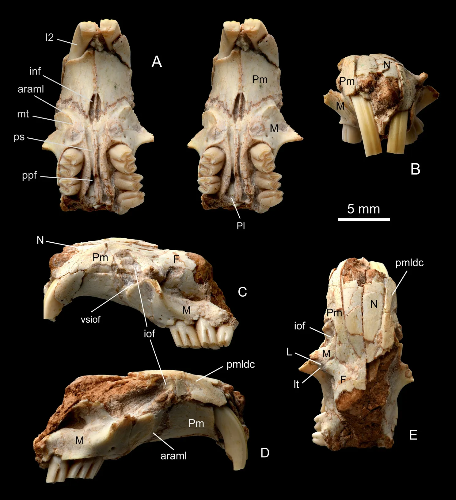
Fig. 1 Skull of Pararhizomys parvulus sp. nov. (IVPP V 26661, holotype)A. ventral view (stereopair); B. anterior view; C. left lateral view; D. right lateral view; E. dorsal view Foramina and other structures: araml. anterior rim of attachment area for m. masseter lateralis 外层咬肌附着区前缘;I2. second upper incisor 第二上门齿;inf. incisive foramen 门齿孔;iof. infraorbital foramen 眶下孔;lt. lacrimal tubercle 泪结节;mt. masseteric tubercle 咬肌结节;pmldc. premaxillary laterodorsal crest 前颌骨侧背嵴;ppf. posterior palatine foramen 腭后孔;ps. palatine sulcus 腭沟;vsiof. ventral slit of infraorbital foramen 眶下孔腹侧裂隙Bones: F. frontal; L. lacrimal; M. maxilla; N. nasal; Pl. palatine; Pm. premaxilla
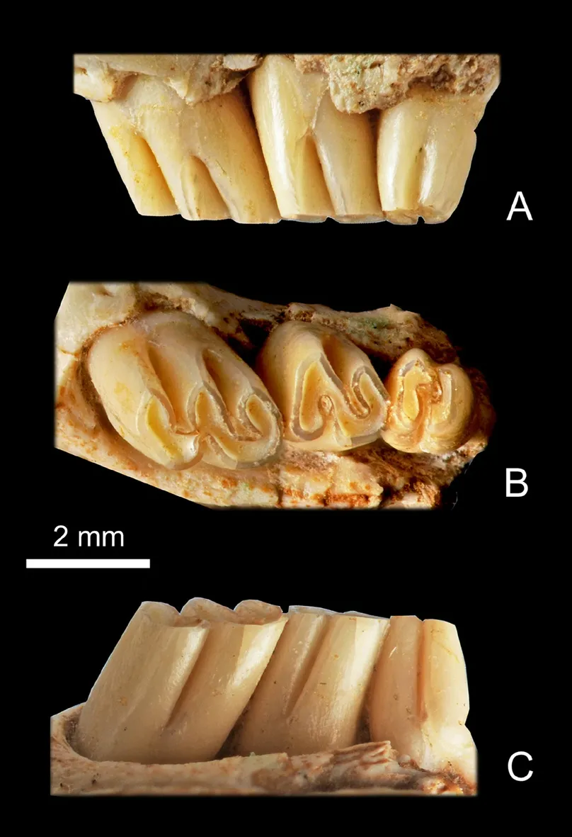
Fig. 2 Left M1–3 of Pararhizomys parvulus sp. nov.(IVPP V 26661, holotype)A. buccal view; B. occlusal view; C. lingual view
Ventral view (Fig. 1A): The left and right upper diastemata are both 10 mm long, longer than those of the molar rows. The two incisive foramina (inf) are both 3 mm long, about 3/10 length of the upper diastemata, and located in the middle of the diastemata. The ventral part of the premaxillo-maxillary suture extends mesioposteriorly, intersecting the posterior part of the incisive foramen. The zygomatic plate is located ventrolateral to the infraorbital foramen, expands anterolaterally and faces anterolaterally. The attachment area for them.masseter lateralison the zygomatic plate is broad and concave and has a distinct arched anterior rim (araml). The masseteric tubercle(mt) is located at the mesial end of araml,nearly in the same transverse line with the posterior end of inf. Between mt and M1 there is a bulge representing the posterior end of the alveolus of I2. The left and right molar rows are divergent posteriorly. Along the intermaxillary and interpalatine sutures the sagittal ridge extends from the space between the incisive foramina to the posterior border of palate. The two palatine sulci (ps)extend from the incisive foramen posteriorly,becoming deeper near the molar rows. A pair of posterior palatal foramina (ppf) is located in the palatine sulci, at the level mesial to M2.
Teeth: The upper dental formula is 1.0.0.3/. The molars are high crowned,mesially and lingually hypsodont with roots.The occlusal surfaces of the molars have 2 or 4 reentrants and 2 or 3 lophs. V 26661 is a young individual because all the molars are slightly worn and all the reentrants are open.
The M1 is the largest of the upper molars. The occlusal surface is round-angled rectangular in outline, longer than wide, with slightly longer buccal side than lingual one.There are two buccal reentrants (anterosinus and mesosinus) and two lingual ones (protosinus and sinus). The mesosinus is the longest reentrant transversely and its lingual end turns slightly posteriorly. The anterosinus extends nearly transversely. The sinus is slightly shorter transversely than the anterosinus and extends slightly antero-buccally towards the anterosinus.On the buccal side the anterosinus is deeper than the mesosinus vertically. The sinus on the lingual side is deeper than both the anterosinus and the mesosinus on the buccal side. On the buccal side the dentine tract (DT) is M-shaped with the two sharp tops reaching to the bottoms of the anterosinus and mesosinus. The protosinus is a very shallow groove on the occlusal view, but vertically deeper than the sinus on the anteromesial wall of M1.
The M2 is trapezoid in occlusal view, slightly wider than long, with a convex posterior side. It has one buccal reentrant (mesosinus) and one lingual reentrant (sinus). The mesosinus is transversely longer than the sinus and extends posterolingually. The sinus extends transversely in front of the mesosinus. It is deeper than the mesosinus on the lateral side. On the buccal side the sharp top of DT reaches to the bottom of the mesosinus.
The M3 is smaller than M2 in size. It is subquadrate in occlusal view, with a slightly narrower posterior side. Like M2, it also has one buccal reentrant (mesosinus) and one lingual one (sinus), and the sinus is also deeper than the mesosinus vertically. On the occlusal view the mesosinus and sinus extend transversely, but the sinus is longer than the mesosinus in width.Because the sinus lies slightly more anterior than the mesosinus, the two reentrants overlap and join each other.
The I2 is orthodont, bending strongly with its anterior end turning ventrally. Its posterior end originates from the maxilla in front of the M1. The cross section of I2 is triangular in form,with a rounded lingual angle and an almost flat labial side. The enamel covers the whole labial side, less so on the medial and lateral sides. On the labial side there is a prominent longitudinal ridge, which is located nearer to the medial side than to the lateral one.
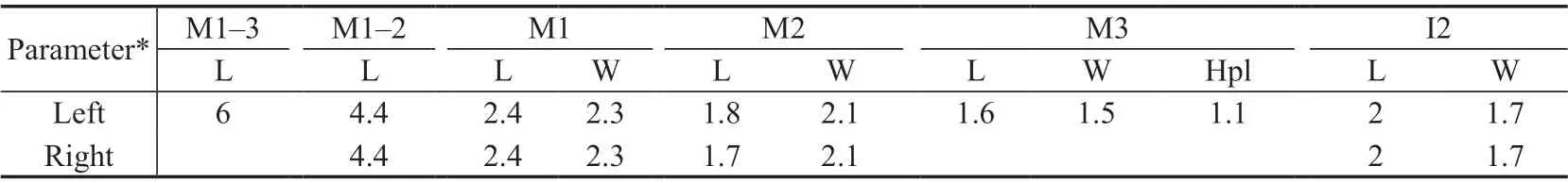
Table 1 Measurements of teeth of Pararhizomys parvulus sp. nov. (IVPP V 26661) (mm)
Comparison and discussion The anterior part of skull of IVPP V 26661 preserves the following diagnostic features of Pararhizomyini: the skull has myomorphous zygomasseteric structure; the nasal has a pointed posterior end, located nearly in the same transverse line with the posterior end of the premaxillo-maxillary suture; the premaxilla has a distinct premaxillary laterodorsal crest (pmldc), which is parallel to the anterior part of naso-premaxillary suture;the upper diastema is longer than the molar row; the zygomatic plate is broad and located ventrolateral to the infraorbital foramen (iof); the attachment area for lateral masseter muscle is confined to maxilla and bordered anteriorly by a distinct curved ridge (araml); upper molars are mesially hypsodont with roots and having simple occlusal feature, lacking posterosinus and M2–3 having one lingual reentrant (sinus), etc.
The Pararhizomyini is known to comprise two genera:PararhizomysTeilhard de Chardin& Young, 1931 andPseudorhizomysWang & Qiu, 2018. As described above, in V 26661 the nasal has a longitudinally convex dorsal side, its anterior end is in the same vertical line as the anterior end of the premaxillary; the dorsal part of the premaxilla bordered by pmdlc is long band-formed; the posterior part of the premaxillo-maxillary suture is straight and extends posteriorly to meet the frontal, forming an obtuse angle with the maxilla-frontal suture and its anterior part intersects the posterior part of the incisive foramen; I2 is orthodont, with triangular cross section and a well-developed longitudinal crest on labial side; three upper molars are mesially hypsodont; M2 and M3 having only one buccal reentrant (mesosinus),without anterosinus, etc. In all these features V 26661 is similar toPararhizomysrather than toPseudorhizomys.
The genusPararhizomysis now known to include four species:P. hipparionum,P.qinensis,P. huaxiaensisandP. longensis. In comparison with them V 26661 differs from all of them in being much smaller in size. The anterior part of the skull and the maxillary diastema are shorter than the half lengths of those ofP. hipparionumandP. huaxiaensis, or subequal to the half length ofP. longensis. The molars are only about 3/5 of the four species mentioned above in length.
Further, V 26661 differs fromP. hipparionum,P. qinensis, andP. longensisin upper molars being higher crowned and lingually hypsodont.
Furthermore, V 26661 differs fromP. hipparionumin M1–3 having sinus deeper than the buccal reentrants vertically; fromP. qinensisin M1–2 having deeper sinus than mesosinus;fromP. huaxiaensisin M3 having sinus deeper than the mesosinus and both reentrants overlap each other on occlusal view; fromP. longensisin M1–3 being more mesially and lingually hypsodont and M3 having transverse sinus and mesosinus.
As a result of the above comparison, it seems quite safe to establish a new species ofPararhizomys, here named asP. parvulus.
Wang and Qiu (2018:fig. 75) discussed the relationships between the four species ofPararhizomys. According to themP. longensismay represent the earliest and the first split branch ofPararhizomys, andP. huaxiaensisis the sister taxon ofP. hipparionum+P. qinensis.
In comparison with the four known species ofPararhizomys,P. parvulusshares withP. huaxiaensisin such apomorpies as M1–3 being both mesially and lingually hypsodont(character 4-2 in Wang and Qiu, 2018:128), and sinus and mesosinus of M3 being transverse(character 5-1). On the other hand, inP. parvulusthe molars are more hypsodont and M3 has overlapped sinus and mesosinus on occlusal surface and vertically deeper sinus than inP. huaxiaensis. All these tend to show thatP. parvulusmay represent a more progressive and specialized branch of theP. huaxiaensis+P. parvulussister group.
The locality where the skull ofPararhizomysparvuluswas collected lies above the locality where the Dashengou Fauna was collected. It seems thatP. parvulusmay be collected from the upper part of the middle Liushu Formation or slightly higher strata. Its age may be middle Late Miocene or slightly later.
AcknowledgmentsThe author would like to express her gratitude to Dr. Xie Kun of Northwest University for entrusting the specimen to IVPP for study, to Prof. Wang Shiqi of IVPP for transferring the skull to the author for further elaboration, and to Drs. Sun Boyang of IVPP and Wei Xiaohao of Northwest University for additional survey of the locality where the studied specimen was collected. Many thanks are given to the two reviewers for their careful review of the manuscript and valuable suggestion to improve the text of the paper. Many thanks are also given to Mr. Gao Wei for the photographs and Miss Si Hongwei for preparing the text pictures.
猜你喜欢
杂志排行

古脊椎动物学报(中英文)的其它文章
- Ulanodon, a new name for the Hyracodontid Ulania Qi,1990 (Perissodactyla, Mammalia)
- First discovery of dinosaur eggs in Nanhu Gebi of Hami,Xinjiang, China
- A Late Pliocene Hipparion houfenense fauna from Yegou,Nihewan Basin and its biostratigraphic significance
- Nothosaurus luopingensis sp. nov. (Sauropterygia)from the Anisian, Middle Triassic of Luoping,Yunnan Province, China